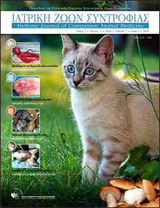
Contents from all issues of the Hellenic Journal of Companion Animal Medicine
- Volume 13 Issue 2 - 2024
- Volume 13 Issue 1 - 2024
- Volume 12 Issue 2 - 2023
- Volume 12 Issue 1 - 2023
- Volume 11 Issue 2 - 2022
- Volume 11 Issue 1 - 2022
- Volume 10 Issue 2 - 2021
- Volume 10 Issue 1 - 2021
- Volume 9 Issue 2 - 2020
- Volume 9 Issue 1 - 2020
- Volume 8 Issue 2 - 2019
- Volume 8 Issue 1 - 2019
- Volume 7 Issue 2 - 2018
- Volume 7 Issue 1 - 2018
- Volume 6 Issue 2 - 2017
- Volume 6 Issue 1 - 2017
- Volume 5 Issue 2 - 2016
- Volume 5 Issue 1 - 2016
- Volume 4 Issue 2 - 2015
- Volume 4 Issue 1 - 2015
- Volume 3 Issue 2 - 2014
- Volume 3 Issue 1 - 2014
- Volume 2 Issue 2 - 2013
- Volume 2 Issue 1 - 2013
- Volume 1 Issue 2 - 2012
- Volume 1 Issue 1 - 2012
-
Volume 13 Issue 2 - 2024
Editorial
Telemedicine as a vital support system for veterinary practice: Enhancing service quality and reducing practitioner stress
Dr. Magda Gerou-Ferriani DVM, CertSAM, DipECVIM-Ca, MRCVS EBVS and RCVS
Full Text HTML - Full Text PDF
Recogised Specialist in Small Animal Medicine Founder and CEO of Veterinary Specialist Advice -VSA-
Review
The gallbladder mucocele in dogs
Trikoili S. DVM, Companion Animal Clinic, School of Veterinary Medicine, Aristotle University of Thessaloniki | Angelou V. DVM, MSc, PhD, Companion Animal Clinic, School of Veterinary Medicine, Aristotle University of Thessaloniki | Konstantinidis A.O. DVM, MSc, PhD, Companion Animal Clinic, School of Veterinary Medicine, Aristotle University of Thessaloniki | Patsikas M. DVM, MD, PhD, Dip ECVDI, Laboratory of Diagnostic Imaging, School of Veterinary Medicine, Aristotle University of Thessaloniki | Papazoglou L.G. DVM, PhD, MRCVS, Companion Animal Clinic, School of Veterinary Medicine, Aristotle University of Thessaloniki
Full Text HTML - Full Text PDF
Review
Orthodontic techniques for the management of mandibular canine lingual displacement
Lorida O. DVM, PhD Candidate, Companion Animal Clinic, School of Veterinary Medicine, Faculty of Health Sciences, Aristotle University of Thessaloniki, Thessaloniki | Kotanidou E. DVM, MSc student, Companion Animal Clinic, School of Veterinary Medicine, Faculty of Health Sciences, Aristotle University of Thessaloniki | Gkiouzelis Ch. Undergraduate student of Veterinary Medicine, Aristotle University of Thessaloniki | Katsakouli P. N. Undergraduate student of Veterinary Medicine, Aristotle University of Thessaloniki | Papadimitriou S. DVM, DDS, PhD, Professor, Companion Animal Clinic, School of Veterinary Medicine, Faculty of Health Sciences, Aristotle University of Thessaloniki
Full Text HTML - Full Text PDF
Review
Interventional procedures for treatment of congenital cardiac disease in small animals
Mavropoulou A. DVM, Ms, PhD, MRCVS, Diplomate of the European College of Internal Medicine (Specialty of Cardiology), RCVS Recognized specialist in Cardiology, Plakentia Veterinary Clinic
Full Text HTML - Full Text PDF
Research/Clinical study
The specialty of Veterinary Dentistry in Greek companion animal practices
Fousekis A. DVM, Private practitioner | Panagiotou A. DVM, Private practitioner | Konstantarou E. DVM, Private practitioner | Kokkinou E. A. DVM, Private practitioner | Rizos S. Student, Faculty of Veterinary Medicine, Aristotle University of Thessaloniki, Greece | Filippitzi M. E. DVM, PhD, Assistant Professor, Faculty of Veterinary Medicine, Aristotle University of Thessaloniki, Greece | Papadimitriou S. DVM, DDS, PhD, Professor, Faculty of Veterinary Medicine, Aristotle University of Thessaloniki, Greece
Full Text HTML - Full Text PDF -
Volume 13 Issue 1 - 2024
Editorial
4th Industrial Revolution, Artificial Intelligence and Companion Animal Medicine
George Mantziaras DVM, PhD, ECAR resident, Private practitioner Athens, Greece
Full Text HTML - Full Text PDF
Οral Communications: Pathology
Association between serum cobalamin and folate concentrations and histopathological findings, duration of clinical signs and dysbiosis index in cats with chronic enteropathy and healthy cats
Moraiti K. PhD Student, Clinic of Medicine, Faculty of Veterinary Science, University of Thessaly, Karditsa | Karra D. PhD Student, Clinic of Medicine, Faculty of Veterinary Science, University of Thessaly, Karditsa | Newman S. DVM, DVSc, DACVP, Newman Specialty VetPath, Hicksville, NY, USA | Suchodolski J. Professor, Texas A&M University, Texas, USA | Steiner J. Distinguished Professor, Texas A&M University, Texas, USA | Xenoulis P. Associate Professor, Clinic of Medicine, Faculty of Veterinary Science, University of Thessaly, Karditsa and Adjunct Professor, Texas A&M University, Texas, USA
Full Text HTML - Full Text PDF
Οral Communications: Pathology
Relationship between serum Spec fPL and triglyceride concentrations in cats
Moraiti K. PhD Student, Clinic of Medicine, Faculty of Veterinary Science, University of Thessaly, Karditsa | Walker L. Undergraduated student, Texas A&M University, Texas, USA | Hung M. DVM, DACVIM, VCA Animal Specialty Group, San Diego, CA, USA | Steiner J. Distinguished Professor, Texas A&M University, Texas, USA | Xenoulis P. Associate Professor, Clinic of Medicine, Faculty of Veterinary Science, University of Thessaly, Karditsa and Adjunct Professor, Texas A&M University, Texas, USA
Full Text HTML - Full Text PDF
Οral Communications: Pathology
Cases report of idiopathic canine hyperlipidemia
Pitropaki M. DVM, MSc, PhD student, Clinic of Medicine, Faculty of Veterinary Medicine, University of Thessaly, Karditsa, Greece | Xenoulis P. Associate Professor, Clinic of Medicine, Faculty of Veterinary Science, University of Thessaly, Karditsa and Adjunct Professor, Texas A&M University, Texas, USA
Full Text HTML - Full Text PDF
Οral Communications: Pathology
Evaluation of fecal microbiota transplantation as adjunct management of cats with chronic enteropathy in a controlled, blinded, randomized clinical trial
Karra D. A. PhD Student, Clinic of Medicine, Faculty of Veterinary Science, University of Thessaly, Karditsa | Lidbury J.A. Associate Professor, Gastrointestinal Laboratory, Department of Small Animal Clinical Sciences, Texas A&M University, College Station, TX, USA | Suchodolski J. S. Professor, Texas A&M University, Texas, USA | Steiner J. Distinguished Professor, Texas A&M University, Texas, USA | Xenoulis P. Associate Professor, Clinic of Medicine, Faculty of Veterinary Science, University of Thessaly, Karditsa and Adjunct Professor, Texas A&M University, Texas, USA
Full Text HTML - Full Text PDF
Οral Communications: Pathology
Transient myocardial thickening in cats: 8 clinical cases
Pavlioudaki D. DVM, Plakentia Veterinary Clinic, Athens, Greece | Mylonidi T. DVM, Plakentia Veterinary Clinic, Athens, Greece | Mavropoulou A. DVM, Ms, PhD, MRCVS, Diplomate ECVIM-CA (Cardiology), RCVS recognised specialist in cardiology, Plakentia Veterinary Clinic, Athens, Greece
Full Text HTML - Full Text PDF
Οral Communications: Pathology
Presumptive secondary immune-mediated pancytopenia in a dog
Kouklaki E. DVM, MSc Student, Companion Animal Clinic, School of Veterinary Medicine, Aristotle University of Thessaloniki, Greece | Ninis S. DVM, Clinic of Companion Animal Medicine, School of Veterinary Medicine, Aristotle University of Thessaloniki, Greece | Ginoudis A. DVM, PhD Student, Diagnostic Laboratory, School of Veterinary Medicine, Aristotle University of Thessaloniki, Greece | Mylonakis M. DVM, PhD, Professor, Clinic of Companion Animal Medicine, School of Veterinary Medicine, Aristotle University of Thessaloniki, Greece
Full Text HTML - Full Text PDF
Οral Communications: Surgery
Arytenoid cartilage lateralization in a cat with laryngeal paralysis
Terzidis C. DVM, Plakentia Veterinary Clinic | Dourdas G. DVM, CSAVP/Soft Tissue Surgery, Plakentia Veterinary Clinic
Full Text HTML - Full Text PDF
Οral Communications: Surgery
Pelvic symphysiotomy for the excision of a colorectal adenocarcinoma in a dog
Vorloka A. DVM, MSc, Plakentia Veterinary Clinic | Dourdas G. DVM, CSAVP/Soft Tissue Surgery, Plakentia Veterinary Clinic
Full Text HTML - Full Text PDF
Οral Communications: Surgery
Clinical case: Intrabdominal testicular torsion in a juvenile dog
Iliadis P. DVM, Plakentia Veterinary Clinic, Athens | Georgiadou E. DVM, DVM,GPCert (US), ISVPS, Plakentia Veterinary Clinic, Athens | Karra K.E. DVM, GPCert (DI) ISVPS, Plakentia Veterinary Clinic, Athens | Kalogirou S. DVM, Plakentia Veterinary Clinic, Athens | Dourdas G. DVM, CSAVP/Soft Tissue, Plakentia Veterinary Clinic, Athens
Full Text HTML - Full Text PDF
Οral Communications: Surgery
Congenital shoulder luxation in dog: Presentation of two clinical cases
Papaefthymiou S.K. DVM, Postgraduate student, Companion Animal Clinic, Faculty of Veterinary Medicine, School of Health Sciences, Aristotle University of Thessaloniki, Thessaloniki, Greece | Krystalli A.A. DVM, PhD, Companion Animal Clinic, Faculty of Veterinary Medicine, School of Health Sciences, Aristotle University of Thessaloniki, Thessaloniki, Greece | Kousi T. DVM, MSc, Research Scientist, The Mathis Lab of Adaptive Intelligence, Swiss Federal Institute of Technology (EPFL), Lausanne, Switzerland | Prassinos N.N. DVM, PhD, Professor Companion Animal Clinic, School of Health Sciences, Faculty of Veterinary Medicine, Aristotle University of Thessaloniki, Thessaloniki, Greece
Full Text HTML - Full Text PDF
Οral Communications: Surgery
CORA based medial distal femoral closing wedge ostectomy in young Akita – inu with developmental 4th grade lateral patellar luxation
Paschalidis G. DVM, MSc Student, Companion Animal Clinic, Faculty of Veterinary Medicine, Aristotle University of Thessaloniki, Thessaloniki, Greece | Kyriazis A. DVM, Private Practitioner, Protypo Iatreio Zoon, Thessaloniki, Greece | Hristov S. DVM, Private Practitioner, AVSB, Cert SAS, United Veterinary Clinic, Varna, Bulgaria | Tsiaras H. DVM, Intern, Laboratory of Diagnostic Imaging, School of Veterinary Medicine Aristotle University of Thessaloniki, Greece
Full Text HTML - Full Text PDF
Οral Communications: Surgery
Femoral head and neck excision. Retrospective study in 108 dogs
Krystalli A.A. DVM, PhD, Companion Animal Clinic, Faculty of Veterinary Medicine, School of Health Sciences, Aristotle University of Thessaloniki, Thessaloniki, Greece | Kazakos G.M. DVM, PhD, Professor Companion Animal Clinic, School of Health Sciences, Faculty of Veterinary Medicine, Aristotle University of Thessaloniki, Thessaloniki, Greece | Sideri A.I. DVM, PhD, Assistant Professor, Clinic of Surgery, School of Veterinary Science, Faculty of Health Sciences, University of Thessaly, Karditsa, Greece | Papaefthymiou S.K. DVM, Postgraduate student, Companion Animal Clinic, Faculty of Veterinary Medicine, School of Health Sciences, Aristotle University of Thessaloniki, Thessaloniki, Greece | Anatolitou A. DVM, MSc, PhD, Companion Animal Clinic, School of Health Sciences, Faculty of Veterinary Medicine, Aristotle University of Thessaloniki, Thessaloniki, Greece | Prassinos N.N. DVM, PhD, Professor Companion Animal Clinic, School of Health Sciences, Faculty of Veterinary Medicine, Aristotle University of Thessaloniki, Thessaloniki, Greece
Full Text HTML - Full Text PDF
Οral Communications: Surgery
Extra-skeletal osteosarcoma arising from mesentery and possible association with retained surgical sponge (gossypiboma)
Dorlis C. DVM, GPcertSAS, MRCVS, Resident of European College of Veterinary Surgeons, University of Edinburgh, Hospital for Small Animals, Roslin, Midlothian EH25 9RG | Pratschke K. MVB MVM MScClinOnc CertSAS DiplECVS FRCVS, Senior Lecturer, Small Animal Soft Tissue Surgery, University of Edinburgh, Hospital for Small Animals, Roslin, Midlothian EH25 9RG
Full Text HTML - Full Text PDF
Οral Communications: Dentistry
Tooth fractures: Clinical presentation, causes, treatment in dogs and cats: 161 cases (2018-2023)
Chountala C. DVM, intern in veterinary dentistry, Companion Animal Clinic, Faculty of Veterinary Medicine, School of Health Sciences, Aristotle University of Thessaloniki | Papadimitriou S. Professor, Companion Animal Clinic, Faculty of Veterinary Medicine, School of Health Sciences, Aristotle University of Thessaloniki
Full Text HTML - Full Text PDF
Οral Communications: Dentistry
Concurrence of a dentigerous cyst with an anaplastic sarcoma in a young male cat
Papadaki C. DVM, Postgraduate student, Companion Animal Clinic, School of Veterinary Medicine Aristotle University of Thessaloniki, Greece | Lorida O. DVM, PhD candidate, Companion Animal Clinic, School of Veterinary Medicine Aristotle University of Thessaloniki, Greece | Brellou G. DVM, PhD, Associate Professor, Laboratory of Pathology, School of Veterinary Medicine, Aristotle University of Thessaloniki, Greece | Papadimitriou S. DVM, PhD, Professor of Veterinary Dentistry, Companion Animal Clinic, School of Veterinary Medicine Aristotle University of Thessaloniki, Greece
Full Text HTML - Full Text PDF
Οral Communications: Dentistry
Comparative study of clinical and radiographic findings in dogs suffering from periodontitis
Anastasiou S. DVM, ΜSc, Companion Animal Clinic, School of Veterinary Medicine, Aristotle University of Thessaloniki | Lorida O. DVM, PhD Student, Companion Animal Clinic, Faculty of Veterinary Medicine, Aristotle University of Thessaloniki, Thessaloniki, Greece | Papadimitriou S. DVM, DDS, Associate Professor, Companion Animal Clinic, Faculty of Veterinary Medicine, Aristotle University of Thessaloniki, Thessaloniki, Greece
Full Text HTML - Full Text PDF
Οral Communications: Dentistry
Surgical endodontic therapy in 3 dogs
Lorida O. DVM, PhD Student, Companion Animal Clinic, Faculty of Veterinary Medicine, Aristotle University of Thessaloniki, Thessaloniki, Greece | Papadimitriou S. DVM, DDS, Associate Professor, Companion Animal Clinic, Faculty of Veterinary Medicine, Aristotle University of Thessaloniki, Thessaloniki, Greece
Full Text HTML - Full Text PDF
Οral Communications: Dentistry
Treatment of mandibular fractures associated with advanced periodontitis in 3 dogs
Lorida O. DVM, PhD Student, Companion Animal Clinic, Faculty of Veterinary Medicine, Aristotle University of Thessaloniki, Thessaloniki, Greece | Nikita G.E. DVM | Papadimitriou S. DVM, DDS, Associate Professor, Companion Animal Clinic, Faculty of Veterinary Medicine, Aristotle University of Thessaloniki, Thessaloniki, Greece
Full Text HTML - Full Text PDF
Οral Communications: Dentistry
Caudal mandibulectomy, due to recurrent odontogenic keratocyst in a dog
Lorida O. DVM, PhD Student, Companion Animal Clinic, Faculty of Veterinary Medicine, Aristotle University of Thessaloniki, Thessaloniki, Greece | Tontis D. DVM, PhD, Associate Professor, Laboratory of Pathology, School of Veterinary Medicine, Aristotle University of Thessaloniki, Thessaloniki, Greece | Patsikas M. Veterinarian, Doctor, PhD, Professor, Imaging Diagnostics Unit, School of Veterinary Medicine, Aristotle University of Thessaloniki | Papadimitriou S. DVM, DDS, Associate Professor, Companion Animal Clinic, Faculty of Veterinary Medicine, Aristotle University of Thessaloniki, Thessaloniki, Greece
Full Text HTML - Full Text PDF
Οral Communications: Dentistry
Patellar fracture and dental anomaly syndrome in cats
Bikos K. DVM, CertAVP, MRCVS, Clinical Director, SurgiVets Veterinary Surgery & Medicine, Moschato, Attica | Kalligeris D. DVM, MLT, Clinical Director, SurgiVets Veterinary Surgery & Medicine, Moschato, Attica | Aivaliotis D. DVM, Associate Veterinarian, SurgiVets Veterinary Surgery & Medicine, Moschato, Attica
Full Text HTML - Full Text PDF
Οral Communications: Anesthesiology
Retrospective study: Variations in heart and respiratory rate after the administration of regional anaesthesia with a nerve locator in limb surgeries in dogs (2016-2021)
Polyzois G. DVM, MSc, Freelancer, Veterinary Imaging Center Alphavet, Kifisia, Greece | Kazakos G. DVM, PhD, Anaesthesiology and Intensive Care Professor, Companion Animal Clinic, Faculty of Veterinary Medicine, Aristotle University of Thessaloniki, Thessaloniki, Greece | Anagnostou T. DVM, PhD, DipECVAA, Anaesthesiology and Intensive Care Associate Professor, Companion Animal Clinic, Faculty of Veterinary Medicine, Aristotle University of Thessaloniki, Thessaloniki, Greece
Full Text HTML - Full Text PDF
Οral Communications: Anesthesiology
A multimodal opioid-sparing anaesthetic approach in a cat forelimb amputation
Georgiou S.G. DVM, MSc in Algology, PhD in Veterinary Anaesthesia, Volos Veterinary Specialists, Volos | Diamantis F. DVM, MSc Surgery, SASTS (SCIVAC), Volos Veterinary Specialists, Volos | Koutina F.A. DVM, GpCert Internal Medicine (candidate), Volos Veterinary Specialists, Volos | Milini T.K. DVM, GpCert Internal Medicine (candidate), Volos Veterinary Specialists, Volos | Papagiorgou E. DVM, GpCert Emergency Medicine and Surgery (candidate), Volos Veterinary Specialists, Volos | Galatos A.D. DVM, PhD, Dipl ECVAA, EVS (in anaesthesia), Professor of animal surgery & anaesthesia, Clinic of Surgery, Faculty of Veterinary Science, University of Thessaly, Karditsa, Greece
Full Text HTML - Full Text PDF
Οral Communications: Anesthesiology
Fatal anaphylactic reaction during canine transfusion with a compatible blood type
Tzortzi I. DVM, MSc, PhD Candidate, Companion Animal Clinic, School of Veterinary Medicine, Faculty of Health Sciences, Aristotle University of Thessaloniki, Thessaloniki, Greece | Sarpekidou E. DVM, PhD Candidate, Companion Animal Clinic, School of Veterinary Medicine, Faculty of Health Sciences, Aristotle University of Thessaloniki, Thessaloniki, Greece | Kouklaki E. DVM, Postgraduate student, Companion Animal Clinic, School of Veterinary Medicine, Faculty of Health Sciences, Aristotle University of Thessaloniki, Thessaloniki, Greece | Tsiaras Ch. DVM, Intern, Laboratory of Diagnostic Imaging, School of Veterinary Medicine, Faculty of Health Sciences, Aristotle University of Thessaloniki, Thessaloniki, Greece | Mylonakis M. DVM, PhD, Professor of Companion Animal Medicine, Companion Animal Clinic School of Veterinary Medicine, Faculty of Health Sciences, Aristotle University of Thessaloniki, Thessaloniki, Greece | Kazakos G. DVM, PhD, Professor of Surgery and Anesthesiology-Critical Care, School of Veterinary Medicine, Faculty of Health Sciences, Aristotle University of Thessaloniki, Thessaloniki, Greece
Full Text HTML - Full Text PDF
Οral Communications: Anesthesiology
The use of dopamine and noradrenaline for restore hypotension in cats undergoing dental procedures with acetylpromazine as pre-anesthetic medication
Kalantzis D. DVM, Intern at the Department of Anaesthesia and Critical care, Companion Animal Clinic, Faculty of Veterinary Medicine, Aristotle University of Thessaloniki, Greece | Tzortzi I. DVM, MSc, PhD Candidate, Companion Animal Clinic, School of Veterinary Medicine, Faculty of Health Sciences, Aristotle University of Thessaloniki, Thessaloniki, Greece | Lorida O. DVM, PhD Candidate, Companion Animal Clinic, Department of Veterinary Medicine, Aristotle University of Thessaloniki, Greece | Papageorgiou V. DVM, MSc, PhD candidate, Companion Animal Clinic, School of Veterinary Medicine, Aristotle University of Thessaloniki | Papadimitriou S. DVM, DDS, PhD, Professor, Companion Animal Clinic, Department of Veterinary Medicine, Aristotle University of Thessaloniki, Greece | Kazakos G. DVM, PhD, Professor of Surgery and Anesthesiology-Critical Care, School of Veterinary Medicine, Faculty of Health Sciences, Aristotle University of Thessaloniki, Thessaloniki, Greece
Full Text HTML - Full Text PDF
Οral Communications: Anesthesiology
Urinary tract infection and septic peritonitis associated with Proteus mirabilis in a paraplegic dog
Papadaki C. DVM, Postgraduate student, Companion Animal Clinic, School of Veterinary Medicine Aristotle University of Thessaloniki, Greece | Sarpekidou E. DVM, PhD student, Companion Animal Clinic, School of Veterinary Medicine Aristotle University of Thessaloniki, Greece | Tsiaras C. DVM, Intern, Laboratory of Diagnostic Imaging, School of Veterinary Medicine Aristotle University of Thessaloniki, Greece | Zapridis V. DVM, ECVΑΑ Resident, Companion Animal Clinic, School of Veterinary Medicine Aristotle University of Thessaloniki, Greece | Tzortzi I. DVM, Msc, PhD Student, Companion Animal Clinic, School of Veterinary Medicine Aristotle University of Thessaloniki, Greece | Kazakos G. DVM, PhD, Professor of Surgery and Anesthesiology-Critical Care, School of Veterinary Medicine Aristotle University of Thessaloniki, Greece
Full Text HTML - Full Text PDF
Οral Communications: Anesthesiology
Α comparative study of two models of metabolic syndrome in rats: preliminary results
Margaritis Ι. DVM, MSc, PhD. Postdoctoral researcher, Laboratory of Animal Physiology, Faculty of Veterinary Medicine, School of Health Sciences, Aristotle University of Thessaloniki | Mavrovouniotis Ι. MD, Biopathologist, PhD candidate, Free lancer, Νea Ιonia, Thessaloniki | Giannenas Ι. DVM, PhD, Associate Professor, Laboratory of Nutrition, Faculty of Veterinary Medicine, School of Health Sciences, Aristotle University of Thessaloniki | Pitaoulis Ι. Undergraduate student, Faculty of Veterinary Medicine, School of Health Sciences, Aristotle University of Thessaloniki | Rekari R. Undergraduate student, Faculty of Veterinary Medicine, School of Health Sciences, Aristotle University of Thessaloniki | Xanthopoulos A. Undergraduate student, Faculty of Veterinary Medicine, School of Health Sciences, Aristotle University of Thessaloniki | Taitzoglou I. DVM, PhD, Professor, Laboratory of Animal Physiology, Faculty of Veterinary Medicine, School of Health Sciences, Aristotle University of Thessaloniki | Zervos I. DVM, PhD, Associate Professor, Laboratory of Animal Physiology, Faculty of Veterinary Medicine, School of Health Sciences, Aristotle University of Thessaloniki
Full Text HTML - Full Text PDF
Οral Communications: Neurology - Neurosurgery - Equine
Modified laryngoplasty in a case of underdeveloped muscular process of the arytenoid cartilage
Zamboulis D.E. DVM, Associate professor, Companion Animal Clinic, School of Veterinary Medicine, Faculty of Health Sciences, A.U.Th., Thessaloniki | Tseliou A. DVM, Postgraduate Student, Companion Animal Clinic, School of Veterinary Medicine, Faculty of Health Sciences, A.U.Th., Thessaloniki | Toutoudaki Z. DVM, Postgraduate Student, Companion Animal Clinic, School of Veterinary Medicine, Faculty of Health Sciences, A.U.Th., Thessaloniki | Diakakis N. DVM, Professor, Companion Animal Clinic, School of Veterinary Medicine, Faculty of Health Sciences, A.U.Th., Thessaloniki
Full Text HTML - Full Text PDF
Οral Communications: Neurology - Neurosurgery - Equine
The use of isoflurane in refractory status epilepticus in dogs. Retrospective study of 10 years
Sarpekidou Ε. DVM, PhD Candidate, Companion Animal Clinic, School of Veterinary Medicine, Faculty of Health Sciences, Aristotle University of Thessaloniki, Thessaloniki, Greece | Polyzois G. DVM, MSc, Companion Animal Clinic, School of Veterinary Medicine, Faculty of Health Sciences, Aristotle University of Thessaloniki, Thessaloniki, Greece | Papageorgiou V. DVM, MSc, PhD Candidate, Companion Animal Clinic, School of Veterinary Medicine, Faculty of Health Sciences, Aristotle University of Thessaloniki, Thessaloniki, Greece | Tzortzi I. DVM, MSc, Companion Animal Clinic, School of Veterinary Medicine, Faculty of Health Sciences, Aristotle University of Thessaloniki, Thessaloniki, Greece | Polyzopoulou Z. DVM, PhD, Professor Companion Animal Clinic, School of Veterinary Medicine, Faculty of Health Sciences, Aristotle University of Thessaloniki, Thessaloniki, Greece | Kazakos G. DVM, PhD, Professor Companion Animal Clinic, School of Veterinary Medicine, Faculty of Health Sciences, Aristotle University of Thessaloniki, Thessaloniki, Greece
Full Text HTML - Full Text PDF
Οral Communications: Neurology - Neurosurgery - Equine
Sacral osteochondrosis in a German Shepherd Dog
Sarpekidou E. DVM, PhD Candidate, Companion Animal Clinic, Veterinary Medicine, Aristotle University of Thessaloniki, Thessaloniki, Greece | Konstantopoulou E. DVM, Hellenic Air Force | Mantziaras G. DVM, PhD, ECHOVET, Marousi, Attiki | Tsoulou K.A. Undergraduate Student, Veterinary Medicine, Aristotle University of Thessaloniki, Thessaloniki, Greece | Mavropoulou E.A. DVM, Radiology Intern, Companion Animal Clinic, Veterinary Medicine, Aristotle University of Thessaloniki, Thessaloniki, Greece | Kazakos G. DVM, PhD, Professor, Companion Animal Clinic, Veterinary Medicine, Aristotle University of Thessaloniki, Thessaloniki, Greece
Full Text HTML - Full Text PDF
Οral Communications: Neurology - Neurosurgery - Equine
Vertebral osteoma in an adult cat
Sarpekidou E. DVM, PhD Candidate, Companion Animal Clinic, Veterinary Medicine, Aristotle University of Thessaloniki, Thessaloniki, Greece | Politis C. DVM, MSc Student, Companion Animal Clinic, School of Veterinary Medicine, Faculty of Health Sciences, Aristotle University of Thessaloniki, Thessaloniki, Greece | Mavropoulou E.A. DVM, Radiology Intern, Companion Animal Clinic, School of Veterinary Medicine, Faculty of Health Sciences, Aristotle University of Thessaloniki, Thessaloniki, Greece | Patsikas M. DVM, PhD, DipECVDI, MD, Professor, Companion Animal Clinic, Veterinary Medicine, Aristotle University of Thessaloniki, Thessaloniki, Greece | Kazakos G. DVM, PhD, Professor, Companion Animal Clinic, Veterinary Medicine, Aristotle University of Thessaloniki, Thessaloniki, Greece
Full Text HTML - Full Text PDF
Οral Communications: Neurology - Neurosurgery - Equine
Lumbosacral disease in 4 dogs with reproductive system neoplasia
Paschalidis G. DVM, MSc Student, Companion Animal Clinic, Faculty of Veterinary Medicine, Aristotle University of Thessaloniki, Thessaloniki, Greece | Sarpekidou E. DVM, PhD Student, Companion Animal Clinic, Faculty of Veterinary Medicine, Aristotle University of Thessaloniki, Thessaloniki, Greece | Politis C. DVM, MSc, Surgery and Obstetrics Unit, Companion Animal Clinic, School of Veterinary Medicine, Aristotle University of Thessaloniki, Greece | Tsiaras H. DVM, Intern, Laboratory of Diagnostic Imaging, School of Veterinary Medicine Aristotle University of Thessaloniki, Greece | Angelou V. DVM, PhD Soft Tissue Surgery, Companion Animal Clinic, Faculty of Veterinary Medicine, Aristotle University of Thessaloniki, Thessaloniki, Greece | Ververidis H. DVM, Associate Professor, Companion Animal Clinic, Faculty of Veterinary Medicine, Aristotle University of Thessaloniki, Thessaloniki, Greece | Kazakos G. DVM, Professor, Anesthesiology and Intensive Care Unit, Companion Animal Clinic, School of Veterinary Medicine, Aristotle University of Thessaloniki
Full Text HTML - Full Text PDF
Οral Communications: Neurology - Neurosurgery - Equine
Effect of dexmedetomidine on hind limb myotatic reflexes in dogs with thoracolumbar syndrome. Preliminary results
Politis C. DVM, MSc, Surgery and Obstetrics Unit, Companion Animal Clinic, School of Veterinary Medicine, Aristotle University of Thessaloniki, Greece | Sarpekidou E. DVM, PhD Student, Companion Animal Clinic, Faculty of Veterinary Medicine, Aristotle University of Thessaloniki, Thessaloniki, Greece | Polyzopoulou Ζ. DVM, Professor, Department of Laboratory Diagnostics, Companion Animal Clinic, School of Veterinary Medicine, Aristotle University of Thessaloniki, Thessaloniki, Greece | Kazakos G. DVM, Professor, Anesthesiology and Intensive Care Unit, Companion Animal Clinic, School of Veterinary Medicine, Aristotle University of Thessaloniki
Full Text HTML - Full Text PDF
Οral Communications: Neurology - Neurosurgery - Equine
Possible reperfusion syndrome in a paraplegic cat after being trapped in a bottom-hung window
Politis C. DVM, MSc, Surgery and Obstetrics Unit, Companion Animal Clinic, School of Veterinary Medicine, Aristotle University of Thessaloniki, Greece | Sarpekidou E. DVM, PhD Student, Companion Animal Clinic, Faculty of Veterinary Medicine, Aristotle University of Thessaloniki, Thessaloniki, Greece | Chaitidis E. DVM, Intern, Laboratory of Pathology, Veterinary School of Thessaloniki | Stylianaki I. DVM, Assistant Professor, Laboratory of Pathology, Veterinary School of Thessaloniki | Poutahidis T. DVM, PhD, Professor, Laboratory of Pathology, School of Veterinary Medicine, Faculty of Health Sciences, Aristotle University of Thessaloniki, Greece | Kazakos G. DVM, Professor, Anesthesiology and Intensive Care Unit, Companion Animal Clinic, School of Veterinary Medicine, Aristotle University of Thessaloniki
Full Text HTML - Full Text PDF
Οral Communications: Diagnostic Imaging
Intervertebral disc herniation in dogs: in which cases surgical treatment is not an option based on the MRI findings
Chrysanthakopoulou F. DVM, Private Practitioner, Alphavet Veterinary Diagnostic Imaging Center, Athens, Greece | Vantsou V.I. DVM, Private Practitioner, Alphavet Veterinary Diagnostic Imaging Center, Athens, Greece | Roumelioti S. DVM, Private Practitioner, Alphavet Veterinary Diagnostic Imaging Center, Athens, Greece | Papadopoulis P. DVM, Private Practitioner, Alphavet Veterinary Diagnostic Imaging Center, Athens, Greece | Panopoulos I. DVM, Private Practitioner, Alphavet Veterinary Diagnostic Imaging Center, Athens, Greece
Full Text HTML - Full Text PDF
Οral Communications: Diagnostic Imaging
Pancreatic abscess in a dog: Diagnosis with Contrast Enhanced Ultrasonography (CEUS) and surgical management
Trikoupi G. DVM, MSc, PhD Student, Companion Animal Clinic, Faculty of Veterinary Medicine, Aristotle University of Thessaloniki, Thessaloniki, Greece | Tzenetidou Z. DVM, MSc, Companion Animal Clinic, Faculty of Veterinary Medicine, Aristotle University of Thessaloniki, Thessaloniki, Greece | Vomva A. DVM, Private Practitioner, Konstantinos Neofitos Veterinary Clinic, Thessaloniki, Greece | Oikonomou A. DVM, MSc, Companion Animal Clinic, Faculty of Veterinary Medicine, Aristotle University of Thessaloniki, Thessaloniki, Greece | Foka X. DVM, Private Practitioner, Konstantinos Neofitos Veterinary Clinic, Thessaloniki, Greece | Neofitos K. DVM, Private Practitioner, Konstantinos Neofitos Veterinary Clinic, Thessaloniki, Greece | Papadopoulou P. DVM, PhD, Associate Professor, Companion Animal Clinic, Faculty of Veterinary Medicine, Aristotle University of Thessaloniki, Thessaloniki, Greece
Full Text HTML - Full Text PDF
Οral Communications: Diagnostic Imaging
Recurrent craniodorsal mediastinal hemorrhage in a healthy dog
Mavropoulou E.A. DVM, Intern at the Laboratory of Diagnostic Imaging, School of Veterinary Medicine, Aristotle University of Thessaloniki | Koutsomani- Sofrona D. DVM, Intern at the Laboratory of Diagnostic Imaging, School of Veterinary Medicine, Aristotle University of Thessaloniki | Nikita G.E. DVM, Private Practitioner, Thessaloniki | Papageorgiou V. DVM, MSc Surgery, PhD student of Anesthesia and Intensive Unit Care, School of Veterinary Medicine, Aristotle University of Thessaloniki | Marinou M. Special Laboratory Teaching Stuff, Laboratory of Diagnostic Imaging, School of Veterinary Medicine, Aristotle University of Thessaloniki | Papadopoulou P. DVM, PhD, Assistant Professor, Laboratory of Diagnostic Imaging, School of Veterinary Medicine, Aristotle University of Thessaloniki | Patsikas M.N. DVM, MD, PhD, DiplECVDI, Professor, Laboratory of Diagnostic Imaging, School of Veterinary Medicine, Aristotle University of Thessaloniki
Full Text HTML - Full Text PDF
Οral Communications: Diagnostic Imaging
Contrast-Enhanced Voiding Urosonography (CE-VUS) for the detection of vesicoureteral reflux in young dogs: Preliminary results
Trikoupi G. DVM, MSc, PhD Student, Companion Animal Clinic, Faculty of Veterinary Medicine, Aristotle University of Thessaloniki, Thessaloniki, Greece | Adamama-Moraitou K. DVM, PhD, Professor, Companion Animal Clinic, Faculty of Veterinary Medicine, Aristotle University of Thessaloniki, Thessaloniki, Greece | Papadopoulou F. DM, PhD, Associate Professor, Radiology department, Faculty of Medicine, Democritus University of Thrace, Greece | Papadopoulou P. DVM, PhD, Associate Professor, Companion Animal Clinic, Faculty of Veterinary Medicine, Aristotle University of Thessaloniki, Thessaloniki, Greece
Full Text HTML - Full Text PDF
Οral Communications: Diagnostic Imaging
Cases of acute respiratory distress in dogs: computed tomography and radiologic findings
Roumelioti S. DVM, ISVPS GpCert (US), ECVDI Resident, Alphavet, Athens, Greece | Papadopoulis P. DVM, ECVDI Resident, Alphavet, Athens, Greece | Vantsou V. DVM, Intern of Diagnostic Imaging, Alphavet, Athens, Greece | Chrysanthakopoulou F. DVM, Intern of Diagnostic Imaging, Alphavet, Athens, Greece | Panopoulos I. DVM, PhD, Dip. ECVDI, EBVS Specialist, Alphavet, Athens, Greece
Full Text HTML - Full Text PDF
Οral Communications: Diagnostic Imaging
A case report of a Ragdoll cat with hypospadias and abnormally developed external genitalia
Roumelioti Κ. DVM, Private Practitioner, MSc, Vetmed Polygonou, Echovet | Flarakos Μ. DVM, Private Practitioner, Vetclinic Flarakos | Kafasis Τ. DVM, Private Practitioner, MSc, Vetmed Polygonou | Dimitrelos Α. VetStudent, Faculty of Veterinary Science, University of Thessaly, Karditsa, Greece | Mantziaras G. DVM, Private Practitioner, PhD, Echovet
Full Text HTML - Full Text PDF
Οral Communications: Diagnostic Imaging
Contrast enhanced voiding urosonography as an alternative diagnostic method of pelvic bladder in a dog
Trikoupi G. DVM, MSc, PhD Student, Companion Animal Clinic, Faculty of Veterinary Medicine, Aristotle University of Thessaloniki, Thessaloniki, Greece | Adamama-Moraitou K. DVM, PhD, Professor, Companion Animal Clinic, Faculty of Veterinary Medicine, Aristotle University of Thessaloniki, Thessaloniki, Greece | Papadopoulou P. DVM, PhD, Associate Professor, Companion Animal Clinic, Faculty of Veterinary Medicine, Aristotle University of Thessaloniki, Thessaloniki, Greece
Full Text HTML - Full Text PDF
Οral Communications: Surgery
Intrapelvic leiomyoma
Tsimpou F. DVM, Intern, Surgery and Obsterics unit, Companion Animal Clinic, Faculty of Veterinary Medicine, Aristotle University of Thessaloniki, Thessaloniki, Greece | Totonidou L. DVM, MSc student, Surgery and Obsterics unit, Companion Animal Clinic, Faculty of Veterinary Medicine, Aristotle University of Thessaloniki, Thessaloniki, Greece | Kefaki M. Veterinary Student, Faculty of Veterinary Medicine, Aristotle University of Thessaloniki, Thessaloniki, Greece | Aggelou V. DVM, MSc, PhD, Surgery and Obsterics unit, Companion Animal Clinic, Faculty of Veterinary Medicine, Aristotle University of Thessaloniki, Thessaloniki, Greece | Papazoglou L. DVM, PhD, MRCVS, Professor, Surgery and Obsterics unit, Companion Animal Clinic, Faculty of Veterinary Medicine, Aristotle University of Thessaloniki, Thessaloniki, Greece
Full Text HTML - Full Text PDF
Οral Communications: Surgery
Pectus excavatum in dogs and cats
Karatzoglou G. DVM, MSc Student, School of Veterinary Medicine, Faculty of Health Sciences, Aristotle University of Thessaloniki | Tsitsilianou A. DVM, MSc Student, School of Veterinary Medicine, Faculty of Health Sciences, Aristotle University of Thessaloniki | Papazoglou L. DVM, Professor Small Animal Surgery, PhD, MRCVS, Head Companion Animal Clinic, School of Veterinary Medicine, Faculty of Health Sciences, Department of Clinical Sciences, Clinic of Companion Animals, Aristotle University of Thessaloniki
Full Text HTML - Full Text PDF
Οral Communications: Surgery
Anterior colpotomy for the excision of a vaginal mass in a dog: a case report
Elekidou E. DVM, MSc, PhD student, Surgery and Obstetrics Unit, Companion Animal Clinic, School of Veterinary Medicine, Faculty of Health Sciences, Aristotle University of Thessaloniki | Iliadis P. DVM, Postgraduate student, Surgery and Obstetrics Unit, Companion Animal Clinic, School of Veterinary Medicine, Faculty of Health Sciences, Aristotle University of Thessaloniki | Brellou G.D. DVM, PhD, Associate Professor, Laboratory of Pathology, School of Veterinary Medicine, Faculty of Health Sciences, Aristotle University of Thessaloniki | Ververidis C. DVM, PhD, Associate Professor, Surgery and Obstetrics Unit, Companion Animal Clinic, School of Veterinary Medicine, Faculty of Health Sciences, Aristotle University of Thessaloniki
Full Text HTML - Full Text PDF
Οral Communications: Surgery
Management of canine Transmissible Venereal Tumour: a case series
Elekidou E. DVM, MSc, PhD student, Surgery and Obstetrics Unit, Companion Animal Clinic, School of Veterinary Medicine, Faculty of Health Sciences, Aristotle University of Thessaloniki | Tsitsilianou A. DVM, Postgraduate student, Surgery and Obstetrics Unit, Companion Animal Clinic, School of Veterinary Medicine, Faculty of Health Sciences, Aristotle University of Thessaloniki | Fragkou F. DVM, PhD, Private Practitioner, Thessaloniki Ververidis C. DVM, PhD, Associate Professor, Surgery and Obstetrics Unit, Companion Animal Clinic, School of Veterinary Medicine, Faculty of Health Sciences, Aristotle University of Thessaloniki
Full Text HTML - Full Text PDF
Οral Communications: Surgery
Unsuccessful post-miscarriage attempt of breeding in a bitch: a case report
Tsiadi-Dimitropoulou C. DVM, MSc student, Surgery and Obstetrics Unit, Companion Animal Clinic, Faculty of Veterinary Medicine, School of Health Sciences, Aristotle University of Thessaloniki | Elekidou E. DVM, MSc, PhD student, Surgery and Obstetrics Unit, Companion Animal Clinic, School of Veterinary Medicine, Faculty of Health Sciences, Aristotle University of Thessaloniki | Ververidis C. DVM, PhD, Associate Professor, Surgery and Obstetrics Unit, Companion Animal Clinic, School of Veterinary Medicine, Faculty of Health Sciences, Aristotle University of Thessaloniki
Full Text HTML - Full Text PDF
Οral Communications: Surgery
Report of 3 cases of “spayed" dogs with suspected οvarian remnant
Roumpas V. DVM, Intern Veterinarian, Surgery and Obstetrics Unit, Companion Animal Clinic, Faculty of Veterinary Medicine, School of Health Sciences, Aristotle University of Thessaloniki | Elekidou E. DVM, MSc, PhD student, Surgery and Obstetrics Unit, Companion Animal Clinic, Faculty of Veterinary Medicine, School of Health Sciences, Aristotle University of Thessaloniki | Aggelou V. DVM, MSc, PhD, Surgery and Obstetrics Unit, Companion Animal Clinic, Faculty of Veterinary Medicine, School of Health Sciences, Aristotle University of Thessaloniki | Brellou G.D. DVM, PhD, Associate Professor, Laboratory of Pathology, Faculty of Veterinary Medicine, School of Health Sciences, Aristotle University of Thessaloniki | Tzortzi I. DVM, MSc, PhD student, Anesthesia and Intensive Unit Care, Companion Animal Clinic, Faculty of Veterinary Medicine, School of Health Sciences, Aristotle University of Thessaloniki | Ververidis C. DVM, PhD, Associate Professor, Surgery and Obstetrics Unit, Companion Animal Clinic, Faculty of Veterinary Medicine, School of Health Sciences, Aristotle University of Thessaloniki
Full Text HTML - Full Text PDF
Οral Communications: Surgery
Mammary fibroadenomatous hyperplasia in a cat treated with flank ovariohysterectomy: a case report
Holeva P. DVM, Intern Veterinarian, Surgery and Obstetrics Unit, Companion Animal Clinic, Faculty of Veterinary Medicine, School of Health Sciences, Aristotle University of Thessaloniki | Elekidou E. DVM, MSc, PhD student, Surgery and Obstetrics Unit, Companion Animal Clinic, Faculty of Veterinary Medicine, School of Health Sciences, Aristotle University of Thessaloniki | Ververidis C. DVM, PhD, Associate Professor, Surgery and Obstetrics Unit, Companion Animal Clinic, Faculty of Veterinary Medicine, School of Health Sciences, Aristotle University of Thessaloniki
Full Text HTML - Full Text PDF
Οral Communications: Anesthesiology
Inadvertent endobronchial intubation in a dog
Diamanti G. DVM, Intern, Unit of Anesthesiology and Critical Care, School of Veterinary Medicine, Aristotle University of Thessaloniki, Greece | Kourmpeti S. DVM, MSc Student, Unit of Anesthesiology and Critical Care, School of Veterinary Medicine, Aristotle University of Thessaloniki, Greece | Zapridis V. DVM, Resident of ECVAA, Unit of Anesthesiology and Critical Care, School of Veterinary Medicine, Aristotle University of Thessaloniki, Greece | Sarpekidou I. DVM, PhD Candidate, Unit of Surgery, School of Veterinary Medicine, Aristotle University of Thessaloniki, Greece | Mavropoulou E. DVM, Intern, Laboratory of Diagnostic Imaging, School of Veterinary Medicine, Aristotle University of Thessaloniki, Greece | Kazakos G. DVM, PhD, Professor of Small Αnimal Surgery and Anesthesiology - Critical care, School of Veterinary Medicine, Aristotle University of Thessaloniki, Greece
Full Text HTML - Full Text PDF
Οral Communications: Anesthesiology
Anesthetic management during laparoscopic ovariectomy without positive ventilation: Report of two cases
Koliou E. DVM, Intern, Anaesthesia and Critical Care Unit, School of Veterinary Medicine, Aristotle University of Thessaloniki, Thessaloniki, Greece | Varkoulis K. DVM, MSc, resident ECVAA, School of Veterinary Medicine, Aristotle University of Thessaloniki, Thessaloniki, Greece | Ververidis C. DVM, PhD, Professor of Obstetrics and Physiopathology of Reproduction of Companion Animals, Companion Animal Clinic, School of Veterinary Medicine, Aristotle University of Thessaloniki, Thessaloniki, Greece | Kazakos G. DVM, PhD, Professor, Clinic of Surgery - Anaesthesia and Intensive Care Unit, School of Veterinary Medicine, University of Thessaloniki, Thessaloniki, Greece
Full Text HTML - Full Text PDF
Οral Communications: Anesthesiology
Intramuscular administration of etomidate in cats as a preanaesthetic medication. Report of 15 clinical cases
Tsandila A. DVM, Post-graduate Veterinarian, Anaesthesia and Intensive Care Unit, Companion Animal Clinic, School of Veterinary Medicine, Aristotle University of Thessaloniki, Thessaloniki, Greece | Argyriou N. DVM, Post-graduate Veterinarian, Anaesthesia and Intensive Care Unit, Companion Animal Clinic, School of Veterinary Medicine, Aristotle University of Thessaloniki, Thessaloniki, Greece | Katsampis S. DVM, MSc, Companion Animal Clinic, School of Veterinary Medicine, Aristotle University of Thessaloniki | Tzortzi I. DVM, MSc, PhD candidate, Companion Animal Clinic, School of Veterinary Medicine, Aristotle University of Thessaloniki | Kazakos G. DVM, PhD, Professor, Clinic of Surgery - Anaesthesia and Intensive Care Unit, School of Veterinary Medicine, University of Thessaloniki, Thessaloniki, Greece
Full Text HTML - Full Text PDF
Οral Communications: Anesthesiology
Anaesthesia in cat with pectus excavatum
Tzirinis C. DVM, Intern in Anaesthesia and Analgesia intern Companion Animal Clinic, Faculty of Veterinary Medicine, Aristotle University of Thessaloniki, Thessaloniki, Greece | Zapridis V. DVM, mRCVS, resident ECVAA, Companion Animal Clinic, Faculty of Veterinary Medicine, Aristotle University of Thessaloniki, Thessaloniki, Greece | Anagnostou T. DVM, PhD, DipECVAA Professor of Anaesthesiology and ICU, Companion Animal Clinic, Faculty of Veterinary Medicine, Aristotle University of Thessaloniki, Thessaloniki, Greece
Full Text HTML - Full Text PDF
Οral Communications: Anesthesiology
Anaphylactic reaction intraoperatively in a dog
Marioridou Ι. DVM, Postgraduate student of Anesthesiology and Intensive Care at the Department of Veterinary Medicine, Aristotle University of Thessaloniki, Greece | Zaprides V. DVM, Resident of ECVAA, Unit of Anesthesiology and Critical Care, School of Veterinary Medicine, Aristotle University of Thessaloniki, Greece | Lorida O. DVM, PhD Candidate, Companion Animal Clinic, Department of Veterinary Medicine, Aristotle University of Thessaloniki, Greece | Papadimitriou S. DVM, Dentist, PhD, Surgery Unit, Companion Animal Clinic, Department of Veterinary Medicine, Aristotle University of Thessaloniki, Greece | Kazakos G. DVM, PhD, Unit of Anesthesiology - Intensive Care, Companion Animal Clinic, Department of Veterinary Medicine, Aristotle University of Thessaloniki, Greece
Full Text HTML - Full Text PDF
Οral Communications: Anesthesiology
Pregabalin administration for the management of chronic neuropathic pain in a cat
Georgiou S.G. DVM, MSc in Algology, PhD in Veterinary Anaesthesia, Volos Veterinary Specialists, Volos | Papagiorgou E. DVM, GPCert Emergency Medicine and Surgery (candidate), Volos Veterinary Specialists, Volos | Diamantis F. DVM, MSc Surgery, SASTS (SCIVAC), Volos Veterinary Specialists, Volos | Milini T.K. DVM, GPCert Internal Medicine (candidate), Volos Veterinary Specialists, Volos | Lafioniatis S. DVM, GPCertSAS (candidate), mGRSK, Small Animal Practice Savvas Lafioniatis | Karra K.E. DVM, GPCertDI ISVPS, Plakentia Veterinary Clinic, Athens
Full Text HTML - Full Text PDF
Οral Communications: Surgery
Ureteral stent placement in 2 dogs with ureterolithiasis
Dourdas G. DVM, CSAVP/Soft Tissue Surgery, Plakentia Veterinary Clinic | Svoronou M. DVM, MSc, Plakentia Veterinary Clinic
Full Text HTML - Full Text PDF
Οral Communications: Surgery
Treatment of 3rd degree lateral patella luxation in two juvenile, large breed dogs, during their rapid growing phase
Vasilakopoulos A. DVM, Private Practitioner, VetSurgery, Cholargos, Greece | Katsara D. DVM, Private Practitioner, VetSurgery, Cholargos, Greece | Parsa A. DVM, Private Practitioner, VetSurgery, Cholargos, Greece | Papasotiriou K. Veterinary medicine student at VFU, University of Veterinary and Pharmaceutical Sciences Brno, Czechia | Tsipianitis E.E. DVM, Private Practitioner, VetSurgery, Cholargos, Greece
Full Text HTML - Full Text PDF
Οral Communications: Surgery
Spontaneous intraoperative recovery of a Torsade de pointes in canine lung lobectomy. A case report
Svania P. DVM, MSc student of Surgery, Companion Animal Clinic, School of Veterinary Medicine, Aristotle University of Thessaloniki, Thessaloniki, Greece | Varkoulis K. DVM, MSc, ECVAA Resident, School of Veterinary Medicine, Aristotle University of Thessaloniki, Thessaloniki, Greece | Aggelou V. DVM, MSc, PhD, Surgery and Obstetrics Unit, Companion Animal Clinic, School of Veterinary Medicine, Aristotle University of Thessaloniki, Thessaloniki, Greece | Sarpekidou E. DVM, PhD Candidate, Companion Animal Clinic, School of Veterinary Medicine, Aristotle University of Thessaloniki, Thessaloniki, Greece | Patsikas Μ. DVM, PhD, Dip ECVI, Professor of Diagnostic Imaging, School of Veterinary Medicine, Aristotle University of Thessaloniki,Thessaloniki, Greece | Kazakos G. DVM, PhD, Associate Professor of Surgery and Anesthesia - Intensive Care, School of Veterinary Medicine, Aristotle University of Thessaloniki, Thessaloniki, Greece
Full Text HTML - Full Text PDF
Οral Communications: Surgery
Angularis oris axial pattern flap. Presentation of 4 cases in dogs and cats
Angelou V. DVM, MSc, PhD, Companion Animal Clinic, Faculty of Veterinary Medicine, Aristotle University of Thessaloniki, Thessaloniki, Greece | Thomas A. DVM, MBS, PhD, Assistant Professor of Small Animal Surgery, Companion Animal Clinic, Faculty of Veterinary Medicine, Aristotle University of Thessaloniki, Thessaloniki, Greece | Iliadis P. DVM, MSc, Small Animal Surgery, Companion Animal Clinic, Faculty of Veterinary Medicine, Aristotle University of Thessaloniki, Thessaloniki, Greece | Papazoglou L. DVM, MRCVS, Associate Professor, Companion Animal Clinic, Faculty of Veterinary Medicine, Aristotle University of Thessaloniki, Thessaloniki, Greece
Full Text HTML - Full Text PDF
Οral Communications: Surgery
Adenoma-adenocarcinoma perianal gland tumors
Tsitsimpikou A. DVM, Veterinarian Intern, Surgery and Obstetrics Unit, Companion Animal Clinic, School of Veterinary Medicine, Faculty of Health Sciences, Aristotle University of Thessaloniki | Daskalaki A. DVM, MSc Student, Surgery and Obstetrics Unit, Companion Animal Clinic, School of Veterinary Medicine, Faculty of Health Sciences, Aristotle University of Thessaloniki | Aggelou V. DVM, MSc, PhD, Surgery and Obstetrics Unit, Companion Animal Clinic, School of Veterinary Medicine, Faculty of Health Sciences, Aristotle University of Thessaloniki | Brellou G. DVM, PhD, Αssociate professor, School of Veterinary Medicine, Faculty of Health Sciences, Aristotle University of Thessaloniki, Greece | Papazoglou L. DVM, PhD, MRCVS, Professor, Surgery and Obstetrics Unit, Companion Animal Clinic, School of Veterinary Medicine, Faculty of Health Sciences, Aristotle University of Thessaloniki, Greece
Full Text HTML - Full Text PDF
Οral Communications: Surgery
Case report of a traumatic pharyngeal rupture in a dog, with extensive skin necrosis of the cervical and anterior sternal region
Thyriakis I. DVM, MSc student of Small Animal Surgery, Surgery and Obstetrics Unit, Companion Animals Clinic, Faculty of Veterinary Medicine, School of Health Sciences, Aristotle University of Thessaloniki, Thessaloniki, Greece | Tsiadi-Dimitropoulou C. DVM, MSc student of Small Animal Surgery, Surgery and Obstetrics Unit, Companion Animals Clinic, Faculty of Veterinary Medicine, School of Health Sciences, Aristotle University of Thessaloniki, Thessaloniki, Greece | Angelou V. DVM, MSc, PhD, Surgery and Obstetrics Unit, Companion Animals Clinic, Faculty of Veterinary Medicine, School of Health Sciences, Aristotle University of Thessaloniki, Thessaloniki, Greece | Tzortzi I. DVM, MSc, PhD Candidate, Anaesthesia and Intensive Care Unit, Companion Animals Clinic, Faculty of Veterinary Medicine, School of Health Sciences, Aristotle University of Thessaloniki, Thessaloniki, Greece | Kazakos G. DVM, PhD, Professor of Small Animal Surgery, Anaesthesia and Intensive Care Medicine, Anaesthesia and Intensive Care Unit, Companion Animals Clinic, Faculty of Veterinary Medicine, School of Health Sciences, Aristotle University of Thessaloniki, Thessaloniki, Greece | Papazoglou L. DVM, PhD, Professor of Small Animal Surgery, Surgery and Obstetrics Unit, Companion Animals Clinic, Faculty of Veterinary Medicine, School of Health Sciences, Aristotle University of Thessaloniki, Thessaloniki, Greece
Full Text HTML - Full Text PDF
Οral Communications: Surgery
Lip-to-nose flap for reconstruction of a nasal planum defect following squamous cell carcinoma tumor resection in a cat
Svoronou M. DVM, CSAVP/Soft Tissue Surgery, Plakentia Veterinary Clinic | Dourdas G. DVM, MSc, Plakentia Veterinary Clinic
Full Text HTML - Full Text PDF
Οral Communications: Pathology
Pleural effusion in cats – retrospective study in 40 cases
Sidira V. DVM, trained in Cardiology at European School for Advanced Veterinary Studies (ESAVS)
Full Text HTML - Full Text PDF
Οral Communications: Pathology
A rare case of feline conjunctival mucoepidermoid carcinoma
Papadopoulou E. DVM, Postgraduate student, Companion Animal Clinic, School of Veterinary Medicine, Faculty of Health Sciences, Aristotle University of Thessaloniki, Thessaloniki, Greece | Kakosimos L. DVM, Postgraduate student, Companion Animal Clinic, School of Veterinary Medicine, Faculty of Health Sciences, Aristotle University of Thessaloniki, Thessaloniki, Greece | Stylianaki I. DVM, Associate Professor, Laboratory of Pathology, School of Veterinary Medicine, Faculty of Health Sciences, Aristotle University of Thessaloniki, Thessaloniki, Greece | Zogka S. DVM, Postgraduate student, Companion Animal Clinic, School of Veterinary Medicine, Faculty of Health Sciences, Aristotle University of Thessaloniki, Thessaloniki, Greece | Chaitidis E. Postgraduate student, Laboratory of Pathology, School of Veterinary Medicine, Faculty of Health Sciences, Aristotle University of Thessaloniki, Thessaloniki, Greece | Thomas A.L. DVM, PhD Associate Professor, Companion Animal Clinic, School of Veterinary Medicine, Faculty of Health Sciences, Aristotle University of Thessaloniki, Thessaloniki, Greece | Komnenou A. DVM, PhD Professor, Companion Animal Clinic, School of Veterinary Medicine, Faculty of Health Sciences, Aristotle University of Thessaloniki, Thessaloniki, Greece
Full Text HTML - Full Text PDF
Οral Communications: Pathology
Establishment and application of a surveillance system for Canine Chronic Kidney Disease (CKD) in Greece
Chortara I. DVM, Asclepius One Health Platform, Greece | Tsipianitis E. DVM, Private Practitioner, VetSurgery Holargos, Greece | Marinos Z. Journalist-Press Office, Asclepius One Health Platform, Greece | Pavlidou E. DVM, MBA, Asclepius One Health Platform, Greece | Trachili A. DVM, Private Practitioner, Patra, Greece | Papadimitriou S. DVM, DDs, PhD, Professor, Faculty of Veterinary Medicine, Aristotle University of Thessaloniki, Greece | Valiakos G. DVM, MSc, PhD, Assistant Professor, Faculty of Veterinary Medicine, University of Thessaly, Greece
Full Text HTML - Full Text PDF
Οral Communications: Pathology
Hyperviscosity Syndrome: A rare complication of dehydration
Papathanasiou A. DVM, Plakentia Veterinary Clinic, Athens | Timiou D. DVM, MSc Companion Animal Medicine, GPcert Oncology, Plakentia Veterinary Clinic, Athens
Full Text HTML - Full Text PDF
Οral Communications: Pathology
Peritoneal, pleural and pericardial effusions in a dog with carcinomatosis
Ziavras A. DVM, Plakentia Veterinary Clinic, Athens | Xatzistylianou M. DVM, Postgraduate Diploma in Veterinary Clinical Pathology ENVT, Plakentia Veterinary Clinic, Athens | Timiou D. DVM, MSc Companion Animal Medicine, GPcert Oncology, Plakentia Veterinary Clinic, Athens | Mylonakis M.E. DVM, PhD, Professor, Companion Animal Clinic, School of Veterinary Medicine, Aristotle University of Thessaloniki, Greece
Full Text HTML - Full Text PDF
Οral Communications: Pathology
Use of eltrombopag and filgrastim in the treatment of E. canisassociated aplastic pancytopenia in a dog
Doulianaki M. DVM, Plakentia Veterinary Clinic, Athens | Timiou D. DVM, MSc Small animal medicine, GPcert Oncology, Plakentia Veterinary Clinic, Athens | Mylonakis M.E. DVM, PhD, Professor, Companion Animal Clinic, School of Veterinary Medicine, Aristotle University of Thessaloniki, Greece
Full Text HTML - Full Text PDF
Οral Communications: Pathology
Intranasal mast cell tumor in a dog
Tsaknaki A.A. DVM, Companion Animal Clinic, School of Veterinary Medicine, Aristotle University of Thessaloniki, Greece | Kouklaki E. DVM, MSc Student, Clinic of Companion Animal Medicine, School of Veterinary Medicine, Aristotle University of Thessaloniki, Greece | Ginoudis A. DVM, PhD Student, Diagnostic Laboratory, School of Veterinary Medicine, Aristotle University of Thessaloniki, Greece | Patsikas M. DVM, PhD, Professor, Laboratory of Diagnostic Imaging, Clinic of Companion Animal Medicine, School of Veterinary Medicine, Aristotle University of Thessaloniki, Greece | Mylonakis M. DVM, PhD, Professor, Clinic of Companion Animal Medicine, School of Veterinary Medicine, Aristotle University of Thessaloniki, Greece
Full Text HTML - Full Text PDF
Οral Communications: Pathology
Successful treatment of feline infectious peritonitis associated myocarditis and immune-mediated hemolytic anemia in a cat
Avliotis G. DVM, MSc Student, Companion Animal Clinic, School of Veterinary Medicine, Aristotle University of Thessaloniki, Greece | Christoforidis T. DVM, MSc Student, Companion Animal Clinic, School of Veterinary Medicine, Aristotle University of Thessaloniki, Greece | Tachmazidou A. DVM, MSc, PhD student, Companion Animal Clinic, School of Veterinary Medicine, Aristotle University of Thessaloniki, Greece | Mylonakis M. DVM, PhD, Professor, Companion Animal Clinic, School of Veterinary Medicine, Aristotle University of Thessaloniki, Greece | Koutinas C. DVM, PhD, Associate Professor, Companion Animal Clinic, School of Veterinary Medicine, Aristotle University of Thessaloniki, Greece
Full Text HTML - Full Text PDF
Οral Communications: Pathology
Cardiovascular dirofilariosis in dogs: does it remain a privilege of Northern Greece?
Sioutas G. DVM, PhD Candidate, Companion Animal Clinic, Veterinary Medicine, Aristotle University of Thessaloniki, Thessaloniki, Greece | Papadopoulos E. DVM, MSc, PhD, Dip. EVPC, Professor, Head of the Laboratory of Parasitology and Parasitic Diseases, School of Veterinary Medicine, Faculty of Health Sciences, Aristotle University of Thessaloniki, Thessaloniki, Greece | Kanaki E. DVM, CEVA HELLAS, Ilioupoli, Attiki
Full Text HTML - Full Text PDF
Οral Communications: Pathology
Aortic thrombosis in a dog with protein losing enteropathy. A case report
Martinis D.E. DVM, PgCertSAM, Plakentia Veterinary Clinic, Athens, Greece | Koutelidou D. DVM, Plakentia Veterinary Clinic, Athens, Greece | Karra K.E. DVM, GPCert(DI) ISVPS, Plakentia Veterinary Clinic, Athens, Greece
Full Text HTML - Full Text PDF
Οral Communications: Pathology
Arterial thromboembolism in a cat with primary lung tumor, without underlying cardiac disease. Report on a case
Karanasiou A. DVM, Plakentia Veterinary Clinic, Athens, Greece | Iliadis P. DVM, Plakentia Veterinary Clinic, Athens, Greece | Chatzistylianou M. DVM, Postgraduate Diploma in Veterinary Clinical Pathology ENVT (Diplôme d’Ecole), Plakentia Veterinary Clinic, Athens, Greece | Karra K. E. DVM, GPCert(DI) ISVPS, Plakentia Veterinary Clinic, Athens, Greece
Full Text HTML - Full Text PDF
Poster Presentations
Simultantenous antebrachial correction and pancarpal arthrodesis with the use of custom 3d printed guides and 3d printed plate
Mitrakos D. DVM PGCertSAS MRCVS, RCVS advanced practitioner small animal surgery, WEST MIDLANDS REFERRALS, UK | Oxley B. MA VETMB DSAS (ORTHO), VET3D founder, UK
Full Text HTML - Full Text PDF
Poster Presentations
Unusual case of renal Ooteosarcoma in a dog
Karadima V. DVM, MRCVS, OV, Internship in Equine Medicine and Surgery AUTh, White Cross Vets, Liverpool, UK | Minelli R. DVM, MRCVS, CertAVP(VC) CertAVP(SAM) CertAVP(VDI) PgDip(VPS), White Cross Vets, UK
Full Text HTML - Full Text PDF
Poster Presentations
Balloon dilation of tracheal stenosis in a polytraumatized cat
Totta Ε. DVM, CertAVP, PgCert VPS, Plakentia Veterinary Clinic, Athens, Greece
Full Text HTML - Full Text PDF
Poster Presentations
Ligation of the jugular vein in a horse
Toutoudaki Z. DVM, Postgraduate Student, Companion Animal Clinic, School of Veterinary Medicine, Faculty of Health Sciences, A.U.Th., Thessaloniki | Tseliou A. DVM, Postgraduate Student, Companion Animal Clinic, School of Veterinary Medicine, Faculty of Health Sciences, A.U.Th., Thessaloniki | Zamboulis D.E. DVM, PhD, Associate professor, Companion Animal Clinic, School of Veterinary Medicine, Faculty of Health Sciences, A.U.Th., Thessaloniki | Diakakis N. DVM, PhD, Professor, Companion Animal Clinic, School of Veterinary Medicine, Faculty of Health Sciences, A.U.Th., Thessaloniki
Full Text HTML - Full Text PDF
Poster Presentations
Congenital cyst in the spermatic cord of a dog. A clinical case report
Zoumpoulidis N. DVM, Postgraduate, Companion Animal Clinic, Faculty of Veterinary Medicine, Aristotle University of Thessaloniki, Thessaloniki, Greece | Karamitros A. DVM, Private Practitioner, Small Animal Clinic, Thessaloniki, Greece | Ververidis Ch. DVM, PhD, Associate Professor, Companion Animal Clinic, Faculty of Veterinary Medicine, Aristotle University, Thessaloniki, Greece
Full Text HTML - Full Text PDF
Poster Presentations
Xenotransfusion in a cat with severe anemia due to gastric ulcer
Dorlis C. DVM, GPcertSAS, MRCVS, Resident of European College of Veterinary Surgeons, University of Edinburgh, Hospital for Small Animals, Roslin, Midlothian EH25 9RG | Arce M.G. Resident in Small Animal Emergency & Critical Care, University of Edinburgh, Hospital for Small Animals, Roslin, Midlothian EH25 9RG | Llewellyn E. BVetMed, DipACVECC, DipECVECC, Senior Lecturer in Small Animal Emergency and Critical Care, University of Edinburgh, Hospital for Small Animals, Roslin, Midlothian EH25 9RG | Ryan J. MVB CertSAS Dipl.ECVS MRCVS RCVS & European Veterinary Specialist in Small Animal Surgery, Lecturer in Small Animal Orthopaedics, University of Edinburgh, Hospital for Small Animals, Roslin, Midlothian EH25 9RG
Full Text HTML - Full Text PDF
Poster Presentations
Third-degree perineal laceration
Tseliou A. DVM, Postgraduate Student, Companion Animal Clinic, School of Veterinary Medicine, Faculty of Health Sciences, A.U.Th., Thessaloniki | Toutoudaki Z. DVM, Postgraduate Student, Companion Animal Clinic, School of Veterinary Medicine, Faculty of Health Sciences, A.U.Th., Thessaloniki | Zambouli D.Ε. DVM, PhD Associate professor, Companion Animal Clinic, School of Veterinary Medicine, Faculty of Health Sciences, A.U.Th., Thessaloniki | Kiosis E. DVM, PhD, Professor, Companion Animal Clinic, School of Veterinary Medicine, Faculty of Health Sciences, A.U.Th., Thessaloniki | Diakakis N. DVM, PhD, Professor, Companion Animal Clinic, School of Veterinary Medicine, Faculty of Health Sciences, A.U.Th., Thessaloniki
Full Text HTML - Full Text PDF
Poster Presentations
The effect of administration of a subsequent dose of dexmedetomidine or other sedatives following an initial dose of dexmedetomidine on the degree of sedation and recovery in cats
Margeti Χ. DVM, PhD student, Surgery Clinic, Faculty of Veterinary Science, University of Thessaly, Karditsa, Greece | Κazakos G. DVM, PhD, Professor, Companion Animal Clinic, Faculty of Veterinary Medicine, Aristotle University of Thessaloniki, Thessaloniki, Greece | Skabardonis V. DVM, PhD, Associate Professor, Department of Epidemiology, Biostatistics and Αnimal Ηealth Economics, Faculty of Veterinary Science, University of Thessaly, Karditsa, Greece | Zacharopoulou T. DVM, PhD student, Surgery Clinic, Faculty of Veterinary Science, University of Thessaly, Karditsa, Greece | Galatos A. DVM, PhD, Professor, Surgery Clinic, Faculty of Veterinary Science, University of Thessaly, Karditsa, Greece | Papatsiros V. DVM, PhD, Professor, Clinic of Internal Medicine, Faculty of Veterinary Science, University of Thessaly, Karditsa, Greece | Loukopoulos E. DVM, Postgraduate Student, Surgery Clinic, Faculty of Veterinary Science, University of Thessaly, Karditsa, Greece | Tsioli V. DVM, Postgraduate Student, Surgery Clinic, Faculty of Veterinary Science, University of Thessaly, Karditsa, Greece | Flouraki E. DVM, PhD, Assistant Professor, Surgery Clinic, Faculty of Veterinary Science, University of Thessaly, Karditsa, Greece
Full Text HTML - Full Text PDF
Poster Presentations
Prediction of the date of parturition in the bitch
Pontikaki A.E. DVM, MSc, Surgery of Companion Animals, Intern in Small Animal Surgery with Anaesthesia, Small Animal Teaching Hospital, University of Liverpool, England | Barkoulis A. DVM, ΜSc, Surgery of Companion Animals, Surgical Intern, Fitzpatrick Referrals, Godalming, England | Elekidou E. DVM, MSc, PhD student, Surgery and Obstetrics Unit, Companion Animal Clinic, Faculty of Veterinary Medicine, School of Health Sciences, Aristotle University of Thessaloniki | Ververidis C. DVM, PhD, Associate Professor, Surgery and Obstetrics Unit, Companion Animal Clinic, Faculty of Veterinary Medicine, School of Health Sciences, Aristotle University of Thessaloniki
Full Text HTML - Full Text PDF
Poster Presentations
Bronchial pulmonary disease in acute canine monocytic ehrlichiosis in a Cane Corso
Pantavou K.T. DVM, GPCert (SAM), ISVPS, Plakentia Veterinary Clinic, Athens | Karra K.E. DVM, GPCertDI, ISVPS, Plakentia Veterinary Clinic, Athens
Full Text HTML - Full Text PDF
Poster Presentations
A rare case of spinal cord nephroblastoma in a pet rabbit
Tsoumanis V. DVM, Postgraduate student, Companion Animal Clinic, School of Veterinary Medicine, Faculty of Health Sciences, Aristotle University of Thessaloniki, Thessaloniki, Greece | Stylianaki I. DVM, PhD, Associate Professor, Laboratory of Pathology, School of Veterinary Medicine, Faculty of Health Sciences, Aristotle University of Thessaloniki, Thessaloniki, Greece | Pandelidou M. DVM, Postgraduate student, Companion Animal Clinic, School of Veterinary Medicine, Faculty of Health Sciences, Aristotle University of Thessaloniki, Thessaloniki, Greece | Sarpekidou E. DVM, PhD Candidate, Companion Animal Clinic, School of Veterinary Medicine, Faculty of Health Sciences, Aristotle University of Thessaloniki, Thessaloniki, Greece | Chaitidis E. DVM, Postgraduate student, Laboratory of Pathology, School of Veterinary Medicine, Faculty of Health Sciences, Aristotle University of Thessaloniki, Thessaloniki, Greece | Patsikas M. DVM, PhD, Dip ECVI, Professor of Diagnostic Imaging, School of Veterinary Medicine, Aristotle University of Thessaloniki, Thessaloniki, Greece | Kazakos G. DVM, PhD, Professor, Companion Animal Clinic, School of Veterinary Medicine, Faculty of Health Sciences, Thessaloniki, Greece | Komnenou A. DVM, PhD, Professor, Companion Animal Clinic, School of Veterinary Medicine, Faculty of Health Sciences, Thessaloniki, Greece
Full Text HTML - Full Text PDF
Poster Presentations
Management of a septic dog after ruptured pyometra and peritonitis
Printezi F. DVM, Plakentia Veterinary Clinic, Athens | Totta E. DVM, CertAVP, PgCert VPS, Plakentia Veterinary Clinic, Athens
Full Text HTML - Full Text PDF
Poster Presentations
A case of gizzard parasitism caused by (Cheilospirura hamulosa) in racing pigeons (Columba livia domestica)
Mantzios T. DVM, PhD, Unit of Avian Medicine, Faculty of Veterinary Medicine, Aristotle University of Thessaloniki, Thessaloniki, Greece | Sioutas G. DVM, PhD student, Laboratory of Parasitology, Faculty of Veterinary Medicine, Aristotle University of Thessaloniki, Thessaloniki, Greece | Papadopoulos I. DVM, PhD, Professor, Laboratory of Parasitology, Faculty of Veterinary Medicine, Aristotle University of Thessaloniki, Thessaloniki, Greece | Renieri T. DVM, Intern, Unit of Avian Medicine, Faculty of Veterinary Medicine, Aristotle University of Thessaloniki, Thessaloniki, Greece | Papathanasiou A. DVM, Intern, Unit of Avian Medicine, Faculty of Veterinary Medicine, Aristotle University of Thessaloniki, Thessaloniki, Greece | Karkas S. DVM, Intern, Unit of Avian Medicine, Faculty of Veterinary Medicine, Aristotle University of Thessaloniki, Thessaloniki, Greece | Simeonidou I. DVM, PhD, Assistant Professor, Laboratory of Parasitology, Faculty of Veterinary Medicine, Aristotle University of Thessaloniki, Thessaloniki, Greece | Tsiouris V. DVM, PhD, Associate Professor, Unit of Avian Medicine, Faculty of Veterinary Medicine, Aristotle University of Thessaloniki, Thessaloniki, Greece
Full Text HTML - Full Text PDF -
Volume 12 Issue 2 - 2023
Editorial
The role of environmental allergen immunotherapy in current management of dogs with atopic dermatitis
Manolis N. Saridomichelakis, DVM, PhD
Full Text HTML - Full Text PDF
Diplomate of the European College of Veterinary Dermatology (ECVD)
Professor of Companion Animal Medicine, University of Thessaly, Greece
Case Report
Perineal urethrostomy in male dogs: A report of 5 (five) cases (2000-2019)
Splinaki C. DVM, ΜSc, Companion Animal Clinic, Department of Veterinary Medicine, Aristotle University of Thessaloniki | Papadopoulou M. DVM, Companion Animal Clinic, Department of Veterinary Medicine, Aristotle University of Thessaloniki | Chatzimisios K. DVM, MSc, MRCVS, Companion Animal Clinic, Department of Veterinary Medicine, Aristotle University of Thessaloniki | Angelou V. DVM, MSc, PhD, Companion Animal Clinic, Department of Veterinary Medicine, Aristotle University of Thessaloniki | Papazoglou L. G. DVM, PhD, MRCVS, Companion Animal Clinic, Department of Veterinary Medicine, Aristotle University of Thessaloniki
Abstract - Full Text HTML - Full Text PDF
Case Report
Chronic coxofemoral luxation in a dog. Non-surgical treatment
Krystalli A. A. DVM, PhD, Surgery & Obstetrics Unit, Companion Animal Clinic, School of Veterinary Medicine, Faculty of Health Sciences, Aristotle University, Thessaloniki, Greece | Papaefthymiou S. K. DVM, Postgraduate student, Surgery & Obstetrics Unit, Companion Animal Clinic, School of Veterinary Medicine, Faculty of Health Sciences, Aristotle University, Thessaloniki, Greece | Prassinos N. N. DVM, PhD, Professor, Surgery & Obstetrics Unit, Companion Animal Clinic, School of Veterinary Medicine, Faculty of Health Sciences, Aristotle University, Thessaloniki, Greece | Patsikas M. N. DVM, MD, PhD, Dipl ECVDI, Professor Laboratory of Diagnostic Imaging, School of Veterinary Medicine, Faculty of Health Sciences, Aristotle University, Thessaloniki, Greece
Abstract - Full Text HTML - Full Text PDF
Review
Spinal walking in dogs and cats
Groutidou A. DVM, Small Animal Clinic, Faculty of Veterinary Medicine, A.U.T.H., Thessaloniki | Nikandrou M. E. DVM, Small Animal Clinic, Faculty of Veterinary Medicine, A.U.T.H., Thessaloniki | Sarpekidou E. DVM, PhD candidate, Small Animal Clinic, Faculty of Veterinary Medicine, A.U.T.H., Thessaloniki | Kazakos G. DVM, PhD, Small Animal Clinic, Faculty of Veterinary Medicine, A.U.T.H., Thessaloniki
Abstract - Full Text HTML - Full Text PDF
Review
Epidural anaesthesia and analgesia in small animal medicine
Tsitsilianou Al. DVM, Companion Animal Clinic, School of Veterinary Medicine, Aristotle University of Thessaloniki, Thessaloniki, Greece | Kazakos G. DVM, PhD, Αssociate Professor of Anesthesia and Intensive Unit Care, Companion Animal Clinic, School of Veterinary Medicine, Aristotle University of Thessaloniki, Thessaloniki, Greece | Kotsidou M. DVM, Companion Animal Clinic, School of Veterinary Medicine, Aristotle University of Thessaloniki, Thessaloniki, Greece
Abstract - Full Text HTML - Full Text PDF
Republication
Osteoarthritis (OA) cases in dogs and cats in Greece
Valiakos G. Assistant Professor, Faculty of Veterinary Science, University of Thessaly, Greece | Tsipianitis E. Asclepius One Health Platform, Greece | Trachili A. Asclepius One Health Platform, Greece | Marinos Z. Asclepius One Health Platform, Greece
Abstract - Full Text HTML - Full Text PDF
Perineal urethrostomy in male dogs: A report of 5 (five) cases (2000-2019Five intact male dogs, four mixed-breed and one French Bulldog, with a median age of 5 years underwent perineal urethrostomy for the treatment of traumatic urethral rupture or recurrent urolithiasis. The clinical presentation of the dogs included symptoms of dysuria and urination from the site of the wound or through a urethrocutaneous fistula. The urethrostomy orifice which was 2-2.5 cm long, ended in the perineum approximately midway between the anus and the scrotum. The postoperative complications included hematuria, with a median duration of six days in all animals, while long-term urinary tract infection was observed in one dog. After a median follow-up of three years, one dog died of unrelated causes while the others are in good condition and free of urinary tract symptoms.Chronic coxofemoral luxation in a dog. Non-surgical treatmentCoxofemoral luxation is a displacement of the femoral head from the acetabulum. Most coxofemoral luxations (75%) are craniodorsal. Ventral and caudal luxations occur less frequently. A 1.5-year-old Greek Hound Dog was presented with right hind limb lameness. Orthopedic examination revealed pain, limited range of motion, and crepitus on passive flexion and extension of the right hip joint. Right gluteal muscle atrophy was also present. Radiographs confirmed the diagnosis of craniodorsal coxofemoral luxation. The owner opted for conservative treatment. Seven years following admission the dog remained free of lameness. Based on the findings of this report not all coxofemoral luxations require surgical treatment.Spinal walking in dogs and catsSpinal walking (SW) is the acquisition of involuntary motor function, in dogs and cats, left paraplegic and without deep pain perception (DPP) in the hind legs, following an acute spinal cord injury concerning the thoracolumbar region. SW is clinically important since it improves the odds of options besides euthanasia in these animals.
This article aims to offer a general view of the animal signalment and the clinical and neurological examination findings that hint at increased probabilities of developing SW, as well as what actions the owner and the veterinarian could take to achieve it.
Studies show that younger and more lightweight animals with increased muscle spasticity are more likely to develop SW. Even though recovery of the motor function occurs spontaneously, the introduction of the animal in a physical rehabilitation program can help to develop a strong musculoskeletal system that can support the acquisition of SW. Multimodal approaches including traditional techniques, such as passive range of motion, kinesiotherapy and massage, as well as contemporary ones, such as epidural stimulation, prove most useful.
Success rates in developing SW in dogs are, according to the present bibliography, 10-59% with the median time needed for its acquisition varying between 75 days to 28 months. Higher success rates and lower median time are observed in animals that followed a physical rehabilitation program. In cats, the success rate ranges from 0 to 45%, and again higher percentages are observed with the introduction of physical rehabilitation. The median time observed in cats is 47 days.
Consequently, an animal suffering an acute thoracolumbar lesion, which is paraplegic and without DPP should not be condemned. SW could, under certain conditions and with the help of a determined owner, provide the animal with an acceptable quality of life.Epidural anaesthesia and analgesia in small animal medicineEpidural anaesthesia - analgesia (EAA) is a frequently practised regional anaesthetic technique in both human and small animal anaesthesia. Epidural anaesthesia refers to the injection of local anaesthetics in the epidural space for perioperative desensitization of a surgical site producing sensory, motor and autonomic block. Epidural analgesia refers to the injection of opioids in the epidural space for perioperative and postoperative pain manage- ment. Its use in small animal clinical practice pro- vides optimum conditions for a plethora of soft tissue and orthopaedic surgeries. EAA may provide a better quality of anaesthesia by decreasing pain and thus anaesthetic and analgesic requirements. Depending on the drug selection EAA can contribute to perioperative stability in anaesthesia, as well as postoperative patient comfort. EAA in dogs and cats is performed in various locations depending on the desired dermatome which needs to be anaesthetized. This can be cervical, thoracic, lumbar, sacro-coccygeal and coccygeal. The most common site for an epidural injection is between the seventh lumbosacral vertebra and the first sacral vertebra. Epidural administration of local anaesthetics and opioids can be implemented by a series of different techniques including the hanging drop technique, the loss of resistance, the detection of extradural pressure waves and more. A minimum volume of 0,2mlkg-1 is required to achieve the cranial spread of the local anaesthetic and the desired epidural blockade. This volume is usually measured based on body weight, but there is also another technique depending on the occipito – coccygeal distance. The commonly administered local anaesthetics include lidocaine, bupivacaine, ropivacaine. Morphine, buprenorphine, methadone, and tramadol, have been used as well. The epidural blockade might induce some side effects depending on the drug selection, including hypoventilation secondary to respiratory depression, cardiovascular depression, neurological complications, opioid-related pruritus and urinary retention. Some important possible complications include technical failure, contamination and hematoma formation. This review aims to provide an update on the EAA technique, the commonly administered local anaesthetics and their combinations, and the possible adverse effects which should be taken into consideration. While initially challenging, is a minimally invasive technique which can be included in a balanced anaesthetic protocol with a perioperative pain management plan.Osteoarthritis (OA) cases in dogs and cats in GreeceThe text of this study is a republication of the project “Osteoarthritis (OA) Cases in Dogs and Cats in Greece”, which was published in poster form at the 27 th FECAVA Eurocongress, (Prague, Czech Republic 8-11 June 2022. The study received the “Best Poster Presentation of the Congress” award.
In this study, a surveillance system was organized an implemented in Greece to collect data on cases of canine and feline osteoarthritis (OA) encountered by veterinarians in daily practice. Moreover, the impact of the addition of a pet owner-completed canine and feline checklist on the diagnosis of OA was assessed. Findings demonstrated that realtime data collection systems on the occurrence of diseases/syndromes in everyday clinical practice, can be an important tool for veterinarians as they can significantly help them adapt their diagnostic and therapeutic approach. It has also been shown that the introduction of a checklist completed by the pet owner contributes to an increase in the number of OA cases recognized by the veterinarian.
Osteoarthritis (OA) Cases in Dogs and Cats in Greece. Valiakos G, Tsipianitis E, Trachili A, Marinos Z., 27 th FECAVA Eurocongress, Prague (Czech Republic), 8-11 June 2022, p. 348.. -
Volume 12 Issue 1 - 2023
Editorial
Antimicrobial resistance and antibiotic use in companion animals: the current knowledge gap and the approach to fill it
George Valiakos Assistant Professor School of Veterinary Medicine University of Thessaly
Full Text HTML - Full Text PDF
Οral Communications: Diagnostic Imaging
Polypoid adenoma in the canine duodenum
Roumelioti S. DVM, ISVPS GpCert (US), Vets4life, Athens | Lyraki M. DVM, MSc, DipECVIM-CA MRCVS, EBVS® European Specialist in Small Animal Internal Medicine, Vets4life, Athens | Panopoulos I. DVM, PhD, DipECVDI, EBVS Specialist, Alphavet, Athens
Full Text HTML - Full Text PDF
Οral Communications: Diagnostic Imaging
Thymoma in feline patients: imaging findings
Roumelioti S. DVM, ISVPS GpCert (US), Alphavet, Athens | Skrekas C. DVM, Alphavet, Athens | Chrysanthakopoulou F. DVM, Alphavet, Athens | Tsioli V. DVM, PhD, Associate Professor, Surgical Clinic, Department of Veterinary Medicine, University of Thessaly, Vets4life, Athens | Matralis D. DVM, PhD, ESAVS IM-Oncology, Vets4life, Athens | Panopoulos I. DVM, PhD, Dip.ECVDI, EBVS Specialist, Alphavet, Athens
Full Text HTML - Full Text PDF
Οral Communications: Diagnostic Imaging
Magnetic resonance imaging findings in canine brain atrophy
Chrysanthakopoulou F. DVM, Alphavet, Athens | Roumelioti S. DVM, ISVPS GpCert (US), Alphavet, Athens | Panopoulos I. DVM, PhD, Dip. ECVDI, EBVS Specialist, Alphavet, Athens
Full Text HTML - Full Text PDF
Οral Communications: Diagnostic Imaging
Pneumopericardium associated with blunt trauma in a dog case report
Angelou V. DVM, MSc, PhD, Surgery & Obstetrics Unit, Companion Animal Clinic, School of Veterinary Medicine, Faculty of Health Sciences, Aristotle University of Thessaloniki, Greece | Svigkas P. DVM, School of Veterinary Medicine, Faculty of Health Sciences, Aristotle University of Thessaloniki, Greece | Papazoglou L. DVM, PhD, Professor, Surgery & Obstetrics Unit, Companion Animal Clinic, School of Veterinary Medicine, Faculty of Health Sciences, Aristotle University of Thessaloniki, Greece | Patsikas M.N. DVM, MD, PhD, DiplECVDI, Professor, Laboratory of Diagnostic Imaging, School of Veterinary Medicine, Faculty of Health Sciences, Aristotle University of Thessaloniki, Greece
Full Text HTML - Full Text PDF
Οral Communications: Diagnostic Imaging
Ultrasonographic findings in 20 dogs and 30 cats with chronic kidney disease
Bourdekas P. DVM, Alphavet, Athens | Panopoulos I. DVM, Phd, DipECVDI, Alphavet, Athens
Full Text HTML - Full Text PDF
Οral Communications: Diagnostic Imaging
Zinc toxicosis related to metallic foreign body ingestion in a dog
Bourdekas P. DVM, Alphavet, Athens | Giannopoulos P. DVM, Dafne Koutropoulou Vet Clinic, Rio Patras | Pappa. A. DVM, MSc, Contemporary Veterinary Centre, Thessaloniki | Polidoro G. DVM, MSc, Contemporary Veterinary Centre, Thessaloniki | Κitkas G. DVM, MSc, PhD, Contemporary Veterinary Centre, Thessaloniki
Full Text HTML - Full Text PDF
Οral Communications: Anaesthesiology
Lidocaine splash on the area of the ovaries in laparoscopic ovariectomies in bitches
Svania P. DVM, MSc student of Surgery, Companion Animal Clinic, School of Veterinary Medicine, Aristotle University of Thessaloniki, Thessaloniki, Greece | Tzortzi I. DVM, MSc student in the field of Anesthesia and Intensive Unit Care, Companion Animal Clinic, School of Veterinary Medicine, Aristotle University of Thessaloniki, Thessaloniki, Greece | Ververidis C. DVM, PhD, Assistant Professor of Obstetrics and Physiopathology of Reproduction of Companion Animals, Companion Animal Clinic, School of Veterinary Medicine, Aristotle University of Thessaloniki, Thessaloniki, Greece | Anagnostou T. DVM, PhD, Dipl.ECVAA, Αssociate Professor of Anesthesia and Intensive Unit Care, Companion Animal Clinic, School of Veterinary Medicine, Aristotle University of Thessaloniki, Thessaloniki, Greece
Full Text HTML - Full Text PDF
Οral Communications: Anaesthesiology
Pulse Pressure Variation (PPV) as an indicator of fluid responsiveness: reference in clinical cases
Tzortzi I. DVM, MSc student in the field of Anesthesia and Intensive Unit Care, Companion Animal Clinic, School of Veterinary Medicine, Aristotle University of Thessaloniki, Thessaloniki, Greece | Varkoulis K. DVM, MSc, Companion Animal Clinic, School of Veterinary Medicine, Aristotle University of Thessaloniki, Thessaloniki, Greece | Zapridis V. DVM, MRCVS, Companion Animal Clinic, School of Veterinary Medicine, Aristotle University of Thessaloniki, Thessaloniki, Greece | Kazakos G. DVM, PhD, Αssociate Professor of Anesthesia and Intensive Unit Care, Companion Animal Clinic, School of Veterinary Medicine, Aristotle University of Thessaloniki, Thessaloniki, Greece | Anagnostou T. DVM, PhD, dipl ECVAA, Αssociate Professor of Anesthesia and Intensive Unit Care, Companion Animal Clinic, School of Veterinary Medicine, Aristotle University of Thessaloniki, Thessaloniki, Greece
Full Text HTML - Full Text PDF
Οral Communications: Anaesthesiology
Effect of phenylephrine as an ophthalmic solution on the median arterial blood pressure in a dog under general anaesthesia: a case report
Totonidou L. DVM, Intern at the Department of Anaesthesia and Critical care, Companion Animal Clinic, Faculty of Veterinary Medicine, Aristotle University of Thessaloniki | Zioga E. DVM, Intern at the Department of Anaesthesia and Critical care, Companion Animal Clinic, Faculty of Veterinary Medicine, Aristotle University of Thessaloniki | Kazakos G. DVM, PhD, Associate Professor, Department of Anaesthesia and Critical care, Companion Animal Clinic, Faculty of Veterinary Medicine, Aristotle University of Thessaloniki
Full Text HTML - Full Text PDF
Οral Communications: Anaesthesiology
The effect of combination of dexmedetomidine, butorphanol and ketamine to the respiratory function of tomcats undergoing orchiectomy
Zouloumis V. DVM, MSc student, Companion Animal Clinic, Faculty of Veterinary Medicine, Aristotle University of Thessaloniki, Thessaloniki, Greece | Papageorgiou V. DVM, PhD student, Companion Animal Clinic, Faculty of Veterinary Medicine, Aristotle University of Thessaloniki, Thessaloniki, Greece | Ververidis C. DVM, PhD, Assistant Professor, Companion Animal Clinic, Faculty of Veterinary Medicine, Aristotle University of Thessaloniki, Thessaloniki, Greece | Kazakos G. DVM, PhD, Associate Professor, Companion Animal Clinic, Faculty of Veterinary Medicine, Aristotle University of Thessaloniki, Thessaloniki, Greece
Full Text HTML - Full Text PDF
Οral Communications: Anaesthesiology
Propofol requirements for anaesthesia induction in dogs that suffer from cervical or thoracolumbar syndrome
Sarpekidou E. DVM, PhD student, Companion Animal Clinic, Faculty of Veterinary Medicine, Aristotle University of Thessaloniki, Thessaloniki, Greece | Koura M. DVM, Intern at the Department of Anaesthesia and Critical care, Companion Animal Clinic, Faculty of Veterinary Medicine, Aristotle University of Thessaloniki | Kazakos G. DVM, PhD, Associate Professor, Companion Animal Clinic, Faculty of Veterinary Medicine, Aristotle University of Thessaloniki, Thessaloniki, Greece | Pavlidou K. DVM, PhD, Post Doc Researcher, Companion Animal Clinic, Veterinary Medicine, Aristotle University of Thessaloniki, Thessaloniki, Greece
Full Text HTML - Full Text PDF
Οral Communications: Anaesthesiology
Management of a case of pneumothorax that did not significantly affect oxygenation in a dog
Papageorgiou V. DVM, MSc, PhD candidate, Companion Animal Clinic, School of Veterinary Medicine, Aristotle University of Thessaloniki | Polizois G. DVM, MSc student, Companion Animal Clinic, School of Veterinary Medicine, Aristotle University of Thessaloniki | Papazoglou L. DVM, PhD, MRCVS, Professor of Surgery, Companion Animal Clinic, School of Veterinary Medicine, Aristotle University of Thessaloniki | Patsikas M. DVM, MD, PhD, Professor of Diagnostic Imaging Unit, Companion Animal Clinic, School of Veterinary Medicine, Aristotle University of Thessaloniki | Kazakos G. DVM, PhD, Associate Professor of Surgery, Anaesthesia and Intensive Care Unit, Companion Animal Clinic, School of Veterinary Medicine, Aristotle University of Thessaloniki
Full Text HTML - Full Text PDF
Οral Communications: Geriatrics - Internal Medicine
Genetic counseling as a tool in the modern Small Animal veterinary practice. Investigation of genetic redisposition in 5 cases
Roumelioti K. DVM, MSc, Medical Genetics: Clinic & Laboratory Direction, Department of Medicine, Private Practitioner, Vet.Center of Reproduction, Athens, Greece | Kafasis T. DVM, MSc, Private Practitioner, Veterinary Center of Polygono, Athens, Greece
Full Text HTML - Full Text PDF
Οral Communications: Geriatrics - Internal Medicine
Aging is not a disease. Presentation of 6 oncological cases in geriatric patients who earned significant lifetime
Karnezi D. DVM, MSc in Internal Medicine of companion animals, EMSAVM (Master in Small Animal Veterinary Medicine) Oncology student (last semester), Vet Center YourPetDoctors, Holargos, Greece | Karnezi G. DVM, MSc in Companion Animal surgery and Anesthesia, trained in veterinary ultrasonography, Vet Center YourPetDoctors, Holargos, Greece
Full Text HTML - Full Text PDF
Οral Communications: Geriatrics - Internal Medicine
A case report of a fungal meningoencephalitis caused by Cryptococcus neoformans in a dog
Athanasiou A.N. DVM, Specialty Intern in Neurology and Neurosurgery, San Marco Veterinary Clinic and Laboratory, Veggiano, Italy | Burbaitė E. DVM, Specialty Intern in Neurology and Neurosurgery, San Marco Veterinary Clinic and Laboratory, Veggiano, Italy | Cocchetto A. DVM, ECVN Resident in Veterinary Neurology and Neurosurgery, San Marco Veterinary Clinic and Laboratory, Veggiano, Italy | Menchetti M. DVM, Ph.D, Dipl. ECVN, EBVS® European Specialist in Veterinary Neurology, Head of Neurology and Neurosurgery Department, San Marco Veterinary Clinic and Laboratory, Veggiano, Italy
Full Text HTML - Full Text PDF
Οral Communications: Geriatrics - Internal Medicine
Prevalence of pancreatitis and exocrine pancreatic insufficiency in cats with diabetes mellitus
Moraiti K. PhD Student, Clinic of Medicine, Faculty of Veterinary Science, University of Thessaly, Karditsa, Greece | Pitropaki M. Veterinarian, Clinic of Medicine, Faculty of Veterinary Science, University of Thessaly, Karditsa, Greece | Del Baldo F. Veterinarian, Clinic of Medicine, Faculty of Veterinary Science, University of Thessaly, Karditsa, Greece | Fracassi F. Junior Assistant Professor, Department of Veterinary Medical Sciences, University of Bologna, Italy | Karra D. PhD Student, Clinic of Medicine, Faculty of Veterinary Science, University of Thessaly, Karditsa, Greece | Vaggeli V. Undergraduate Student, Faculty of Veterinary Science, University of Thessaly, Karditsa, Greece | Steiner J.M. Regents Professor and University Distinguished Professor, GI Lab, Texas A&M University, College Station, Texas, USA | Xenoulis P. Associate Professor, Clinic of Medicine, Faculty of Veterinary Science, University of Thessaly, Karditsa, Greece and Adjunct Assistant Professor, Texas A&M University, College Station, Texas, USA
Full Text HTML - Full Text PDF
Οral Communications: Geriatrics - Internal Medicine
Prevalence of pancreatitis and exocrine pancreatic insufficiency in dogs with diabetes mellitus
Moraiti K. PhD Student, Clinic of Medicine, Faculty of Veterinary Science, University of Thessaly, Karditsa, Greece | Del Baldo F. Junior Assistant Professor, Department of Veterinary Medical Sciences, University of Bologna, Italy | Fracassi F. Associate Professor, Department of Veterinary Medical Sciences, University of Bologna, Italy | Vaggeli V. Undergraduate Student, Faculty of Veterinary Science, University of Thessaly, Karditsa, Greece | Steiner J.M. Regents Professor and University Distinguished Professor, GI Lab, Texas A&M University, College Station, Texas, USA | Xenoulis P. Associate Professor, Clinic of Medicine, Faculty of Veterinary Science, University of Thessaly, Karditsa, Greece and Adjunct Assistant Professor, Texas A&M University, College Station, Texas, USA
Full Text HTML - Full Text PDF
Οral Communications: Equine - Reproduction
A case of sialolithiasis in a Haflinger horse
Boufis P. DVM, PhD student, Faculty of Veterinary Science, University of Thessaly, Karditsa, Greece | Tyrnenopoulou P. DVM, PhD, Assistant Professor, Clinic of Surgery, Faculty of Veterinary Science, University of Thessaly, Karditsa, Greece
Full Text HTML - Full Text PDF
Οral Communications: Equine - Reproduction
Application of a prosthetic splint on a donkey
Vathi N. Veterinarian, Postgraduate Student, Companion Animal Clinic, School of Veterinary Medicine, Faculty of Health Sciences, A.U.Th., Thessaloniki | Tseliou A. Veterinarian, Postgraduate Student, Companion Animal Clinic, School of Veterinary Medicine, Faculty of Health Sciences, A.U.Th., Thessaloniki | Kolios H. Industrial Designer, Founder of bio3Dvet | Ifantis A. Veterinarian, Co-Founder of bio3Dvet | Diakakis N. Veterinarian, Professor, Companion Animal Clinic, School of Veterinary Medicine, Faculty of Health Sciences, A.U.Th., Thessaloniki
Full Text HTML - Full Text PDF
Οral Communications: Equine - Reproduction
Segmental posthectomy (‘reefing’) in the horse. Retrospective study of 7 cases
Tyrnenopoulou P. DVM, PhD, Assistant Professor, Faculty of Veterinary Science, University of Thessaly, Karditsa | Vathi N. DVM, Post graduate student, Faculty of Veterinary Science, Aristotle University of Thessaloniki, Thessaloniki | Tseliou A. DVM, Post graduate student, Faculty of Veterinary Science, Aristotle University of Thessaloniki, Thessaloniki | Diakakis N. DVM, PhD, Professor, Faculty of Veterinary Science, Aristotle University of Thessaloniki, Thessaloniki
Full Text HTML - Full Text PDF
Οral Communications: Equine - Reproduction
Diestrus, anestrus and gestation. How does the haematological picture of the bitch change?
Margaritis Ι. DVM, MSc, PhD, Scientific co-worker, Laboratory of Animal Physiology, Faculty of Veterinary Medicine, School of Health Sciences, Aristotle University of Thessaloniki | Pardali D. DVM, PhD, Associate Professor, Diagnostics Laboratory, Faculty of Veterinary Medicine, School of Health Sciences, Aristotle University of Thessaloniki | Theodosiadis I. DVM, MRCVS, Vets4pets, Halesowen, UK | Ververidis C. DVM, PhD, Assistant Professor, Surgery & Obstetrics Unit, Companion Animal Clinic, Faculty of Veterinary Medicine, School of Health Sciences, Aristotle University of Thessaloniki
Full Text HTML - Full Text PDF
Οral Communications: Equine - Reproduction
Endoscopic vaginoscopy in bitches during the ovarian cycle and gestation
Papadopoulou Μ. DVM, Companion Animal Clinic, School of Veterinary Medicine, Aristotle University of Thessaloniki, Thessaloniki, Greece | Splinaki C. DVM, Companion Animal Clinic, School of Veterinary Medicine, Aristotle University of Thessaloniki, Thessaloniki, Greece | Ververidis C. DVM, PhD, Companion Animal Clinic, School of Veterinary Medicine, Aristotle University of Thessaloniki, Thessaloniki, Greece
Full Text HTML - Full Text PDF
Οral Communications: Dentistry - Maxillofacial Surgery
Clinical signs and therapeutic approach in twenty dogs with an acquired oronasal fistula (2021-2022)
Chortara I. DVM, Companion Animal Clinic, Department of Veterinary Medicine, Aristotle University of Thessaloniki | Lorida O. DVM, PhD Candidate, Companion Animal Clinic, Department of Veterinary Medicine, Aristotle University of Thessaloniki | Kyrkou S. Undergraduate Student, Department of Veterinary Medicine, Aristotle University of Thessaloniki | Mamatsiou V. Undergraduate Student, Department of Veterinary Medicine, Aristotle University of Thessaloniki | Papadimitriou S. DVM, Dentist, PhD, Professor, Companion Animal Clinic, Department of Veterinary Medicine, Aristotle University of Thessaloniki
Full Text HTML - Full Text PDF
Οral Communications: Dentistry - Maxillofacial Surgery
Condylar fractures in eleven cats treated with interarcade bonding or condylectomy
Chortara I. DVM, Companion Animal Clinic, Department of Veterinary Medicine, Aristotle University of Thessaloniki | Lampraki S. Undergraduate Student, Department of Veterinary Medicine, Aristotle University of Thessaloniki | Maroullaki A. Undergraduate Student, Department of Veterinary Medicine, Aristotle University of Thessaloniki | Papadimitriou S. DVM, Dentist, PhD, Professor, Companion Animal Clinic, Department of Veterinary Medicine, Aristotle University of Thessaloniki
Full Text HTML - Full Text PDF
Οral Communications: Dentistry - Maxillofacial Surgery
Temporomandibular joint ankylosis in three dogs
Oikonomou A. DVM, MSc Student, Companion Animals Clinic, School of Veterinary Medicine, Aristotle University of Thessaloniki | Lorida O. DVM, PhD Candidate, Companion Animals Clinic, School of Veterinary Medicine, Aristotle University of Thessaloniki | Tsitsimpikou A. DVM | Patsikas M. DVM, MD, PhD, DiplECVD, Professor, Imaging Diagnostics Unit, School of Veterinary Medicine, Aristotle University of Thessaloniki | Papadimitriou S. DVM, DDS, PhD, Professor, Companion Animals Clinic, School of Veterinary Medicine, Aristotle University of Thessaloniki
Full Text HTML - Full Text PDF
Οral Communications: Dentistry - Maxillofacial Surgery
Dentigerous cysts of the right and left first mandibular premolar in a young dog
Lorida O. DVM, PhD Candidate, Companion Animal Clinic, Faculty of Veterinary Medicine, Aristotle University of Thessaloniki, | Barkoulis A. DVM, MSc student, Companion Animal Clinic, Faculty of Veterinary Medicine, Aristotle University of Thessaloniki, | Patsikas M. DVM, MD, PhD, DiplECVD, Professor, Imaging Diagnostics Unit, School of Veterinary Medicine, Aristotle University of Thessaloniki | Papadimitriou S. DVM, DDS, PhD, Professor, Companion Animals Clinic, School of Veterinary Medicine, Aristotle University of Thessaloniki | Maragos G. DVM, Private Practitioner
Full Text HTML - Full Text PDF
Οral Communications: Dentistry - Maxillofacial Surgery
Βilateral condylectomy in a brachycephalic cat with caudoventral temporomandibular joint luxation
Paschalidis G. DVM, MSc Student, Companion Animal Clinic, Faculty of Veterinary Medicine, Aristotle University of Thessaloniki | Lorida O. DVM, PhD Candidate, Companion Animal Clinic, Faculty of Veterinary Medicine, Aristotle University of Thessaloniki | Karatzoglou G. DVM | Patsikas M. DVM, MD, PhD, DiplECVD, Professor, Imaging Diagnostics Unit, School of Veterinary Medicine, Aristotle University of Thessaloniki | Papadimitriou S. DVM, DDS, PhD, Professor, Companion Animals Clinic, School of Veterinary Medicine, Aristotle University of Thessaloniki
Full Text HTML - Full Text PDF
Οral Communications: Internal Medicine
Sequential immune-mediated thrombocytopenia and immunemediated hemolytic anemia (evans syndrome) in a dog
Smyroglou Ε. DVM, MSc student, Diagnostic Laboratory, Companion Animal Clinic, School of Veterinary Medicine, Faculty of Health Sciences, Aristotle University of Thessaloniki, Greece | Fasoula S. DVM, MSc student, Diagnostic Laboratory, Companion Animal Clinic, School of Veterinary Medicine, Faculty of Health Sciences, Aristotle University of Thessaloniki, Greece | Karakitsou V. DVM, MSc, Companion Animal Clinic, School of Veterinary Medicine, Faculty of Health Sciences, Aristotle University of Thessaloniki, Greece | Giapitzoglou S. DVM, MSc, Companion Animal Clinic, School of Veterinary Medicine, Faculty of Health Sciences, Aristotle University of Thessaloniki, Greece | Mylonakis M.E. DVM, PhD, Professor, Companion Animal Clinic, School of Veterinary Medicine, Faculty of Health Sciences, Aristotle University of Thessaloniki, Greece
Full Text HTML - Full Text PDF
Οral Communications: Internal Medicine
Prevalence of pancreatitis and exocrine pancreatic insufficiency in cats with chronic enteropathies
Karra D. PhD Student, Clinic of Medicine, Faculty of Veterinary Science, University of Thessaly, Karditsa, Greece | Moraiti K. PhD Student, Clinic of Medicine, Faculty of Veterinary Science, University of Thessaly, Karditsa, Greece | Lidbury J. Associate Professor, Gastrointestinal Laboratory, Department of Small Animal Clinical Sciences, Texas A&M University, College Station, TX, USA | Steiner J. Regents Professor and University Distinguished Professor, Texas A&M University, College Station, Texas, USA | Xenoulis P. Associate Professor, Clinic of Medicine, Faculty of Veterinary Science, University of Thessaly, Karditsa, Greece and Adjunct Assistant Professor, Texas A&M University, College Station, Texas, USA
Full Text HTML - Full Text PDF
Οral Communications: Internal Medicine
Hypervitaminosis D due to over supplementation: a case report
Timiou D. DVM, MSc (small animal medicine), Plakentia Veterinary Clinic, Athens | Stathopoulou V. DVM, GPCertSAM, GPCertEndo (ISVPS), PgCertSAM (HAU) | Liapis I. DVM, Cert (Ophth), Plakentia Veterinary Clinic, Athens
Full Text HTML - Full Text PDF
Οral Communications: Internal Medicine
Primary hyperlipidemia in a Miniature Schnauzer: a case report
Christoforaki S. Companion Animal Clinic, Unit of Medicine, School of Veterinary Medicine, Faculty of Health Sciences, Aristotle University of Thessaloniki. | Konstantinidis A.O. Companion Animal Clinic, Unit of Medicine, School of Veterinary Medicine, Faculty of Health Sciences, Aristotle University of Thessaloniki | Pardali D. Diagnostic Laboratory, School of Veterinary Medicine, Faculty of Health Sciences, Aristotle University of Thessaloniki | Soubasis N. Companion Animal Clinic, Unit of Medicine, School of Veterinary Medicine, Faculty of Health Sciences, Aristotle University of Thessaloniki | Adamama-Moraitou K.K. Companion Animal Clinic, Unit of Medicine, School of Veterinary Medicine, Faculty of Health Sciences, Aristotle University of Thessaloniki
Full Text HTML - Full Text PDF
Οral Communications: Internal Medicine
Alterations in platelet indices in dogs with naturally occurring canine hyperadrenocorticism: a retrospective study of 23 cases (2010-2022)
Daravigka Al. Companion Animal Clinic, Unit of Medicine, School of Veterinary Medicine, Faculty of Health Science, Aristotle University of Thessaloniki | Oikonomidis I.L. DVM, PhD, DipACVP, DipECVCP, MRCVS | Konstantinidis A.O. Companion Animal Clinic, Unit of Medicine, School of Veterinary Medicine, Faculty of Health Science, Aristotle University of Thessaloniki | Dimitraki V. Companion Animal Clinic, Unit of Medicine, School of Veterinary Medicine, Faculty of Health Science, Aristotle University of Thessaloniki | Katsiampoulas El. Companion Animal Clinic, Unit of Medicine, School of Veterinary Medicine, Faculty of Health Science, Aristotle University of Thessaloniki | Soubasis N. Companion Animal Clinic, Unit of Medicine, School of Veterinary Medicine, Faculty of Health Science, Aristotle University of Thessaloniki
Full Text HTML - Full Text PDF
Οral Communications: Diagnostic Imaging - Neurosurgery
Urethral disruption due to prostatic abscess in a dog diagnosed by contrast-enhanced voiding urosonography (CE-VUS)
Trikoupi G. DVM, MSc, PhD Student, Companion Animal Clinic, Faculty of Veterinary Medicine, Aristotle University of Thessaloniki, Thessaloniki, Greece | Papadopoulou P. DVM, PhD, Assistant Professor, Companion Animal Clinic, Faculty of Veterinary Medicine, Aristotle University of Thessaloniki, Thessaloniki, Greece | Adamama-Moraitou A. DVM, PhD, Professor, Companion Animal Clinic, Faculty of Veterinary Medicine, Aristotle University of Thessaloniki, Thessaloniki, Greece | Patsikas M. DVM, DM, PhD, Professor, Companion Animal Clinic, Faculty of Veterinary Medicine, Aristotle University of Thessaloniki, Thessaloniki, Greece
Full Text HTML - Full Text PDF
Οral Communications: Diagnostic Imaging - Neurosurgery
Cervical myelopathy due to extradural synovial cysts in a Cane Corso with generalized calcinosis cutis, secondary to chronic corticosteroid therapy
Politis C. DVM, MSc student, Surgery and Obstetrics Unit, Companion Animal Clinic, School of Veterinary Medicine, Aristotle University of Thessaloniki | Sarpekidou E. DVM, PhD student, Surgery and Obstetrics Unit, Companion Animal Clinic, School of Veterinary Medicine, Aristotle University of Thessaloniki | Kakaroumba P. DVM, Plakentia Veterinary Clinic, Ag. Paraskevi, Attiki, Greece | Farmaki R. DVM, PhD, DipECVD, Medicine Unit, Companion Animal Clinic, School of Veterinary Medicine, Aristotle University of Thessaloniki & Plakentia Veterinary Clinic, Ag. Paraskevi, Attiki, Greece | Kazakos G. DVM, Associate Professor, Anesthesiology and Intensive Care Unit, Companion Animal Clinic, School of Veterinary Medicine, Aristotle University of Thessaloniki
Full Text HTML - Full Text PDF
Οral Communications: Diagnostic Imaging - Neurosurgery
Subarachnoid diverticulum between the 5th and 6th lumbar vertebrae in a cat
Sarpekidou E. DVM, PhD Student, Companion Animal Clinic, Veterinary Medicine, Aristotle University of Thessaloniki, Thessaloniki, Greece | Politis C. DVM, MSc Student, Companion Animal Clinic, Veterinary Medicine, Aristotle University of Thessaloniki, Thessaloniki, Greece | Tzortzi I. DVM, MSc Student, Companion Animal Clinic, Veterinary Medicine, Aristotle University of Thessaloniki, Thessaloniki, Greece | Pavlidou K. DVM, PhD, Post Doc Researcher, Companion Animal Clinic, Veterinary Medicine, Aristotle University of Thessaloniki, Thessaloniki, Greece | Kazakos G. DVM, PhD, Associate Professor, Companion Animal Clinic, Veterinary Medicine, Aristotle University of Thessaloniki, Thessaloniki, Greece
Full Text HTML - Full Text PDF
Οral Communications: Diagnostic Imaging - Neurosurgery
Ventriculoperitoneal shunt placement in a cat with congenital hydrocephalus
Sarpekidou E. DVM, PhD student, Companion Animal Clinic, Faculty of Veterinary Medicine, Aristotle University of Thessaloniki, Thessaloniki, Greece | Aggelou V. DVM, MSc, PhD, Companion Animal Clinic, Faculty of Veterinary Medicine, Aristotle University of Thessaloniki, Thessaloniki, Greece | Patsikas M. DVM, MD, DipECVDI, Professor of Veterinary Radiology, Companion Animal Clinic, Faculty of Veterinary Medicine, Aristotle University of Thessaloniki, Thessaloniki, Greece | Pavlidou K. DVM, PhD, Post Doc Researcher, Companion Animal Clinic, Veterinary Medicine, Aristotle University of Thessaloniki, Thessaloniki, Greece | Kazakos G. DVM, PhD, Associate Professor, Companion Animal Clinic, Faculty of Veterinary Medicine, Aristotle University of Thessaloniki, Thessaloniki, Greece
Full Text HTML - Full Text PDF
Οral Communications: Diagnostic Imaging - Neurosurgery
Traumatic myelopathy caused by air riffle projectile in cats: A retrospective study of 20 cats (1999-2014)
Sarpekidou E. DVM, PhD student, Companion Animal Clinic, Faculty of Veterinary Medicine, Aristotle University of Thessaloniki, Thessaloniki, Greece | Krystalli A. DVM, PhD student, Companion Animal Clinic, Faculty of Veterinary Medicine, Aristotle University of Thessaloniki, Thessaloniki, Greece | Mpaka O. DVM, MSc, PhD, Companion Animal Clinic, Faculty of Veterinary Medicine, Aristotle University of Thessaloniki, Thessaloniki, Greece | Patsikas M. DVM, MD, DipECVDI, Professor of Veterinary Radiology, Companion Animal Clinic, Faculty of Veterinary Medicine, Aristotle University of Thessaloniki, Thessaloniki, Greece | Kazakos G. DVM, PhD, Associate Professor, Companion Animal Clinic, Faculty of Veterinary Medicine, Aristotle University of Thessaloniki, Thessaloniki, Greece
Full Text HTML - Full Text PDF
Οral Communications: Surgery
A rare case of bipartite patella in a dog
Krystalli A.A. DVM, PhD student, Surgery & Obstetrics Unit, Companion Animal Clinic, School of Veterinary Medicine, Faculty of Health Sciences, Aristotle University of Thessaloniki, Greece, Scholar of the State Scholarships Foundation | Panopoulou K.Ch. DVM, Postgraduate student, Orthopaedic Department, Surgery & Obstetrics Unit, Companion Animal Clinic, School of Veterinary Medicine, Faculty of Health Sciences, Aristotle University of Thessaloniki, Greece | Patsikas M.N. DVM, MD, PhD, DipECVDI, Professor, Laboratory of Diagnostic Imaging, School of Veterinary Medicine, Faculty of Health Sciences, Aristotle University of Thessaloniki, Greece | Prassinos N.N. DVM, PhD, Professor, Surgery & Obstetrics Unit, Companion Animal Clinic, School of Veterinary Medicine, Faculty of Health Sciences, Aristotle University of Thessaloniki, Greece
Full Text HTML - Full Text PDF
Οral Communications: Surgery
Perinephric pseudocyst in cats: a case report
Daniil I.L. DVM, Postgraduate training in Companion Animal Clinic, Faculty of Veterinary Medicine, School of Health Sciences, Aristotle University of Thessaloniki, Greece, Private Practitioner, CityVets Thessaloniki | Drapetis G.I. DVM, Private Practitioner, CityVets Thessaloniki | Kouki M.I. DVM, MSc, PhD, Private Practitioner
Full Text HTML - Full Text PDF
Οral Communications: Surgery
Humeral osteochondrosis in dogs: Surgical or conservative treatment? Α report of 10 clinical cases
Papaefthymiou S.K. DVM, Postgraduate student, Surgery & Obstetrics Unit, Companion Animal Clinic, School of Veterinary Medicine, Faculty of Health Sciences, Aristotle University of Thessaloniki, Greece | Krystalli A.A. DVM, PhD student, Scholar of the State Scholarships Foundation, Surgery & Obstetrics Unit, Companion Animal Clinic, School of Veterinary Medicine, Faculty of Health Sciences, Aristotle University of Thessaloniki, Greece | Bairamis T. DVM, Small Animal Rotating Intern, School of Veterinary Medicine, University College Dublin, Ireland | Patsikas M.N. DVM, MD, PhD, DipECVDI, Professor, Laboratory of Diagnostic Imaging, School of Veterinary Medicine, Faculty of Health Sciences, Aristotle University of Thessaloniki, Greece | Prassinos N.N. DVM, PhD, Professor, Surgery & Obstetrics Unit, Companion Animal Clinic, School of Veterinary Medicine, Faculty of Health Sciences, Aristotle University of Thessaloniki, Greece
Full Text HTML - Full Text PDF
Οral Communications: Surgery
Use of biomaterials in the healing of long bone defects
Pappa Ε. Ι. DVM, PhD student, Department of Veterinary Clinics Study, Faculty of Veterinary Science, University of Thessaly, Karditsa, Greece | Athanasiou L. V. DVM, PhD, Professor, Department of Veterinary Clinics Study, Faculty of Veterinary Science, University of Thessaly, Karditsa, Greece | Georgiou S. DVM, PhD, Adjunct teaching staff, Department of Veterinary Clinics Study, Faculty of Veterinary Science, University of Thessaly, Karditsa, Greece | Barbagianni M. DVM, PhD, Assistant Professor, Department of Veterinary Clinics Study, Faculty of Veterinary Science, University of Thessaly, Karditsa, Greece | Psalla D. DVM, PhD, Associate Professor, Laboratory of Pathology, School of Veterinary Medicine, Faculty of Health Sciences, Aristotle University of Thessaloniki, Greece | Fthenakis G. DVM, PhD, Professor, Department of Veterinary Clinics Study, Faculty of Veterinary Science, University of Thessaly, Karditsa, Greece | Tsioli V. DVM, PhD, Associate Professor, Laboratory of Pathology, School of Veterinary Medicine, Faculty of Health Sciences, Aristotle University of Thessaloniki, Greece | Sideri A. DVM, PhD, Assistant Professor, Department of Veterinary Clinics Study, Faculty of Veterinary Science, University of Thessaly, Karditsa, Greece
Full Text HTML - Full Text PDF
Οral Communications: Surgery
Semitendinosus muscle transposition for large perineal hernia repair in a dog
Trianti Ε. DVM, Vetcare Veterinary Clinic, Preveza | Kotromagia D. DVM, Animal Dogtors, Athens | Christinis E. DVM, Vetcare Veterinary Clinic, Preveza | Dermisiadou E. DVM, PhD, Freelancer Small Animal Surgeon, Thessaloniki
Full Text HTML - Full Text PDF
Οral Communications: Surgery
Perineal urethrostomy in the male dog: a report of 5 clinical cases
Splinaki C. DVM, MSc student, Companion Animal Clinic, School of Veterinary Medicine, Aristotle University of Thessaloniki, Thessaloniki, Greece | Angelou V. DVM, MSc, Companion Animal Clinic, School of Veterinary Medicine, Aristotle University of Thessaloniki, Thessaloniki, Greece | Hatzimistios K. DVM, MSc, Companion Animal Clinic, School of Veterinary Medicine, Aristotle University of Thessaloniki, Thessaloniki, Greece | Papadopoulou M. DVM, MSc student, Companion Animal Clinic, School of Veterinary Medicine, Aristotle University of Thessaloniki, Thessaloniki, Greece | Papadopoulou P. DVM, PhD, Laboratory of Diagnostic Imaging, School of Veterinary Medicine, Aristotle University of Thessaloniki, Thessaloniki, Greece | Papazoglou L.G. DVM, PhD, MRCVS, Companion Animal Clinic, School of Veterinary Medicine, Aristotle University of Thessaloniki, Thessaloniki, Greece
Full Text HTML - Full Text PDF
Οral Communications: Surgery
Modified cranial advancement of the prepuce in dogs with idiopathic chronic penile protrusion: a report of 3 cases
Papadopoulou Μ. DVM, Companion Animal Clinic, School of Veterinary Medicine, Aristotle University of Thessaloniki, Thessaloniki, Greece | Angelou V. DVM, MSc, PhD, Companion Animal Clinic, School of Veterinary Medicine, Aristotle University of Thessaloniki, Thessaloniki, Greece | Chatzimisios K. DVM, MSc, Companion Animal Clinic, School of Veterinary Medicine, Aristotle University of Thessaloniki, Thessaloniki, Greece | Anastasiou S. DVM, Companion Animal Clinic, School of Veterinary Medicine, Aristotle University of Thessaloniki, Thessaloniki, Greece | Papazoglou L.G. DVM, MSc, Companion Animal Clinic, School of Veterinary Medicine, Aristotle University of Thessaloniki, Thessaloniki, Greece
Full Text HTML - Full Text PDF
Οral Communications: Surgery
Clinical case: Diaphragmatic advancement and use of polypropylene mesh for the repair of thoracic wall deficit after mass removal in a dog
Iliadis P. DVM, MSc Student, Companion Animals Clinic, Veterinary School of Thessaloniki | Angelou V. DVM, MSc, PhD in Small Animals Surgery, Companion Animals Clinic, Veterinary School of Thessaloniki | Stylianaki I. DVM, Assistant Professor in Veterinary Pathology, Veterinary School of Thessaloniki | Papazoglou L. DVM, Professor in Small Animals Surgery, Companion Animals Clinic, Veterinary School of Thessaloniki
Full Text HTML - Full Text PDF
Οral Communications: Internal Medicine - Cardiology
T-zone lymphoma in a dog
Karagkouni M. DVM, Msc Student, School of Veterinary Medicine, Faculty of Health Sciences, Aristotle University of Thessaloniki | Psaralexi E. DVM, MSc, Private Practitioner, Veterinary Center of Thessaloniki, Pulaia | Mylonakis E. M. DVM, Professor, School of Veterinary Medicine, Faculty of Health Sciences, Aristotle University of Thessaloniki
Full Text HTML - Full Text PDF
Οral Communications: Internal Medicine - Cardiology
Subclavian artery thrombosis in a dog with hyperadrenocorticism
Christoforidis Th. Medicine Unit, Companion Animals Clinic, School of Veterinary Medicine, Faculty of Health Sciences, Aristotle University of Thessaloniki | Sevastoudi St. Medicine Unit, Companion Animals Clinic, School of Veterinary Medicine, Faculty of Health Sciences, Aristotle University of Thessaloniki | Karafylia An. Medicine Unit, Companion Animals Clinic, School of Veterinary Medicine, Faculty of Health Sciences, Aristotle University of Thessaloniki | Kontidis D. Medicine Unit, Companion Animals Clinic, School of Veterinary Medicine, Faculty of Health Sciences, Aristotle University of Thessaloniki | Farmaki R. Medicine Unit, Companion Animals Clinic, School of Veterinary Medicine, Faculty of Health Sciences, Aristotle University of Thessaloniki | Patsikas M. Laboratory of Diagnostic Imaging, School of Veterinary Medicine, Faculty of Health Sciences, Aristotle University of Thessaloniki | Soubasis N. Medicine Unit, Companion Animals Clinic, School of Veterinary Medicine, Faculty of Health Sciences, Aristotle University of Thessaloniki
Full Text HTML - Full Text PDF
Οral Communications: Internal Medicine - Cardiology
Retrospective evaluation of the morphology of mitral valve in dogs with Myxomatous Mitral Valve Disease
Tachmazidou A.O. DVM, PhD candidate, Small Animal Internal Medicine, Faculty of Veterinary Medicine, Aristotle University of Thessaloniki | Koutinas C.K. DVM, Associate Professor, Small Animal Internal Medicine, Faculty of Veterinary Medicine, Aristotle University of Thessaloniki
Full Text HTML - Full Text PDF
Οral Communications: Internal Medicine - Cardiology
Aspiration pneumonia in Irish Wolfhound dog: A case report
Aivaliotis D. DVM, Plakentia Veterinary Clinic, Athens | Xandra I. DVM, Plakentia Veterinary Clinic, Athens | Timiou D. DVM, MSc (small animal medicine), Plakentia Veterinary Clinic, Athens | Stathopoulou V. DVM, GPCertSAM, GPCertEndo (ISVPS), PgCertSAM (HAU) | Liapis I. DVM, Cert (Ophth), Plakentia Veterinary Clinic, Athens
Full Text HTML - Full Text PDF
Οral Communications: Internal Medicine - Cardiology
The intestinal bacterial flora of feline triaditis: correlations with clinicopathological parameters
Fragkou F. DVM, PhD, Postdoctoral Researcher, Scholar of the State Scholarships Foundation (ΙΚΥ). Laboratory of Pathology, School of Veterinary Medicine, Faculty of Health Sciences, Aristotle University of Thessaloniki, Greece | Kyritsi M. Research Assistant, MSc, Institute of Applied Biosciences, Centre for Research and Technology, Greece | Michailidou S. Researcher, Institute of Applied Biosciences, Centre for Research and Technology, Thessaloniki, Greece | Afaloniati H. DVM, PhD Candidate, Laboratory of Biochemistry and Toxicology, School of Veterinary Medicine, Faculty of Health Sciences, Aristotle University of Thessaloniki, Greece | Prassinos N.N. DVM, PhD, Professor, Surgery Unit, Companion Animal Clinic, School of Veterinary Medicine, Faculty of Health Sciences, Aristotle University of Thessaloniki, Greece | Kritsepi-Konstantinou M. DVM, PhD, Professor, Diagnostic Laboratory, School of Veterinary Medicine, Faculty of Health Sciences, Aristotle University of Thessaloniki, Greece | Xenoulis P. DVM, PhD, Αssociate Professor, Clinic of Medicine, Faculty of Veterinary Medicine, University of Thessaly, Karditsa, Greece and Gastrointestinal Laboratory, Department of Small Animal Clinical Sciences, Texas A&M University, College Station, TX, USA | Steiner J.M. University Distinguished Professor, Gastrointestinal Laboratory, Department of Small Animal Clinical Sciences, Texas A&M University, College Station, TX, USA. | Argiriou A. Researcher, Institute of Applied Biosciences, Centre for Research and Technology, Thessaloniki, Greece. Professor, Department of Food Science and Nutrition, University of the Aegean, Lemnos, Greece | Poutahidis T. DVM, PhD, Professor, Laboratory of Pathology, School of Veterinary Medicine, Faculty of Health Sciences, Aristotle University of Thessaloniki, Greece | Adamama-Moraitou K.K. DVM, PhD, Professor, Medicine Unit, Companion Animal Clinic, School of Veterinary Medicine, Faculty of Health Sciences, Aristotle University of Thessaloniki, Greece | Rallis T. † Deceased, July 2018
Full Text HTML - Full Text PDF
Οral Communications: Internal Medicine - Parasitology - Endoscopy
Treatment of urinary bladder bleeding using Holmium Laser
Ferlemis D. DVM, Freelancer, MoVet Animal Hospital, Athens | Bogogiannidou M. DVM, Trained at the Department of Ophthalmology, Surgery and Obstetrics Unit, Companion Animal Clinic, Department of Veterinary Medicine, Faculty of Health Sciences, A.U.Th., Freelancer, MoVet Animal Hospital, Athens | Pantelaki A.K. DVM, Freelancer, MoVet Animal Hospital, Athens | Karavanis E. DVM, PhD, Histopathologist, Freelancer, Thessaloniki
Full Text HTML - Full Text PDF
Οral Communications: Internal Medicine - Parasitology - Endoscopy
Feline toxoplasmosis in Greece: A countrywide seroprevalence study and associated risk factors
Sioutas G. DVM, PhD student, EVPC Resident, Laboratory of Parasitology and Parasitic Diseases, School of Veterinary Medicine, Faculty of Health Sciences, Aristotle University of Thessaloniki, Thessaloniki, Greece | Symeonidou I. DVM, PhD, Assistant Professor, Laboratory of Parasitology and Parasitic Diseases, School of Veterinary Medicine, Faculty of Health Sciences, Aristotle University of Thessaloniki, Thessaloniki, Greece | Gelasakis Α.Ι. DVM, MPH, PhD, Dip.ECSRHM, Assistant Professor, Laboratory of Anatomy and Physiology of Farm Animals, Department of Animal Science, School of Animal Biosciences, Agricultural University of Athens, Athens, Greece | Tzirinis C. DVM student, School of Veterinary Medicine, Faculty of Health Sciences, Aristotle University of Thessaloniki, Thessaloniki, Greece | Papadopoulos E. DVM, MSc, PhD, Dip.EVPC, Professor, Head of the Laboratory of Parasitology and Parasitic Diseases, School of Veterinary Medicine, Faculty of Health Sciences, Aristotle University of Thessaloniki, Thessaloniki, Greece
Full Text HTML - Full Text PDF
Οral Communications: Internal Medicine - Parasitology - Endoscopy
Canine phenobarbital responsive sialadenosis: a report of 3 cases
Karoulia E. Unit of Medicine, Companion Animal Clinic, School of Veterrinary Medicine, Faculty of Health Sciences, Aristotle University of Thessaloniki | Karagkouni M. Unit of Medicine, Companion Animal Clinic, School of Veterrinary Medicine, Faculty of Health Sciences, Aristotle University of Thessaloniki | Konstantinidis A.O. Unit of Medicine, Companion Animal Clinic, School of Veterrinary Medicine, Faculty of Health Sciences, Aristotle University of Thessaloniki | Tachmazidou A. Unit of Medicine, Companion Animal Clinic, School of Veterrinary Medicine, Faculty of Health Sciences, Aristotle University of Thessaloniki | Kitkas G. Private Practitioner, Thessaloniki | Koutinas C. Unit of Medicine, Companion Animal Clinic, School of Veterrinary Medicine, Faculty of Health Sciences, Aristotle University of Thessaloniki | Adamama-Moraitou K.K. Unit of Medicine, Companion Animal Clinic, School of Veterrinary Medicine, Faculty of Health Sciences, Aristotle University of Thessaloniki | Soubasis N. Unit of Medicine, Companion Animal Clinic, School of Veterrinary Medicine, Faculty of Health Sciences, Aristotle University of Thessaloniki | Mylonakis M.E. Unit of Medicine, Companion Animal Clinic, School of Veterrinary Medicine, Faculty of Health Sciences, Aristotle University of Thessaloniki
Full Text HTML - Full Text PDF
Οral Communications: Internal Medicine - Parasitology - Endoscopy
Etiology, diagnosis, treatment, and outcome in 60 dogs with megaesophagus: a retrospective study
Dimitraki V. Medicine Unit, Companion Animals Clinic, School of Veterinary Medicine, Faculty of Health Sciences, Aristotle University of Thessaloniki | Konstantinidis A.O. Medicine Unit, Companion Animals Clinic, School of Veterinary Medicine, Faculty of Health Sciences, Aristotle University of Thessaloniki | Katsiampoulas E. Medicine Unit, Companion Animals Clinic, School of Veterinary Medicine, Faculty of Health Sciences, Aristotle University of Thessaloniki | Patsikas M.N. Laboratory of Diagnostic Imaging, Companion Animals Clinic, School of Veterinary Medicine, Faculty of Health Sciences, Aristotle University of Thessaloniki | Soubasis N. Medicine Unit, Companion Animals Clinic, School of Veterinary Medicine, Faculty of Health Sciences, Aristotle University of Thessaloniki | Adamama-Moraitou K.K. Medicine Unit, Companion Animals Clinic, School of Veterinary Medicine, Faculty of Health Sciences, Aristotle University of Thessaloniki
Full Text HTML - Full Text PDF
Οral Communications: Internal Medicine - Parasitology - Endoscopy
Could ophthalmic solutions of carbonic anhydrase inhibitors be the cause of hypokalemia in a cat?
Katsiampoulas E. Medicine Unit, Companion Animals Clinic, School of Veterinary Medicine, Faculty of Health Sciences, Aristotle University of Thessaloniki | Dimitraki V. Medicine Unit, Companion Animals Clinic, School of Veterinary Medicine, Faculty of Health Sciences, Aristotle University of Thessaloniki | Konstantinidis A.O. Medicine Unit, Companion Animals Clinic, School of Veterinary Medicine, Faculty of Health Sciences, Aristotle University of Thessaloniki | Soubasis N. Medicine Unit, Companion Animals Clinic, School of Veterinary Medicine, Faculty of Health Sciences, Aristotle University of Thessaloniki | Komnenou A. Surgery-Comparative Opthalmology Unit, Companion Animals Clinic, Companion Animals Clinic, School of Veterinary Medicine, Faculty of Health Sciences, Aristotle University of Thessaloniki | Thomas A. L. Surgery-Comparative Opthalmology Unit, Companion Animals Clinic, Companion Animals Clinic, School of Veterinary Medicine, Faculty of Health Sciences, Aristotle University of Thessaloniki | Makri K. Medicine Unit, Companion Animals Clinic, School of Veterinary Medicine, Faculty of Health Sciences, Aristotle University of Thessaloniki | Zikopoulou I. Surgery-Comparative Opthalmology Unit, Companion Animals Clinic, Companion Animals Clinic, School of Veterinary Medicine, Faculty of Health Sciences, Aristotle University of Thessaloniki | Kakosimos L. Surgery-Comparative Opthalmology Unit, Companion Animals Clinic, Companion Animals Clinic, School of Veterinary Medicine, Faculty of Health Sciences, Aristotle University of Thessaloniki | Adamama-Moraitou K.K. Medicine Unit, Companion Animals Clinic, School of Veterinary Medicine, Faculty of Health Sciences, Aristotle University of Thessaloniki
Full Text HTML - Full Text PDF
Οral Communications: Internal Medicine - Parasitology - Endoscopy
Iatrogenic hypoaldosteronism following treatment with trilostane for hyperadrenocorticism in a dog
Pozoukidou E. DVM, Medicine Unit, Companion Animals Clinic, School of Veterinary Medicine, Faculty of Health Sciences, Aristotle University of Thessaloniki | Papadimas K. DVM, Medicine Unit, Companion Animals Clinic, School of Veterinary Medicine, Faculty of Health Sciences, Aristotle University of Thessaloniki | Konstantinidis A.O. DVM, Medicine Unit, Companion Animals Clinic, School of Veterinary Medicine, Faculty of Health Sciences, Aristotle University of Thessaloniki | Soumpasis N. DVM, Medicine Unit, Companion Animals Clinic, School of Veterinary Medicine, Faculty of Health Sciences, Aristotle University of Thessaloniki
Full Text HTML - Full Text PDF
Poster Presentations
MRI-guided focused ultrasound system for veterinary oncology
Damianou C. PhD, Professor, University of Technology, Limassol, Cyprus | Spanoudes Κ. DVM, PhD student, VET EX MACHINA LTD, Nicosia, Cyprus
Full Text HTML - Full Text PDF
Poster Presentations
T1 and T2 values of an Agar-based phantom with inclusion of tumour
Damianou C. PhD, Professor, University of Technology, Limassol, Cyprus | Spanoudes Κ. DVM, PhD student, VET EX MACHINA LTD, Nicosia, Cyprus
Full Text HTML - Full Text PDF
Poster Presentations
Doppler examination in a dog with prostate gland abscess
Barbagianni M.S. DVM, PhD, MS, Assistant Professor, Department of Surgery, Faculty of Veterinary Science, University of Thessaly, Karditsa, Greece | Karatrantos A.T. DVM, trainee in Department of Surgery, Faculty of Veterinary Science, University of Thessaly, Karditsa, Greece | Gouletsou P.G. DVM, PhD, Associate Professor, Department of Reproduction, Faculty of Veterinary Science, University of Thessaly, Karditsa, Greece
Full Text HTML - Full Text PDF
Poster Presentations
Eosinophilic plaque in a cat with Flea Allergic Dermatitis (FAD): a case report
Kakaroumpa P. DVM, Plakentia Veterinary Clinic, Athens
Full Text HTML - Full Text PDF
Poster Presentations
A case of a right cervical mass in a dog
Skrekas C. DVM, Alphavet, Athens, Greece | Bourdekas P. DVM, Alphavet, Athens, Greece | Grigoropoulos C. DVM, Vet Alimos, Athens, Greece | Panopoulos I. DVM, Phd, DipECVDI, Alphavet, Athens, Greece
Full Text HTML - Full Text PDF
Poster Presentations
Treatment of nasal mucinous adenocarcinoma in a cat with Holmium Laser
Ferlemis D. DVM, Freelancer, MoVet Animal Hospital, Athens | Bogogiannidou M. DVM, Trained at the Department of Ophthalmology, Surgery and Obstetrics Unit, Companion Animal Clinic, Department of Veterinary Medicine, Faculty of Health Sciences, A.U.Th., Freelancer, MoVet Animal Hospital, Athens | Pantelaki A.K. DVM, Freelancer, MoVet Animal Hospital, Athens | Stabina M. DVM, Freelancer, MoVet Animal Hospital, Athens | Karavanis E. DVM, PhD, Histopathologist, Freelancer, Thessaloniki
Full Text HTML - Full Text PDF
Poster Presentations
Fungal hepatitis in a dog: a case report
Ferlemis D. DVM, Freelancer, MoVet Animal Hospital, Athens | Bogogiannidou M. DVM, Trained at the Department of Ophthalmology, Surgery and Obstetrics Unit, Companion Animal Clinic, Department of Veterinary Medicine, Faculty of Health Sciences, A.U.Th., Freelancer, MoVet Animal Hospital, Athens | Pantelaki A.K. DVM, Freelancer, MoVet Animal Hospital, Athens | Daravigka A. DVM, GpCert SAM, Postgraduate student, Companion Animal Clinic, Unit of Pathology, Department of Veterinary Medicine, Faculty of Health Sciences, A.U.Th. | Karavanis E. DVM, PhD, Histopathologist, Freelancer, Thessaloniki | Panopoulos I. DVM, PhD in Diagnostic Imaging, Alphavet Animal Hospital, Athens
Full Text HTML - Full Text PDF
Poster Presentations
A case report of histomoniasis in peafowls (Pavo cristatus) in Greece
Mantzios T. DVM, PhD student, Unit of Avian Medicine, Faculty of Veterinary Medicine, Aristotle University of Thessaloniki, Thessaloniki, Greece | Tsiouris V. DVM, PhD, Associate Professor, Unit of Avian Medicine, Faculty of Veterinary Medicine, Aristotle University of Thessaloniki, Thessaloniki, Greece | Sioutas G. DVM, PhD student, Laboratory of Parasitology, Faculty of Veterinary Medicine, Aristotle University of Thessaloniki, Thessaloniki, Greece | Apostolopoulou E.P. DVM, PhD student, Laboratory of Pathology, Faculty of Veterinary Medicine, Aristotle University of Thessaloniki, Thessaloniki, Greece | Kiskinis K. DVM, PhD student, Unit of Avian Medicine, Faculty of Veterinary Medicine, Aristotle University of Thessaloniki, Thessaloniki, Greece | Tsiokas E. DVM, Intern, Unit of Avian Medicine, Faculty of Veterinary Medicine, Aristotle University of Thessaloniki, Thessaloniki, Greece | Βrellou G. D. DVM, PhD, Associate Professor, Laboratory of Pathology, Faculty of Veterinary Medicine, Aristotle University of Thessaloniki, Thessaloniki, Greece | Papadopoulos E. DVM, PhD, Professor, Laboratory of Parasitology, Faculty of Veterinary Medicine, Aristotle University of Thessaloniki, Thessaloniki, Greece
Full Text HTML - Full Text PDF
Poster Presentations
Aelurostrongylosis in a young adult cat
Christoforidis Th. Medicine Unit, Companion Animals Clinic, School of Veterinary Medicine, Faculty of Health Sciences, Aristotle University of Thessaloniki | Konstantinidis A.Ο. Medicine Unit, Companion Animals Clinic, School of Veterinary Medicine, Faculty of Health Sciences, Aristotle University of Thessaloniki | Vergou M. Medicine Unit, Companion Animals Clinic, School of Veterinary Medicine, Faculty of Health Sciences, Aristotle University of Thessaloniki | Papadopoulou P. Laboratory of Diagnostic Imaging, Companion Animals Clinic, School of Veterinary Medicine, Faculty of Health Sciences, Aristotle University of Thessaloniki | Diakou A. Laboratory of Parasitology and Parasitic Diseases, School of Veterinary Medicine, Faculty of Health Sciences, Aristotle University of Thessaloniki | Adamama-Moraitou K.Κ. Medicine Unit, Companion Animals Clinic, School of Veterinary Medicine, Faculty of Health Sciences, Aristotle University of Thessaloniki
Full Text HTML - Full Text PDF
Poster Presentations
Evaluation of toxicity of a silver-nanoparticle cream in healthy rats’ skin
Kouki M.I. PLiN Nanotechnology A.Ε., 57001, Thessaloniki, Greece | Tsouknidas A. PLiN Nanotechnology A.Ε., 57001, Thessaloniki, Greece | Vlachou A. PLiN Nanotechnology A.Ε., 57001, Thessaloniki, Greece | Kaldeli-Kerou A. PLiN Nanotechnology A.Ε., 57001, Thessaloniki, Greece | Karamanidou Th. PLiN Nanotechnology A.Ε., 57001, Thessaloniki, Greece | Brellou G. Associate Professor, Laboratory of Pathology, School of Veterinary Medicine, Aristotle University, Thessaloniki, Greece | Lampropoulou D. Associate Professor, Department of Chemistry, Environmental Pollution Control Laboratory, Aristotle University, Thessaloniki, Greece | Papadimitriou S.A. Professor, Companion Animal Clinic, School of Veterinary Medicine, Aristotle University Thessaloniki, Greece
Full Text HTML - Full Text PDF
Poster Presentations
Evaluation of Silver Nanoparticles (AgNPs) for the control of the canine tick Rhipicephalus sanguineus
Sioutas G. DVM, PhD student, EVPC Resident, Laboratory of Parasitology and Parasitic Diseases, School of Veterinary Medicine, Faculty of Health Sciences, Aristotle University of Thessaloniki, Thessaloniki, Greece | Tsouknidas A. Mechanical Engineer, PhD, Assistant Professor, PLiN Nanotechnology S.A., Thessaloniki, Greece | Kouki M. DVM, MSc, PhD, PLiN Nanotechnology S.A., Thessaloniki, Greece | Vlachou A. Chemist, MSc, PLiN Nanotechnology S.A., Thessaloniki, Greece | Karamanidou T. Chemical Engineer, PhD, PLiN Nanotechnology S.A., Thessaloniki, Greece | Kaldeli K.A. Chemist, MSc, PLiN Nanotechnology S.A., Thessaloniki, Greece | Papadopoulos E. DVM, MSc, PhD, Dip. EVPC, Professor, Head of the Laboratory of Parasitology and Parasitic Diseases, School of Veterinary Medicine, Faculty of Health Sciences, Aristotle University of Thessaloniki, Thessaloniki, Greece
Full Text HTML - Full Text PDF
Poster Presentations
Secondary osteomyelitis of the 3rd phalanx of the equine after hoof abscess
Tseliou A. DVM, Postgraduate Student, Companion Animal Clinic, School of Veterinary Medicine, Faculty of Health Sciences, A.U.Th., Thessaloniki | Vathi N. DVM, Postgraduate Student, Companion Animal Clinic, School of Veterinary Medicine, Faculty of Health Sciences, A.U.Th., Thessaloniki | Tyrnenopoulou P. DVM, Assistant Professor, School of Veterinary Medicine, Faculty of Health Sciences, University of Thessaly, Karditsa | Diakakis N. DVM, Professor, Companion Animal Clinic, School of Veterinary Medicine, Faculty of Health Sciences, A.U.Th., Thessaloniki
Full Text HTML - Full Text PDF -
Volume 11 Issue 2 - 2022
Editorial
Complications in small animal soft tissue surgery
Lysimachos G. Papazoglou
Professor of Surgery, School of Veterinary Medicine Aristotle University of ThessalonikiFull Text HTML - Full Text PDF
Research - Clinical study
Dimensions of normal feline kidneys using computed tomography
Natsiopoulos T. DVM, MRCVS,
Dick White Referrals, Station Farm, London Road, Six Mile Bottom, CB80UH, Cambridgeshire, UK
Tappin S. MA VetMB, CertSAM, Dip ECVIM-CA, FRCVS,
Dick White Referrals, Station Farm, London Road, Six Mile Bottom, CB80UH, Cambridgeshire, UK
Shing H. 3BVSc, MRCVS, GradCert (SAUA), MANZCVS (Small Animal Radiology),
Dick White Referrals, Station Farm, London Road, Six Mile Bottom, CB80UH, Cambridgeshire, UK
Mantis P. DVM, FHEA, DipECVDI, FRCVS,
Dick White Referrals, Station Farm, London Road, Six Mile Bottom, CB80UH, Cam- bridgeshire, UKAbstract - Full Text HTML - Full Text PDF
Research - Clinical study
Prepubic urethrostomy in dogs:
a study of 7 cases (2000-2021)
Chioti E. DVM, MSc,
Companion Animal Clinic, Department of Veterinary Medicine, Aristotle University of Thessaloniki
Angelou V. DVM, MSc, PhD,
Companion Animal Clinic, Department of Veterinary Medicine, Aristotle University of Thessaloniki
Chatzimisios K. DVM, MSc, PhD, MRCVS,
Companion Animal Clinic, Department of Veterinary Medicine, Aristotle University of Thessaloniki
Papadopoulou P. DVM, PhD,
Companion Animal Clinic, Department of Veterinary Medicine, Aristotle University of Thessaloniki
Papazoglou L.G. DVM, PhD, MRCVS,
Companion Animal Clinic, Department of Veterinary Medicine, Aristotle University of ThessalonikiAbstract - Full Text HTML - Full Text PDF
Case report
The use of temporary colostomy for the management of rectal perforation associated with rectocutaneous fistulas in a dog
Svoronou M. Veterinarian, MSc
Dourdas G. Veterinarian, CSAVP/Soft Tissue Surgery
Totta E. Veterinarian, CertAVP, PgCert VPS
Liapis I. Veterinarian, Cert. OphthalmologyAbstract - Full Text HTML - Full Text PDF
Review
Tumor ablation in veterinary oncology
Murphy K.R. Biomedical and Veterinary Sciences, Virginia-Maryland College of Veterinary Medicine, Blacksburg, VA, USA
Hay A.N. Department of Small Animal Clinical Sciences, Virginia-Maryland College of Veterinary Medicine, Blacksburg, VA, USA
Rossmeisl Jr. J.H. Department of Small Animal Clinical Sciences, Virginia-Maryland College of Veterinary Medicine, Blacksburg, VA, USA
Klahn S. Department of Small Animal Clinical Sciences, Virginia-Maryland College of Veterinary Medicine, Blacksburg, VA, USA
Vlaisavljevich E. Department of Biomedical Engineering and Mechanics, Virginia Polytechnic Institute and State University, Blacksburg, VA, USA
Tuohy J. Department of Small Animal Clinical Sciences, Virginia-Maryland College of Veterinary Medicine, Blacksburg, VA, USA
Davalos R.V. Department of Biomedical Engineering and Mechanics, Virginia Polytechnic Institute and State University, Blacksburg, VA, USA / Department of Mechanical Engineering, Virginia Polytechnic Institute and State University, Blacksburg, VA, USA / Virginia Tech-Wake Forest University, School of Biomedical Engineering and Sciences, Blacksburg, VA, USA
Coutermarsh-Ott S. Department of Biomedical Sciences and Pathobiology, Virginia-Maryland College of Veterinary Medicine, Blacksburg, VA, USA
Dervisis N.G. Department of Small Animal Clinical Sciences, Virginia-Maryland College of Veterinary Medicine, Blacksburg, VA, USA / Department of Internal Medicine, Virginia Tech Carilion School of Medicine, Roanoke, VA, USAAbstract - Full Text HTML - Full Text PDF
Review
Feline chronic gingivostomatitis, systemic review of the literature
Kouki Μ.Ι. Veterinarian, MSc, PhD, Private PractitionerAbstract - Full Text HTML - Full Text PDF
This is a single-centre, retrospective study aimed to determine the renal length, width, height, and the thickness of the renal cortex of normal functioning kidneys in a population of eleven domestic shorthair and three domestic longhair cats in the UK. A total of 14 cats, ten female neutered and four male neutered cats, with normal renal function were included in the study. The normal renal function was considered based on normal renal serum biochemical values, normal urine specific gravity (when available), and the absence of renal and other abdominal diseases. The mean pre-contrast dimensions of the right kidney were 3.99 ± 0.33 cm in length, 2.76 ± 0.33 cm in width, 2.31 ± 0.21cm in height, whereas those of the left kidney were 3.91 ± 0.34 cm, 2.79 ± 0.29 cm, 2.44 ± 0.20 cm, respectively. The mean post-contrast renal cortical thickness for the right kidney was 0.49 ± 0.13 cm and for the left 0.49 ± 0.14 cm. Additionally, the renal length (RK or LK) was compared with the length of the second lumbar vertebra (L2) and the abdominal aorta diameter (Ao). The mean RK/L2 and LK/L2 ratios were 2.29 ± 0.19 and 2.24 ± 0.18, respectively, and the mean RK/Ao and LK/Ao ratios were 10.25 ± 1.25 and 10.04 ± 1.22, respectively. There was no statistically significant difference identified in the measurements between the kidneys. This study provides references for the feline renal size. Further studies are warranted to evaluate the application of these reference values in the clinical practice.Dimensions of normal feline kidneys using computed tomography
Seven dogs belonging to different breeds, 6 males and 1 female, with a median age of 4 years, underwent prepubic urethrostomy for the treatment of traumatic rupture, stenosis, or obstruction of the urethra by calculi. The dogs presented with symptoms of stranguria and anuria. The diagnostic investigation included abdominal radiography and retrograde urethrography. The urethrostomy orifice ended laterally to the prepuce in 6 animals and in the midline, in 1 animal. Postoperative complications included hematuria, urinary incontinence, urinary tract infection, peristomal dermatitis, and bladder atony. During a median postoperative follow-up of 4 years, 2 dogs died of other causes while the remaining animals are in very good clinical condition.Prepubic urethrostomy in dogs: a study of 7 cases (2000-2021)
Introduction: This report describes the use of a temporary end on colostomy to treat rectal perforation associated with rectocutaneous fistulas in a dog.The use of temporary colostomy for the management of rectal perforation associated with rectocutaneous fistulas in a dog
Description: The dog was presented with bite wounds in the perineal area. On physical examination an extensive cutaneous deficit in the perineal area and rectal perforation were identified. A primary repair of the rectal rupture was attempted and when dehiscence occurred, a temporary end on colostomy was performed to address fecal contamination of the wound. The wound was managed with tie over bandages until the formation of a healthy granulation bed, and reconstruction was performed with the use of a lateral caudal axial flap. Four months after surgery the perineal region had healed. The colostomy was closed and an end-to-end anastomosis of the colon was performed. One year after the final surgery, the dog showed no fecal incontinence.
Discussion: Perineal wounds are challenging and the use of a temporary colostomy provides fecal diversion and facilitates healing.Cancer in companion animals remains the largest contributor to mortality, with cancer incidence increasing as companion animal longevity continues to improve. Chemotherapy, radiotherapy, and surgery are the main treatment modalities for cancer in veterinary medicine, but these therapies harbor significant limitations. As a result of the limitations of current treatment modalities, significant attention is being paid to the development of minimally-invasive therapeutics for the treatment of companion animal cancers in order to limit morbidity, improve tumor control, and prevent metastasis. In particular, energy-based tumor ablation represents an attractive means of minimally-invasively eradicating tumor tissue, via both thermal and nonthermal mechanisms. Herein, we explore the principles, methodology, and applications of various thermal and nonthermal ablation therapeutics in the treatment of cancer in companion animals. Specifically, we review thermal ablation modalities including cryoablation, high-intensity focused ultrasound (HIFU), laser ablation, micro-wave ablation, and radiofrequency ablation, as well as nonthermal ablation modalities including histotripsy, irreversible electroporation (IRE), and high-frequency irreversible electroporation (H-FIRE), and evaluate the clinical applications of each in small animal oncology.Tumor ablation in veterinary oncology
Feline chronic gingivostomatitis (FCG) is a particularly painful disease of the oral cavity of the cat. Affected cats exhibit erosive, ulcerative or hyperplastic lesions at any site of the mucosa that can be accompanied by other dental diseases. Feline gingivostomatitis should be differentiated by other inflammatory conditions of the mouth, such as other types of stomatitis or neoplasms. Aetiology remains elusive, but is considered to arise from an inappropriate immune response to antigenic stimulation. Feline calicivirus also seems to play a role. Therapeutically, various protocols with diverse results have been pursued. Immunosuppressive, immunomodulatory and antinflammatory agents have been tried in clinical practice. However, the clinical picture of cats affected by FCG, is impressively improved after extended extractions. Medical treatment should be adminis- tered concomitantly for extended periods, even for life in specific cases. Feline chronic gingivostomatitis (FCG) is a particularly painful disease of the oral cavity. It is characterized by extended inflammation of the mucosa and can be accompanied by other dental diseases. Feline chronic gingivostomatitis has been previously described as plasmacytic gingivitis-pharyngitis of the cat, plasmacytic stomatitis-pharyngitis, lymphocytic-plasmacytic gingivitis (Williams & Aller 1992), gingivitis-stomatitis-pharyngitis complex, chronic gingivitis-stomatitis (Diehl & Rosychuk 1993), and also as gingivostomatitis (Lyon 2005). The disease was originally described based on the infiltration of the oral mucosa by lymphocytes and plasmacytes as well as on the high concentration of γ-immunoglobulines in the blood of the affected animals (Johnessee & Huritz 1983). Modern literature describes the disease as feline chronic gingivostomatitis (FCG) (Camy et al. 2010). Histological examination constitutes a useful diagnostic tool, but the characteristic clinical appearance combined with medical history should suffice for an accurate diagnosis.Feline chronic gingivostomatitis, systemic review of the literature
-
Volume 11 Issue 1 - 2022
-
Volume 10 Issue 2 - 2021
-
Volume 10 Issue 1 - 2021
-
Volume 9 Issue 2 - 2020
Editorial
Companion Animal Ethics: a neglected field of bioethics
Chrysanthi Sardeli MD, PhD, Associate Professor
Laboratory of Clinical Pharmacology, School of Medicine, Aristotle University of Thessaloniki, Greece
Laboratory for the Study of Medical Law and Bioethics, Faculty of Law, Aristotle University of Thessaloniki, Greece
Directorate-General for Research and Innovation, European Commission
Ioannis Savvas DVM, PhD, Professor
Companion Animal Clinic, School of Veterinary Medicine, Aristotle University of Thessaloniki, Greece
Research Ethics & Deontology Committee, University of IoanninaFull Text HTML - Full Text PDF
Subtotal colectomy for the treatment of megacolon in cats
Vasileia Angelou DVM, MSc, Kyriakos Chatzimisios DVM, MSc, MRCVS, Tzeni Seltsa* DVM, MSc, Lysimachos Papazoglou DVM, PhD, MRCVS
Companion Animal Clinic, School of Veterinary Medicine, Aristotle University of Thessaloniki, Thessaloniki, Greece
*Veterinary Centre of West ThessalonikiAbstract - Full Text HTML - Full Text PDF
Magnesium in veterinary clinical practice
Eirini Sarpekidou DVM, Athanasios Grammenopoulos DVM, George Kazakos DVM, PhD
Companion Animal Clinic, School of Veterinary Medicine, Aristotle University of Thessaloniki, Thessaloniki, GreeceAbstract - Full Text HTML - Full Text PDF
Surgical management of screw tail and vulva skin folds in dogs
Kyriakos Chatzimisios DVM, MSc, MRCVS, Vasileia Angelou DVM, MSc, Lysimachos Papazoglou DVM, PhD, MRCVS
Companion Animal Clinic, School of Veterinary Medicine, Aristotle University of Thessaloniki, Thessaloniki, GreeceAbstract - Full Text HTML - Full Text PDF
Remifentanil constant rate infusion reduces propofol and isoflurane requirements in spontaneously breathing dogs undergoing orthopaedic surgery
Ioannis Savvas DVM, PhD, George Kazakos DVM, PhD, Tilemahos Anagnostou DVM, PhD, Nikitas Prassinos DVM, PhD
Companion Animal Clinic, School of Veterinary Medicine, Aristotle University of Thessaloniki, Thessaloniki, GreeceAbstract - Full Text HTML - Full Text PDF
The use of epidural puncture pressure profile to identify the epidural space in cats
Eleni Elekidou DVM, MSc, Ioannis Savvas DVM, PhD, Alexia Bourgazli DVM, MSc, PhD
Companion Animal Clinic, School of Veterinary Medicine, Aristotle University of Thessaloniki, Thessaloniki, GreeceAbstract - Full Text HTML - Full Text PDF
Megacolon is a disease characterized by colonic dilatation and hypomotility and can be acquired or congenital. Surgical treatment, including subtotal colectomy, is considered the treatment of choice in cats with episodes of constipation unresponsive to medical management. The aim of this review was to assess all studies of subtotal colectomy as the treatment of choice in cats with megacolon. A systematic literature search was performed using U.S National Library of Medicine (PubMed), Mendeley and Google Scholar databases to identify all studies reporting data on cats with megacolon where a subtotal colectomy was performed. From the 354 studies that were assessed for eligibility, 13 studies finally chosen reporting subtotal colectomy for treatment of megacolon in cats. All these studies were abstract- ed for the following data: demographics (age, sex), cause of megacolon (idiopathic or secondary), type of subtotal colectomy (colocolostomy, ileocolostomy, jejunocolostomy), technique of anastomosis (sutured or stapled), preservation of ileocolic junction, postoperative complications and outcomes. In conclusion this systematic review suggests that subtotal colectomy is a safe technique, with rare postoperative complications and is considered as the treatment of choice for idiopathic megacolon in cats.Subtotal colectomy for the treatment of megacolon in cats
Magnesium (Mg2+) is an intracellular cation with a pivotal role in multiple vital functions. Its systemic importance is a common subject of research in medicine, although in companion animals, clinical and experimental data are still limited. Mg2+ homeostasis is achieved through absorption and reabsorption by the intestine and kidneys, respectively. Mg2+ disorders have been associated with prolonged hospitalisation and higher mortality rates, with neurological and cardiovascular clinical signs prevailing at presentation. Hypermagnesaemia is rarely diagnosed in clinical practice and is associated with impaired renal function or excessive Mg2+ administration, commonly manifesting as diminished to absent tendon reflexes and hypotension. In contrast, Mg2+ deficiency occurs more frequently in clinical practice and is associated with underlying comorbidities. Hypomagnesaemia with or without other concurrent electrolyte disorders, may result in systemic hypotension and cardiac arrhythmias due to a disruption in myocardial action potential (depolarisation and repolarisation phase). Mg2+ deficiency can also account for epileptic seizures or tetanic contractions.Magnesium in veterinary clinical practice
Objective measuring and interpreting Mg2+ concentration are difficult because of its intracellular localisation, when blood serum Mg2+ measurements are the only tests available for clinicians. New techniques have been developed and used in experimental studies, but they are not yet available in the clinical setting. The beneficial properties of Mg2+ salts could be used in systemic diseases, but their administration is often avoided due to ambiguity of concentration measurements. Nevertheless, adverse effects can be avoided with careful administration of Mg2+ in underlying disorders in which the ion’s contribution could lead to more successful results.The aim of this study was to assess the effectiveness of the surgical intervention for the treatment of tail and vulvar skin-fold dermatitis in dogs. Vulvar fold dermatitis occurs in older obese female spayed dogs or in younger dogs that have juvenile vulvar conformation, while screw tail (ingrown tail or corkscrew tail) is a condition encountered frequently in brachycephalic breeds. Lesions that have become refractory to conservative management necessitate surgical treatment.Episioplasty/vulvoplasty is used to remove the excess folds that surround the vulva, while with caudectomy the skin folds are removed along with the screw tail. Data concerning canine episioplasty and caudectomy were retrieved fromthe U.S. National Library of Medicine (PubMed) and the Google Scholar and Elsevier’s Scopus databases. All data were evaluated and only studies, relevant and meeting the inclusion criteriawere included for evaluation. Only a few of the 316 articles were related to canine intertriginous dermatitis, and only 4 retrospective studies onsurgical treatment of tail and vulvar skin foldswere retrieved. Complete resolution of preoperative clinical signs with minimal complications occurred following the recommended surgical treatment. Most of the dog owners were satisfied post-operatively. However, according to this review, more specifically targeted studies with further statistical analysis are required in order to verify this conclusion.Surgical management of screw tail and vulva skin folds in dogs
Opioids are used in orthopaedic surgery in humans for their antinociceptive effects. Remifentanil has not been used in dogs extensively. The objectives of this study were to evaluate the sparing effects of remifentanil constant rate infusion on propofol and isoflurane, in spontaneously breathing dogs.Remifentanil constant rate infusion reduces propofol and isoflurane requirements in spontaneously breathing dogs undergoing orthopaedic surgery
Fifty-four client-owned dogs, scheduled for orthopaedic surgery were premedicated with acepromazine (0.05 mg kg-1 IM), pethidine (3 mg kg-1 IM), and carprofen (4 mg kg-1 IM). They were randomly allocated into two groups: Group R (n=37) received a bolus of remifentanil (3 μg kg-1 ) followed by a constant rate infusion (0.4 μg kg-1 min-1 ). Group P (n=17) received normal saline at the same volume rates. Five minutes after the bolus administration of remifentanil, 0.5 mg kg-1 propofol was administered, at incremental doses, to induce anaesthesia and the trachea was intubated. Isoflurane in 100% oxygen was used to maintain anaesthesia. During surgery, arterial blood pressure, heart rate, arterial saturation of haemoglobin, inspired and end-tidal carbon dioxide (CO2), isoflurane and oxygen (O2) concentrations, and respiratory rate were recorded every 5 min. For each dog, the area under the curve was calculated and standardized by the duration of anaesthesia for the end-tidal isoflurane concentrations and the end-tidal CO2. The total propofol dose for induction was significantly (p<0.0005) lower [0.71 (0-1.5) mg kg-1] in group R compared to group P [3 (0.5-8.7) mg kg-1 ]. Group R required significantly (p<0.0005) less isoflurane for maintenance of anaesthesia [ETiso 0.7% (0.3-1.1%)] compared to group P [1.3% (1.2-1.6%)].
Remifentanil administration may be a useful adjunct to propofol induction and isoflurane inhalant anaesthesia, providing analgesia and reduction of doses of these anaesthetics.Ιn cats, the identification of the epidural space is challenging, because of its small size. The objective of this prospective clinical study was to investigate the presence of distinct epidural puncture pressure profiles that confirm the correct needle placement in the epidural space in cats.The use of epidural puncture pressure profile to identify the epidural space in cats
Twenty-two female adult cats were used in the study, scheduled for ovariohysterectomy. Mean bodyweight was 3.37 kg (ranging from 2.5 to 4.5 kg). After lumbosacral epidural puncture, the epidural needle was connected to a pressure transducer and then to a computer, where the epidural puncture pressure profile was recorded. Then, local anaesthetic was administered through the epidural needle. Correct placement of the needle was evaluated by “lack of resistance to injection of saline” technique.
In 20 out of 22 animals (91%), epidural anaesthesia was proved to be effective. A pressure drop was recorded in 13 cats, in all of which epidural anaesthesia was successful. In the remaining 9 cats, the pressure drop was not clear, with many artefacts, while in two of them the epidural anaesthesia was not successful.
The presence of characteristic epidural puncture pressure profiles could confirm the correct needle placement in the epidural space and may be used in a clinical setting in cats. -
Volume 9 Issue 1 - 2020
Editorial
Small Animal Anaesthesia: Are we doing good (enough)?
Peter W. Kronen, DVM, Dipl ECVAA, Dr med vet
Veterinary Anaesthesia Services - International & Center for Applied Biomolecular Medicine, University of Zürich, SwitzerlandFull Text HTML - Full Text PDF
Food-associated toxicoses in dogs and cats
Elisavet Panagopoulou DVM, Ageliki Vorloka DVM, George Kazakos DVM, PhD
Companion Animal Clinic, School of Veterinary Medicine, Aristotle University of Thessaloniki, Thessaloniki, GreeceAbstract - Full Text HTML - Full Text PDF
Perioperative anaphylaxis in dogs and cats
Vasileia Angelou DVM, MSc, Dimitra Karatzia, George Kazakos DVM, PhD
Companion Animal Clinic, School of Veterinary Medicine, Aristotle University of Thessaloniki, Thessaloniki, GreeceAbstract - Full Text HTML - Full Text PDF
Perioperative management of rabbits and domestic rodents. Part one: sedation and anaesthesia
Panagiota Karamichali DVM, MSc, Kiriaki Ftergioti DVM, George Kazakos DVM, PhD
Companion Animal Clinic, School of Veterinary Medicine, Aristotle University of Thessaloniki, Thessaloniki, GreeceAbstract - Full Text HTML - Full Text PDF
Perioperative management of rabbits and domestic rodents. Part two: fluid therapy, monitoring, analgesia
Panagiota Karamichali DVM, MSc, Kiriaki Giamoustari DVM, Anastasia Komnenou DVM, PhD
Companion Animal Clinic, School of Veterinary Medicine, Aristotle University of Thessaloniki, Thessaloniki, GreeceAbstract - Full Text HTML - Full Text PDF
Systematic review of the prevalence of gastro-esophageal reflux in brachycephalic dogs during anaesthesia
Giulia Polidoro DVM, Virginia Papageorgiou DVM, Emmanouela Sakkadaki DVM, Elisavet Panagopoulou DVM
Companion Animal Clinic, School of Veterinary Medicine, Aristotle University of Thessaloniki, Thessaloniki, GreeceAbstract - Full Text HTML - Full Text PDF
The effect of dexmedetomidine combined with butorphanol on cardiovascular and respiratory system in cats
Konstantinos Varkoulis DVM, Kiriaki Pavlidou DVM, PhD, Ioannis Savvas DVM, PhD
Companion Animal Clinic, School of Veterinary Medicine, Aristotle University of Thessaloniki, Thessaloniki, GreeceAbstract - Full Text HTML - Full Text PDF
An update on the anaesthetic management of gastric dilatation - volvulus in dogs
Kiriaki Pavlidou DVM, PhD, Georgios Polizois DVM, Ioannis Savvas DVM, PhD
Companion Animal Clinic, School of Veterinary Medicine, Aristotle University of Thessaloniki, Thessaloniki, GreeceAbstract - Full Text HTML - Full Text PDF
Aspiration of gastric contents following a gastro-oesophageal reflux episode during anaesthesia in a dog
Eugenia Flouraki* DVM, PhD, Ioannis Savvas DVM, PhD, George Kazakos DVM, PhD, Tilemahos Anagnostou DVM, PhD, Alexia Bourgazli DVM, MSc, PhD
Companion Animal Clinic, School of Veterinary Medicine, Aristotle University of Thessaloniki, Thessaloniki, Greece
*Current address: Surgery Clinic, School of Veterinary Medicine, University of Thessaly, Karditsa, GreeceAbstract - Full Text HTML - Full Text PDF
Gastric dilatation during sedation in a Doberman with intranasal malignancy
Evmorfia Totta DVM, Kyriaki Pavlidou DVM, PhD, Ioannis Savvas DVM, PhD
Companion Animal Clinic, School of Veterinary Medicine, Aristotle University of Thessaloniki, Thessaloniki, GreeceAbstract - Full Text HTML - Full Text PDF
Parasites and BARF: The raw truth
Elias Papadopoulos DVM, MSc, PhD, Dip. EVPC, Georgios Sioutas DVM
Laboratory of Parasitology and Parasitic Diseases, School of Veterinary Medicine, Aristotle University of Thessaloniki, Thessaloniki, GreeceFull Text HTML - Full Text PDF
When toxic substances are ingested by a living organism, they may disrupt normal function, resulting in irreparable damage. Several types of food which are safely consumed by humans, can be proven highly dangerous if ingested by companion animals. Such foods are mainly chocolate, coffee, tea, onion, garlic, grapes, raisins, products containing xylitol, avocado, macadamia nuts and dough (ethanol). Each of the resulting food-associated toxicosis develop through a different aetiopathogenetic mechanism, has various consequences upon the affected organism, multiple clinical signs, and requires special management and treatment.Food-associated toxicoses in dogs and cats
In this report the normal particular characteristics of each species are explained as well as the perioperative risks that may arise. Furthermore, the preanaesthetic management, potential anaesthetic protocols, and the selection of the appropriate anaesthetic regimen for the induction and maintenance of anaesthesia are also described and finally the article is concluded with the principles of postoperative care. Only drug substances that are available in the Greek market to the veterinary clinicians are reported. The aim of this study is to minimise complications that may prove fatal during anaesthesia in a companion animal clinic.Perioperative management of rabbits and domestic rodents. Part one: sedation and anaesthesia
A 4-year-old, female, English bulldog, referred for elective surgery developed gastro-oesophageal reflux and gastric content regurgitation, soon after induction of anaesthesia. After the end of surgery, the oesophagus was flushed with saline 0.9% and the animal was treated with prokinetics, proton pump inhibitors and mucosal protectants. Radiographic evaluation of the thorax was consistent with aspiration pneumonia in the cranial lung lobes and the patient was transferred to the Intensive Care Unit. The dog was treated with antibiotics and oxygen administration. After three days the animal was discharged from the Intensive Care Unit, on the basis of normal clinical examination and blood gases. Two months later, the dog was clinically healthy without any symptoms of cough, dyspnoea or dysphagia. Despite proper preoperative fasting, aspiration of gastric contents and/or pharyngeal secretions may still occur intraoperatively, as long as tracheal intubation may not fully protect the lungsAspiration of gastric contents following a gastro-oesophageal reflux episode during anaesthesia in a dog
The aim of this study was to assess whether brachycephalic dogs are more prone to develop gastro- esophageal reflux (GOR) during anaesthesia. Brachycephalic airway syndrome is a common pathologic condition that affects dogs with certain anatomical abnormalities of the upper airway. Specifically, brachycephalic dogs may present stenotic nares, hypoplastic trachea and other secondary changes such as everted laryngeal saccules, redundant oropharyngeal soft tissue and laryngeal collapse. These conditions increase the negative intrathoracic pressure and provoke GOR, which causes oesophagitis, oesophageal stenosis and aspiration pneumonia. However, during anaesthesia, many factors, like anaesthetic drugs or the patient’s body position, can increase the likelihood of GOR. Data were retrieved from the U.S. National Library of Medicine (PubMed) and the Mendeley databases. All data were evaluated and only articles, relevant and meeting the inclusion criteria, were kept for processing. The majority of 310 articles was related to GOR during anaesthesia in companion animals; some of them took into consideration such an event in dog population, but only two, recently published articles,considered the brachycephalic sub-population. According to these two articles, brachycephalic dogs (symptomatic or not) did not show a higher tendency to GOR during anaesthesia, in comparison to non-brachycephalic dogs. The prevalence of GOR in brachycephalic dogs is an issue that has drawn the attention of research in the last years. This systematic review suggests that, actually, there is not enough evidence to support an increased likelihood of GOR in brachycephalic dogs compared with non-brachycephalic ones, since more specifically targeted studies and a larger population must be examined, in order to allow such a conclusion.Systematic review of the prevalence of gastro-esophageal reflux in brachycephalic dogs during anaesthesia
The article describes the appropriate handling during the pre-anaesthetic physical examination, the administration of fluids, the monitoring of vital functions during surgery and finally the evaluation and management of perioperative pain in rabbits and domestics rodents.Perioperative management of rabbits and domestic rodents. Part two: fluid therapy, monitoring, analgesia
The combination of α2-adrenergic agonists with opioids provides good sedation and analgesia in clinical practice. Much lower doses of an α2-agonist may attain these effects when combined with opioids, because of the synergistic actions of the two agents. To our knowledge, this is the first systematic review, which evaluates the cardiorespiratory effects of dexmedetomidine combined with butorphanol in cats. The authors searched for relevant literature in PubMed, Google Scholar and Scopus using keywords. The exclusion of non-relevant studies to the topic under investigation lead to 9 eligible abstracts for critical evaluation. Studies, which did not evaluate two or more cardiorespiratory parameters, were excluded from the systematic review, resulting in two articles finally being included for analysis. The two studies were assessed for bias based on specific aspects. Specifically, the random sequence generation as well as the study design in regard to blindness of participants and objective outcome evaluation, were analysed. Both articles were categorized as of moderate/strong quality. Six factors were identified as influenced by this drug combination. These factors were: heart rate, respiratory rate, arterial blood pressure, oxygen saturation (SpO2), capillary refill time and colour of mucous membranes. The limitations of this study are the small sample size of the included articles and the inability to search published literature in languages other than English. All procedures in these studies were non-surgical. The combination of dexmedetomidine with butorphanol resulted in a decrease of heart rate and blood pressure. The respiratory rate was reduced significantly in one study but remained similar to the baseline in the other. Mean SpO2 did not differ significantly from baseline. Further studies are required in order to draw safe conclusions.The effect of dexmedetomidine combined with butorphanol on cardiovascular and respiratory system in cats
Gastric dilatation - volvulus (GDV) is a complex and multifactorial condition that requires urgent treatment. The aim of this study is to assess the steps for the anaesthetic management of GDV in dogs. The first step of the GDV management is the stabilization of the patient, so that the surgery can be performed with the less possible risk. Resuscitation with isotonic crystalloid or colloid fluids in order to correct the electrolyte abnormalities, the administration of lidocaine or other antiarrhythmic drugs to treat ventricular arrhythmias and the administration of analgesics are essential in the treatment. Oxygen therapy is also needed. After stabilization, gastric decompression should be performed through an orogastric tube or by gastrocentesis. There are few drugs that can be used in the anaesthetic protocol, with minimal effects on the cardiovascular and respiratory system. Electrolyte and acid-base disorders, electrocardiographic abnormalities, and haemodynamic changes must be closely monitored.An update on the anaesthetic management of gastric dilatation - volvulus in dogs
A 7–year–old Doberman pincher dog was presented for the second scheduled chemotherapy session due to intranasal squamous cell carcinoma. The dog was sedated with dexmedetomidine and butorphanol due to an aggressive behaviour. During sedation, the patient was presented with gastric dilatation which was confirmed radiographically. Emergency gastric decompression was performed and following the stabilization of the patient, chemotherapy was administered. The dog recovered uneventfully.Gastric dilatation during sedation in a Doberman with intranasal malignancy
Anaphylaxis is a severe life-threatening hypersensitivity reaction, which can be triggered by exposure to a variety of antigens. In anaphylaxis, inflammatory mediators are released resulting in the onset of clinical signs. In small animal veterinary practice, the main clinical manifestations are observed in the skin, gastrointestinal tract, cardiovascular system, respiratory system, nervous system and eyes. This variety of clinical signs depends on the route of antigen exposure and on how abrupt the onset of clinical signs was. The diagnosis of anaphylaxis is not based on laboratory methods but on cautious observation of clinical signs and detailed history, which will assist in locating the initiating cause. However, identification of the initiating cause during the perioperative period can be particularly challenging (i.e. when a combination of drugs has been administered). Emergency treatment is frequently based on timely administration of adrenaline in the appropriate dose and by the appropriate route. Furthermore, supportive treatment may include the administration of antihistamines, corticosteroids and bronchodilators whereas oxygen and intravenous fluids are a mainstay of treatment especially when anaphylactic shock has already occurred. Prognosis depends on this variety of clinical signs and the time elapsed from the onset of symptoms to the initiation of treatment. Prevention is not only challenging but unattainable in a clinical setting, based only on allergen avoidance. Even though systemic anaphylaxis can prove fatal even within minutes, studies regarding this subject are scarce. Still nowadays, management of systemic anaphylaxis remains a challenge for the veterinary clinician.Perioperative anaphylaxis in dogs and cats
-
Volume 8 Issue 2 - 2019
Editorial
Evidence based medicine: new tool or new enemy?
Kiriaki Pavlidou, DVM, PhD HJCAM co-editorFull Text HTML - Full Text PDF
Cats are not small dogs, even in orthopaedics
Nikitas Ν. Prassinos DVM, PhD, Miltiadis Τ. Dentsas DVM, Androniki Α. Krystalli DVM
Companion Animal Clinic, School of Veterinary Medicine, Aristotle University of Thessaloniki, Thessaloniki, GreeceAbstract - Full Text HTML - Full Text PDF
Femoral head and neck excision in dogs and cats
Androniki A. Krystalli1 DVM, Nikitas N. Prassinos1 DVM, PhD, Aikaterini I. Sideri2 DVM, PhD
1 Companion Animal Clinic, School of Veterinary Medicine, Aristotle University of Thessaloniki, Thessaloniki, Greece
2 Surgery Clinic, School of Veterinary Medicine, University of Thessaly, Karditsa, GreeceAbstract - Full Text HTML - Full Text PDF
Αnal sacculectomy in dogs and cats
Ageliki Vorloka DVM, Vasilia Aggelou DVM, MSc, Kyros Chatzimisios DVM, MSc, Lysimachos G. Papazoglou DVM, PhD, MRCVS
Companion Animal Clinic, School of Veterinary Medicine, Aristotle University of Thessaloniki, Thessaloniki, GreeceAbstract - Full Text HTML - Full Text PDF
Management of aural haematoma with Penrose drainage in dogs and cats: a retrospective study of 53 cases (1996-2016)
Kyros Chadzimisios1 DVM, MSc, Lysimachos G. Papazoglou1 DVM, PhD, MRCVS, Vasiliki Tsioli2 DVM, PhD, Vasilia Kouti DVM, PhD, Εleni Basdani3 DVM, PhD, Ourania Farmaki1 DVM, PhD, Dip. ECVD, Tilemahos Anagnostou1 DVM, PhD
1 Companion Animal Clinic, School of Veterinary Medicine, Aristotle University of Thessaloniki, Thessaloniki, Greece
2 Surgery Clinic, School of Veterinary Medicine, University of Thessaly, Karditsa, Greece
3 Bessy’s Kleintierklinik, Regensdorf, SwitzerlandAbstract - Full Text HTML - Full Text PDF
Perioperative arrhythmias in small animal patients with no heart disease
Eleni Prastiti DVM, Zoi Tzenetidou DVM, Christos Κoutinas DVM, PhD, George Kazakos DVM, PhD
Companion Animal Clinic, School of Veterinary Medicine, Aristotle University of Thessaloniki, Thessaloniki, GreeceAbstract - Full Text HTML - Full Text PDF
The measurement of trans-diaphragmatic pressure in critically ill dogs in the Intensive Care Unit: preliminary results
Kiriaki Pavlidou DVM, PhD, Ioannis Savvas DVM, PhD
Companion Animal Clinic, School of Veterinary Medicine, Aristotle University of Thessaloniki, Thessaloniki, GreeceAbstract - Full Text HTML - Full Text PDF
Fibrinopurulent pericarditis in a Brittany spaniel dog
Sofia Gouzouma DVM, Vasileia Angelou DVM, MSc, Christos Koutinas DVM, PhD, Dimitra Psalla DVM, PhD, Kyriakos Chatzimisios DVM, MSc, MRCVS, Petros Biskas DVM, Lysimachos G. Papazoglou DVM, PhD, MRCVS
Companion Animal Clinic, School of Veterinary Medicine, Aristotle University of Thessaloniki, Thessaloniki, GreeceAbstract - Full Text HTML - Full Text PDF
A brief history of Greek veterinary surgery
Lysimachos G. Papazoglou DVM, PhD, MRCVS, Vasileia Angelou DVM, MSc
Companion Animal Clinic, School of Veterinary Medicine, Aristotle University of Thessaloniki, Thessaloniki, GreeceAbstract - Full Text HTML - Full Text PDF
Corticosteroids in acute traumatic injury of the central nervous system
George Kazakos DVM, PhD, Eirini Sarpekidou DVM
Companion Animal Clinic, School of Veterinary Medicine, Aristotle University of Thessaloniki, Thessaloniki, GreeceAbstract - Full Text HTML - Full Text PDF
Musculoskeletal disorders in cats are less frequent compared to dogs. However, feline orthopaedics is nowadays an independent field, proving the words “cats are not small dogs”. As a matter of fact, between the two animal species there are several differences, (e.g. anatomical, functional), of which the clinician should be aware, and they should be taken into consideration in order to succeed in diagnosing and managing various orthopaedic conditions. The independent nature of the cat often leads to risky situations and for that reason most of feline orthopaedic disorders originate from trauma. The orthopaedic examination of cats is more difficult compared to dogs and for that reason diagnosing orthopaedic problems in cats can be challenging. In conclusion, there is a trend for most of feline orthopaedic problems to be managed conservatively, however treatment should be individualised for each case.Cats are not small dogs, even in orthopaedics
Femoral head and neck excision is a salvage surgical procedure performed in both dogs and cats. During this procedure the entire femoral head and part of the femoral neck are removed at an angle starting proximal to the greater trochanter and following a direction towards the central ridge of the lesser trochanter. This procedure is intended to alleviate pain in the coxofemoral joint, which is obtained through eliminating the abnormal friction of the articular surfaces and replacing the coxofemoral joint with a functional pseudarthrosis comprised of dense fibrous connective tissue. Remodelling of the acetabulum and the proximal part of the femoral shaft may continue for several years after the procedure.Femoral head and neck excision in dogs and cats
Anal sac disorders are commonly encountered in dogs, but they are less frequent in cats. They include impaction, anal sacculitis, abscessation and neoplasia. Indications for anal sac surgical removal (anal sacculectomy) include chronic recurring anal sacculitis, chronic recurring inflammation due to impaction, abscessation and perianal fistula, anal sac neoplasia and additionally, anal furunculosis when the anal sacs are involved. Τhere is an open and closed surgical technique for anal sacculectomy. Post-surgical complications are more commonly encountered after the open surgical technique. The complication rate is low and the prognosis following surgery is very good.Αnal sacculectomy in dogs and cats
The medical records of 53 dogs and cats with aural haematoma treated with Penrose drainage were retrospectively reviewed. Mean age of the dogs and the cats was 7.6 and 5 years respectively. Otitis externa was diagnosed in 23 dogs and 3 cats and skin disease including atopic dermatitis or pruritic dermatitis, associated with Sarcoptes scabiei, fleas, Malassezia spp. or dermatophytes was diagnosed in 11 dogs and 1 cat. Recurrence of haematoma following Penrose removal was reported in 7 dogs and 1 cat. Those animals were treated with placement of a new Penrose tube. Mild pinna thickening or poor ear carriage was diagnosed in 16 dogs and 2 cats. Cosmetic outcome of the pinna was rated by the owners as good in 26 dogs and 2 cats, as average in 20 dogs and 3 cats and as poor in 3 dogs. No signs of haematoma were reported after a follow-up time of 3.5 years for dogs and 4.5 years for cats.Management of aural haematoma with Penrose drainage in dogs and cats: a retrospective study of 53 cases (1996-2016)
Disorders of the heart rate are frequently observed in the perioperative period. Companion animals with cardiac or non-cardiac disorders, as well as healthy animals undergoing surgery may develop cardiac arrhythmias during the perianaesthetic period. Moreover, in specific surgical disorders affecting organs such as the spleen, the lower urinary tract and the canine gastric dilatation-volvulus syndrome, perioperative cardiac arrhythmias may develop. Identification of the type of arrhythmia and recognition of the cause are based on the continuous monitoring, visual examination of the mucous membranes, peripheral pulse palpation, assessment of temperature, electrocardiography (ECG), capnography, pulse oximetry, and blood pressure measurement. Findings resulting from arrhythmias, including hypotension, dyspnoea, mucosal congestion, hypothermia, decreased level of consciousness, decreased urine output, and increased levels of lactic acid, are indicators of haemodynamic instability and necessitate immediate management of the underlying disorder and often simultaneous symptomatic treatment of the emerging arrhythmia. Moreover, in cases when clinical signs have not yet manifested but certain “critical” characteristics of arrhythmias are observed on the ECG, it is recommended to administer appropriate antiarrhythmic drugs in order to prevent the manifestation of clinical signs or even sudden death. The type (tachyarrhythmia, bradyarrhythmia) as well as the origin (supraventricular, ventricular) of the arrhythmia determine the selection of antiarrhythmics that are appropriate for each case.Perioperative arrhythmias in small animal patients with no heart disease
Patients in the Intensive Care Unit (ICU) are frequently exhibit marked alterations in organ function. The ICU is fully equipped to monitor the critically ill patient and the respiratory function is one of the vital functions monitored. However, the respiratory muscles are poorly monitored in the ICU, although there is evidence that respiratory muscle dysfunction develops in critically ill patients and can cause respiratory failure. There are many available tools to monitor the respiratory muscle function both in humans and animals. The best indicator of diaphragmatic contractility seems to be the measurement of trans-diaphragmatic pressure (Pdi) in medical and veterinary clinical practice. Trans-diaphragmatic pressure is the difference between the intra-gastric pressure (Pgast) and the intra-oesophageal pressure (Poes). The measurements are made using two air-filled balloon catheters, which are placed in the midthird of the oesophagus and into the stomach. Although the technique of Pdi measurement has been investigated in normal healthy dogs under general anaesthesia with the application of Mueller’s manoeuvre, the diaphragmatic function has not been studied in critically ill dogs in a non-fatigued diaphragm in a clinical setting. Twenty-seven client-owned dogs, status ASA II-IV, were enrolled in this cohort study. Trans-diaphragmatic pressure was measured with the application of the Mueller’s manoeuvre within the first 24 hours of hospitalization in the ICU under general anaesthesia. The mean±standard deviation of Pdi was 11.2±5.7 mmHg and that of blood lactate concentration was 2.4±1.2 mmol L-1. In conclusion, the technique of Pdi measurement with balloon catheters can be successfully applied in dogs in the ICU. Trans-diaphragmatic pressure measurement can be a useful tool for the assessment of diaphragmatic function in critically ill dogs at admission and/or during their hospitalization in the ICU and its application seems to be feasible.The measurement of trans-diaphragmatic pressure in critically ill dogs in the Intensive Care Unit: preliminary results
A 6-year-old, sexually intact female Brittany spaniel dog was admitted for further investigation and treatment of anorexia and lethargy. Thoracic radiographs revealed a markedly globoid cardiac silhouette, compatible with pericardial effusion. Echocardiographic examination demonstrated a thickened pericardium and a large volume of effusion leading to cardiac tamponade. Cytology of the pericardial effusion obtained by pericardiocentesis was compatible with neutrophilic inflammation. Inoculation of the obtained fluid was negative. A subtotal pericardiectomy was performed under isoflurane anaesthesia. Histopathology of the excised pericardium was compatible with fibrinopurulent pericarditis. The exact cause of pericardial effusion could not be determined. Two years after surgery the dog was in good clinical condition and free of clinical signs. This is a unique case of fibrinopurulent pericarditis in a small breed dog with an unknown aetiology, that was successfully treated with pericardiectomy.Fibrinopurulent pericarditis in a Brittany spaniel dog
The origins of veterinary surgery in Greece can be traced in mid-20th century, when professor I. Vikelidis of Aristotle University of Thessaloniki organized and developed academic veterinary surgery. This article attempts to describe briefly and review historical, scientific developments and practices of Greek veterinary surgery in the 20th century.A brief history of Greek veterinary surgery
Acute traumatic injury of the central nervous system, both of the spinal cord and the brain, can lead to permanent damage in both organs. Despite the theoretical benefits and the inclusion of corticosteroids in the management of such cases for many years, substantiated and clinical value from these drugs has not been identified thus far, therefore their usefulness has been questioned (Alderson & Roberts 1997).Corticosteroids in acute traumatic injury of the central nervous system
-
Volume 8 Issue 1 - 2019
Editorial
I. Savvas, K. Pavlidou
Canine oral neoplasms treated by surgical excision: Retrospective study of 63 cases
N.D. Tsimitris, M.Ι. Kouki, G.D. Brellou, L.G. Papazoglou, T. Slini, P. Papadopoulou, S.A. PapadimitriouAbstract - Full Text HTML - Full Text PDF
Neoplasia of lacrimal gland origin in a dog and surgical management by transfrontal orbitotomy: A case report
I.K. Liapis, Ι. Panopoulos, D. PsallaAbstract - Full Text HTML - Full Text PDF
Αnaesthesia for dogs and cats with liver disease
T. Αnagnostou, P. ΚaramichaliAbstract - Full Text HTML - Full Text PDF
Oesophageal strictures associated with oral antibiotics in cats-a report of three cases
Β. Σταθοπούλου, Ι. ΛιαπήςAbstract - Full Text HTML - Full Text PDF
Medical records of 63 dogs admitted with oral neoplasms at the Companion Animal Clinic, School of Veterinary Medicine of the Aristotle University of Thessaloniki over a 10-year-period (2005-2014) were reviewed. The objective of this retrospective study was to describe the long-term outcome associated with curative-intent surgery, and the prevalence of the most common oral neoplasms in relation to the population characteristics of dogs presented at the clinic. Fifteen different types of neoplasms were recognised. The majority of the population was male adult, purebred dogs. Male predilection was found for fibrosarcomas, peripheral odontogenic fibromas, acanthomatous ameloblastomas, and oral papillomas.The most commonly occurring malignant neoplasms were malignant melanomas, fibrosarcomas, squamous cell carcinomas and undifferentiated pleomorphic sarcomas. Τhe most frequently encountered benign neoplasms were canine acanthomatous ameloblastomas, peripheral odontogenic fibromas and viral papillomatosis. The majority of tumors occurred on the mandibular and maxillary gingiva. Bone invasion was radiographically observed in 65.71% of the cases. Regional lymph node metastasis was very common among malignant melanomas, but for the rest type of the tumours data is not sufficient. Mandibulectomy or maxillectomy were the treatment of choice for the malignant infiltrating types and for acanthomatous ameloblastomas. Complications were recorded in 34.9% of all cases and were classified either as major or minor. Local recurrence was mostly observed in fibrosarcomas. Curative-intent surgery was able to provide a sufficient cause specific survival, even though a large number of animals were initially presented with masses of substantial size, according to WHO staging.Canine oral neoplasms treated by surgical excision Retrospective study of 63 cases
The aim of the present report is to describe the clinical signs, diagnostic evaluation, surgical management and follow-up of a tumour of lacrimal gland origin in a dog. The dog was admitted due to mild exophthalmos and upper eyelid oedema. The physical and ultrasonographic examination of the eye, as well as the cytological examination findings of aspirate obtained with ultrasonographic guidance, led to the diagnosis of orbital neoplasia. During computed tomography of the orbit, it was revealed that the mass was located in the superorostral aspect of the orbit, in the anatomical location of the lacrimal gland. The mass was surgically excised via extensive transfrontal orbitotomy, as described by Hakansson. Histopathology of the mass revealed lacrimal gland adenoma. The dog remains asymptomatic 3 years after surgery. The primary tumours of the orbit are uncommon in dogs and lacrimal gland tumours are rare. In cases of timely diagnosis, surgical excision is the treatment of choice. The surgical techniques that provide extensive exposure of the orbit offer the possibility of excising these tumours in such a way as to preserve the integrity of the eye and optical nerve, thus ensuring that vision is maintained.Neoplasia of lacrimal gland origin in a dog and surgical management by transfrontal orbitotomy. A case report
Veterinarians sometimes face cases of animals with liver disease that they should anaesthetise to perform surgery unrelated to the liver disease. This can also occur in surgeries that is considered necessary to establish a diagnosis for the liver disease (e.g. liver biopsy) or surgery to treat the liver disease (e.g. partial hepatectomy, ligation of portosystemic shunt). After performance of surgery on an animal with liver disease has been decided, every effort must be made to stabilise the condition of the animal pre-operatively. Special attention must be paid to pathology stemming from the liver disease and potentially adversely affecting anaesthesia and surgery. With regard to the pre-anaesthetic medication and the anaesthetic protocol, use of drugs largely dependent on hepatic metabolism and elimination should be avoided. Furthermore, the fact that all central nervous system depressant drugs have an exaggerated effect, when administered to animals with hepatic dysfunction, should be taken into consideration. Almost all injectable anaesthetics are subject to hepatic metabolism and/or elimination, while the novel inhalant anaesthetics are not extensively metabolised by the liver. In high risk cases of animals with liver disease, the anaesthetic goal is to minimise the use of injectable anaesthetics, which are subject to hepatic metabolism, and to use inhalational anaesthetics for induction and maintenance of anaesthesia instead. Peri-operative analgesia for animals with liver disease undergoing surgery can be accomplished via the administration of opioids (a constant rate infusion of remifentanil is an excellent choice) or via the epidural administration of local anaesthetics and/ or preservative-free morphine. An epidural injection should be attempted only if potential bleeding disorders have been ruled out or treated.Αnaesthesia for dogs and cats with liver disease
Drug-induced oesophagitis (DO) resulting in benign oesophageal strictures (BOS) is a pathological condition with limited reports in cats, dogs and human in the literature. In this case series, three feline cases of oesophageal stricture subsequent to doxycycline, clindamycin and amoxicillin/clavulanic acid administration are reported. In all cases, the predominant clinical signs included salivation, regurgitation of dry food and weight loss with normal appetite. Diagnosis was reached by standard or contrast-enhanced radiographic evaluation and confirmed by oesophagoscopy, during which typical oesophagitis lesions were absent. The stricture site in all three cases was located in the cranial intrathoracic oesophagus. Balloon dilation under endoscopic guidance was undertaken in order to resolve the BOS with a favourable outcome in two cats after 12 and 3 balloon dilation sessions respectively, performed at 3 to7-day intervals, whereas, in the third cat the severity of the stricture resulted in dilation failure and oesophageal perforation. The two cats remained asymptomatic for 4 years and 14 months respectively, while the third case was euthanised at the owner’s request. The present case series includes the first report of oesophageal stricture subsequent to amoxicillin/clavulanic acid administration and underlines the need for food or water consumption to follow any oral administration of medicines to cats. In addition, it is demonstrated that the number of balloon dilation sessions necessary to resolve the strictures may exceed what is commonly reported in the literature.Oesophageal strictures associated with oral antibiotics in cats. A report of three cases
-
Volume 7 Issue 2 - 2018
Editorial
Serafim Ath. Papadimitriou
Canine thyroid tumours: diagnosis and treatment
Prastiti Ε., Τzenetidou Ζ., Papazoglou L.G.Abstract - Full Text HTML - Full Text PDF
Transpelvic urethrostomy in three cats
Dourdas G., Liapis I.Abstract - Full Text HTML - Full Text PDF
Exposure of a cat to human-edible mushrooms: were they toxic?
Liatis T., Adamama – Moraitou K., Pardali D., Kavarnos I., Bates N., Rallis T.Abstract - Full Text HTML - Full Text PDF
Anaesthesia in pregnant dogs and cats
Τompoulidou Ε., Ververidis H., Κazakos G.Abstract - Full Text HTML - Full Text PDF
Τhyroid neoplasia accounts for 1-3,8% of all canine tumours, and the most common types include adenoma and carcinoma, the latter being identified in 90% of cases. Exposure to radiation, iodine deficiency, hypothyroidism, or even genetic mutations, are included among the triggering factors. Benign adenomas are usually small and movable, without infiltration of surrounding tissues and the presence of metastasis. In contrast, carcinomas are larger, with extensive infiltrating tendencies, primarily in the lungs and regional lymph nodes. Diagnosis is based on clinical signs emerging due to compression of adjacent organs (oesophagus, trachea, larynx, pharynx), endocrine testing and diagnostic imaging. To reach a definitive diagnosis biopsies for histopathological examination need to be obtained, which will confirm the origin of the mass and will differentiate benign from malignant neoplastic tissue. In cases of thyroid adenoma, surgical excision of the tumour is the treatment of choice and leads to clinical cure. The treatment of choice and the prognosis for thyroid carcinoma depend on tumour size, the extent of infiltration of surrounding tissues, the presence of metastases and the possibility of using alternative treatments. Surgical management is indicated in cases of small and movable carcinomas or with superficial infiltration of surrounding tissues, but it is not recommended in cases of extensively infiltrative and fixed carcinomas.Canine thyroid tumours: diagnosis and treatment
This study describes the application of transpelvic urethrostomy in three male, domestic, shorthair cats. Two male cats, aged one and three years old, presented with micturition disorders, due to stoma stenosis, following perineal urethrostomy, indicated to treat recurrent obstruction of the lower urinary tract. The first cat had undergone a second procedure to restore the stoma stricture. The third cat, aged three, presented with urethral obstruction attributed to lithiasis at the level of the bulbourethral glands, and was admitted with micturition inability, following a failed urinary catheterisation. Transpelvic urethrostomy was selected in all three cases, as this surgical technique allows urethrostomy to be performed approximately 1 centimetre anterior to the bulbourethral glands. Post-operatively, cat No 1 occasionally presented with urinary tract infections, treated with appropriate antimicrobial medication. Cat No 2 revealed pyuria and suppuration of the surgical wound, treated with the placement of a Foley catheter and appropriate antimicrobial medication. Cat No 3 was free of symptoms post-operatively. Stoma stenosis did not occur in any of the cases.Transpelvic urethrostomy in three cats
The owners of a 3-month-old female DSH cat witnessed her eating raw mushrooms of the species Boletus edulis, Boletus aereus and Amanita caesarea. These mushrooms are edible for humans and highly prized in various cuisines. Vomiting, hypersalivation, horizontal head oscillation and limb muscle tremor were developed within 6 hours. Two days later the cat was admitted due to depression and anorexia, while the neurologic signs had subsided. Dehydration, depression, lymphopenia, increased serum urea nitrogen concentration, proteinuria and bilirubinuria were detected. During the 5-day-hospitalisation period, treatment comprised of intravenous fluids, and per os vitamin E and hepatoprotectants (SAMe – vitamin Ε – vitamin C – silibinin complex). Due to mucohaemorrhagic diarrhoea present on the first day of hospitalisation, ampicillin and sucralfate were subsequently added. The kitten recovered completely a week later and was still healthy 8 months later. Mushrooms in general, are classified as edible or poisonous; the latter could be hepatotoxic, neurotoxic, nephrotoxic, gastroenterotoxic, muscarinic or coprinoid. This basic classification based on human experience may not apply to other species, and consequently “edible” mushroom species may be potentially toxic for animals. In addition, in many cases of mushroom ingestion in animals, the species involved remained unidentified. Thus, this case report describes presumed poisoning from three identified mushrooms, Boletus edulis, Boletus aereus and/or Amanita caesarea, which are considered edible for humans, but caused gastrointestinal, hepatic and neurologic signs in a cat. Prognosis in these cases may be favourable, if early supportive care is instituted.Exposure of a cat to human-edible mushrooms: were they toxic?
Anaesthesia during pregnancy in companion animals is necessary in cases of programmed or emergency Caesarean section or in cases when surgical intervention is required. During pregnancy, there are physiological changes in pregnant animals that should be taken into consideration during anaesthesia. These changes mainly affect the cardiovascular and respiratory system and influence to a lesser extent other systems, such as the digestive tract. In case of a Caesarean section, the preanaesthetic agents are usually either avoided or opioids are selected, although recent data support the use of alpha2-adrenergic agonists. Preparation for surgery occurs prior to the induction of anaesthesia, while oxygen is already being inhaled. The standard intravenous anaesthetics can be used for the induction of anaesthesia. In dogs, propofol is superior to thiopental, whereas etomidate is a very good choice in severely debilitated patients. For cats, the use of an α2-adrenergic agonist with ketamine is an adequate choice in order to ensure anaesthetic maintenance. The latter can be achieved in dogs with inhaled anaesthetic agents such as isoflurane, or with injectable anaesthetics like propofol. Epidural anaesthesia is a viable option in some cases. Postsurgical analgesia is based on opioids and/or nonsteroidal anti-inflammatory drugs and it is necessary so that the mother can take care of the neonates. In cases where the pregnant animal undergoes anaesthesia for a non-obstetric surgical procedure, the risk of embryotoxicity must be prevented, e.g. by avoiding benzodiazepines during the first stages of pregnancy, and accurately assessing anaesthetic depth.Anaesthesia in pregnant dogs and cats
-
Volume 7 Issue 1 - 2018
Editorial
George Mantziaras
Protein-losing enteropathy in the dog: a report of two clinical cases
Τrikoupi G., Τompoulidou Ε., Μazaraki Ν., Didagellou C., Lakafossi Ι.Abstract - Full Text HTML - Full Text PDF
Study of the bacterial population of the duodenum and presence of bacteria in the bile of cats with chronic inflammatory bowel disease, cholangitis, pancreatitis, triaditis and small intestinal lymphoma, in comparison to healthy cats
Fragkou F.C., Dimitrakopoulou F., Αdamama-Moraitou Κ.Κ., Poutachidis T., Prassinos Ν.Ν., Κostantinidis Α.Ο., Κritsepi-Konstantinou Μ., Rallis Τ.S.Abstract - Full Text HTML - Full Text PDF
Diagnostic dilemma: neurological or orthopaedic case?
Papageorgiou V., Prassinos Ν.Ν., Κazakos G.Abstract - Full Text HTML - Full Text PDF
Canine anal furunculosis: is there a place for surgery?
Sakkadaki Ε., Farmaki R., Papazoglou L.G.Abstract - Full Text HTML - Full Text PDF
Protein-losing Enteropathy (PLE) stems from multiple causes and it manifests as malabsorption syndrome. In the present report two canine cases of PLE are described, due to eosinophilic, lymphoplasmacytic enteritis and intestinal lymphangiectasia. History and physical examination findings included chronic intermittent diarrhoea, weight loss, subcutaneous oedema and ascites. Biochemistry revealed hypoalbuminaemia and hypoproteinaemia. Both cases underwent exploratory laparotomy in order for multiple histopathology samples from the small intestine to be collected. Treatment was administered in both cases for the primary cause. Response to treatment was satisfactory, despite complications due to long-term administration of medications. These specific cases are of particular interest due to comparatively long-term survival times and satisfactory clinical responses following initiation of treatment, despite the poor prognosis usually reported in the literature.Protein-losing enteropathy in the dog: a report of two clinical cases
The etiopathogenetic relationship between the intestinal flora and the presence of bacteria in bile in feline gastrointestinal disorders has not been studied previously. The aim of the study was the bacteriological analysis of duodenal juice and bile in cats with chronic inflammatory bowel disease (IBD), cholangitis, pancreatitis, and their combinations (triaditis), as well as in cats with intestinal lymphoma. In this prospective study 49 sick cats were included, 45 (25 symptomatic, 20 asymptomatic) with histopathological evidence of IBD, and/or cholangitis, and/or pancreatitis and four with intestinal lymphoma, as well as eight healthy cats. Samples of duodenal juice and bile were collected during exploratory laparotomy and cultured under aerobic, anaerobic and microaerobic conditions in order to isolate, enumerate and identify bacteria following standard microbiological guidelines. Comparisons of the bacterial populations of the duodenum among the groups of cats of the study regarding the growth of aerobic (P=0,831), anaerobic (P=0,406) and the total population of bacteria (P=0,752) did not outline any statistically significant differences. A statistically significant difference was noted in cats with triaditis regarding the growth of anaerobic Clostridiumspp. (P=0,055). Τhe bile samples of the normal and most (48/49, 98%) of the sick cats were bacteriologically negative. However, growth of a strain of Enterobacter cloacae was noted in a bile sample of a cat with IBD and pancreatitis. Inflammatory disorders of the small intestine, the liver, and the pancreas are not related to bacterial growth in the bile. In order to confirm the possibility of triaditis being correlated with an overgrowth of anaerobic intestinal, such as Clostridium spp., further research using sensitive molecular diagnostics will be necessary.Study of the bacterial population of the duodenum and presence of bacteria in the bile of cats with chronic inflammatory bowel disease, cholangitis, pancreatitis, triaditis and small intestinal lymphoma, in comparison to healthy cats
A diagnostic dilemma commonly encountered by the clinician is whether the abnormal gait of a dog can be attributed to a neurological or an orthopaedic origin. In general, lameness is considered to result from orthopaedic disorders, whereas ataxia is expected to be neurological in origin. However, it is not uncommon to encounter cases with uncoordinated gait due to orthopaedic disorders (e.g. hip dysplasia) or lameness due to neurological disorders (e.g. nerve root signature). In order to solve this diagnostic riddle, the careful collection of data from the physical examination and, especially, from the orthopaedic and neurological examination, as well as from several diagnostic tests including imaging and electrophysiology testing are necessary.Diagnostic dilemma: neurological or orthopaedic case?
Anal furunculosis is a chronic progressive canine inflammatory disorder affecting the anus and the perianal region, resulting in ulcers and blind fistula in the skin and subcutaneous tissues of the perianal region. German shepherds are predisposed to this condition. The origins of the disorder are unknown but it can be immune- mediated. Treatment can be medical or surgical, but complete cure is uncommon. Medical treatment with immunomodulatory drugs, mostly cyclosporine or cyclosporine and ketoconazole usually provides satisfactory results. Surgical treatment is employed in cases with no response to medical management, due to increased cost or prolonged duration of the latter and includes complete anoplasty and simultaneous anal sacculectomy.Canine anal furunculosis: is there a place for surgery?
-
Volume 6 Issue 2 - 2017
Editorial
Timoleon Rallis
Fractures of the mandible in cats. Retrospective study of 23 cases
Kleftouri S., Panagopoulou E., Kouki M.I., Papadimitriou S.A.Abstract - Full Text HTML - Full Text PDF
Hypothermia in companion animals
Kleftouri S., Monastiridis A., Kazakos G.Abstract - Full Text HTML - Full Text PDF
Osteoma of the mandible in a cat
Alexopoulou A., Kouki M.I., Papadopoulou P.L., Psalla D., Patsikas M.N., Papadimitriou S.A.Abstract - Full Text HTML - Full Text PDF
Ovarian teratoma with inflammatory components in a bitch
Rabidi S., Papazoglou L.G., Brellou G.D., Patsikas M.N., Ververidis H., Papageorgiou V.Abstract - Full Text HTML - Full Text PDF
Fractures of the mandible in cats often occur due to falls from heights, car accidents or human abuse. The current retrospective study describes 23 cases of feline mandibular fractures, which were treated at the Companion Animal Clinic, at the School of Veterinary Medicine of the Aristotle University of Thessaloniki, during 2008-2015. Young male animals were mostly affected. Eight fractures were caused by car accidents, one after falling from great height, one by human abuse and thirteen were of unknown cause. However, thirty-three fractures are discussed in total, as four cats exhibited multiple fractures. In particular, eleven fractures were located in the mandibular symphysis, seventeen in the rammus and five in the body of the mandible. Mandibular symphyseal separation was treated either with metal wire introduced circumferentially behind the canines or with a figure-of-eight wire placed intraorally between the canines, bonded with acrylic resin. Fractures of the body were repaired with wire, placed either interdentally reinforced with acrylic resin or intrabony combined with screws. Eighteen animals were hospitalised postoperatively. Finally, repetitive surgery was required in one case, while recovery was uncomplicated or minimally complicated for the rest.Fractures of the mandible in cats. Retrospective study of 23 cases
Companion animals maintain their body temperature through thermoregulatory mechanisms located in the hypothalamus that stimulate the production or reduction of heat accordingly in the event of a sudden fall or increase in environmental temperature. Hypothermia occurs when the body temperature falls below 37.5οC. Young, aged, malnourished and small animals are more susceptible. Hypothermia can be caused by exposure to low ambient temperatures or through the disruption of thermoregulatory mechanisms due to various factors including surgical procedures, trauma, systemic disorders, drug administration etc. A drop in temperature results in a lower basal metabolic rate and abnormal function of the cardiovascular, respiratory, urinary and central nervous systems, as well as acid-base balance and haemostasis. Bradycardia, hypotension, bradypnoea, pulmonary hypoventilation and deterioration of consciousness, shivering, hypovolaemia, electrolyte disorders, hyperglycaemia, as well as respiratory and metabolic acidosis are exhibited. Treatment includes rewarming therapy and stabilization via oxygen and fluid administration. The rewarming techniques may be external or internal. External passive techniques include wrapping the animal in blankets and heating its environment. Active external rewarming entails placing a heat source around the animal’s body, such as warm iv solution bags and heating pads, whereas active internal rewarming involves intravenous therapy of warm fluids and more invasive techniques such as abdominal or pleural lavage with warmed solutions.Hypothermia in companion animals
A seven-year-old cat was admitted due to dysphagia caused by a mandibular tumor. Upon examination, a round, non-painful, firm mass which was attached to the underlying bone was revealed. Radiography and computed tomography of the mandible were performed. The mass involved the area between the last premolar tooth and the angular process, up to the neck of the condyloid process. Based on the findings of the computed tomography, a tentative diagnosis of a peripheral osteoma was made. Caudal mandibulectomy was performed and the mass was excised en bloc. Histopathological examination confirmed the diagnosis of an osteoma. Recovery was uneventful; the animal was able to eat quite normally a week after surgery and is symptom-free one year later.Osteoma of the mandible in a cat
A 2-year-old female mixed breed dog was presented with a history of abdominal distention of 2 month duration. Physical examination revealed a non-painful firm abdominal mass. Diagnostic imaging demonstrated a mid-abdominal mass with numerous mineralized densities. An exploratory coeliotomy revealed a mass associated with the left ovary, which was removed through a complete ovariohysterectomy. Histopathological examination of the mass was compatible with a benign ovarian teratoma with inflammatory components. Three years after surgery the dog was reported to be well.Ovarian teratoma with inflammatory components in a bitch
-
Volume 6 Issue 1 - 2017
Editorial
Vlemmas C. Ioannis
Congenital esophageal diverticulum in a dog. A case report
Theochari F.-Αn., Τsikala Κ., Pardali D., Rallis Τ.Abstract - Full Text HTML - Full Text PDF
Elbow hygroma in the dog. Which treatment works better
Kousi T., Angelou V. , Psalla D., Papazoglou L.G.Abstract - Full Text HTML - Full Text PDF
Pleural effusion in the cat: a focus on laboratory diagnosis
Kritsepi-Konstantinou Maria, Tsouloufi Theodora K.Abstract - Full Text HTML - Full Text PDF
Full-thickness mesh skin grafts in dogs and cats. Indications, pathophysiology of graft taking, surgical techniques and complications
Metzinkof L., Kampouri P., Kariki A., Papazoglou L.G.Abstract - Full Text HTML - Full Text PDF
A rare case of congenital esophageal diverticulum in a male, intact, 2-month-old Yorkshire terrier is described in the present report. This is a congenital malformation of the esophageal wall that occurred during fetal development, resulting in incomplete formation of the lamina muscularis and, consequently, herniation of the esophageal wall and distension of the esophageal lumen. The main causes of admission included vomiting and regurgitation. This congenital defect was diagnosed based on esophageal endoscopy findings. Treatment was conservative and included the administration of metoclopramide and dietary guidelines.Congenital esophageal diverticulum in a dog. A case report
Elbow hygroma is a serous fluid accumulation over the olecranon caused by repetitive trauma in dogs lying on hard surfaces during housing. Diagnosis is made by physical examination and fi ne needle aspiration of the hygroma cavity. Treatment may be conservative or surgical. Padded bandages and soft bedding are provided for small hygromas until the formation of a callus. Surgical intervention in reserved for recurrent, large or complicated hygromas and includes drainage or surgical excision.Elbow hygroma in the dog. Which treatment works better
Pleural effusions constitute a common entity in feline medicine. Laboratory evaluation of pleural fluids remains the cornerstone of a proper diagnosis. Total nucleated cell count, total protein concentration and haematocrit values are the most important indices, which, along with cytological findings, are used for the classification of an effusion (transudate, modified transudate, exudate, haemorrhagic effusion). Occasionally, the biochemical examination of effusions is necessary in order to determine the presence of fluid of a more specific aetiology (chyle, feline infectious peritonitis effusions, septic exudates), while microbiological examination remains a standard procedure in suspected septic effusions. After having considered the obtained information, clinicians are usually able to understand the aetiology behind cavitary fluid accumulation and thus make the best therapeutic decision for the cat patient.Pleural effusion in the cat: a focus on laboratory diagnosis
Skin grafts are comprised of the epidermis and dermis. They are harvested from the donor site and transported to the recipient site of the same animal where they undergo adhesion, osmotic fl ow of plasma into the graft (plasmatic imbibition), vascular anastomosis and revascularization in order to be accepted by the recipient site. Full- thickness mesh skin grafts are most commonly used in the veterinary clinical setting in both dogs and cats for the reconstruction of skin defects located mainly on the limbs, where other reconstruction methods are not available. Grafts are placed on skin defects with healthy granulation tissue or on surgical wounds with adequate blood perfusion. Survival of feline grafts is far superior to that of canine grafts.Full-thickness mesh skin grafts in dogs and cats. Indications, pathophysiology of graft taking, surgical techniques and complications
-
Volume 5 Issue 2 - 2016
Editorial
Lykourgos Klamarias
Effect of Enterococcus faecium SF68® (FortiFlora®) administration in dogs with antibiotic responsive or small intestinal bacterial overgrowth diarrhοea
Rallis T.S., Pardali D., Adamama-Moraitou K.K., Kavarnos I.Abstract - Full Text HTML - Full Text PDF
The interpretation of erythrogram in dog and cat
Kritsepi-Konstantinou Maria, Oikonomidis Ioannis L.Abstract - Full Text HTML - Full Text PDF
Simple acid-base disorders (management at the clinic without blood gas analysis)
Kyriakidou A., Kazakos G., Savvas I.Abstract - Full Text HTML - Full Text PDF
The interpretation of leukogram in dog and cat
Kritsepi-Konstantinou Maria, Oikonomidis Ioannis L.Abstract - Full Text HTML - Full Text PDF
The objective of this study was to assess whether dogs suff ering from small intestinal diarrhoea due to small intestinal bacterial overgrowth or antibiotic responsive diarrhoea would benefi t from a combination of Purina Veterinary Diets® EN Gastroenteric® Canine Formula and Purina Veterinary Diets® FortiFlora® Canine Probiotic Nutritional Supplement (Enterococcus faecium SF68®). The study involved twenty-six adult dogs presenting with symptoms compatible with chronic small intestinal diarrhoea that could not be attributed to any specifi c cause. The dogs were randomly divided into two groups: A (EN Gastroenteric plus FortiFlora sachet) and B (EN Gastroenteric plus placebo sachet). Reassessment of symptoms was scheduled every seven days over a 20-day period. Faeces were characterised based on their consistency. Biochemical and haematological parameters of all dogs participating in the study were within normal limits throughout the entire study period. During the fi rst week of the study, no statistically signifi cant diff erences were found between groups A and B in the characteristics of the diarrhoea. After a 14-day period, comparisons between groups showed that there was a statistically signifi cant diff erence (p = 0.0002) regarding resolution of diarrhoea in group A. During the third week of administration, group A maintained the positive outcome (p = 0.0001).Effect of Enterococcus faecium SF68® (FortiFlora®) administration in dogs with antibiotic responsive or small intestinal bacterial overgrowth diarrhοea
Εrythrogram is part of the complete blood count and includes the number of erythrocytes, hemoglobin concentration, hematocrit, erythrocyte indices (mean corpuscular volume, mean corpuscular hemoglobin, mean corpuscular hemoglobin concentration), red cell distribution width, the number and percentage of reticulocytes and erythrocyte morphology. Appropriate blood sampling and handling are essential for the validity of the erythrogram interpretation. Erythrogram major abnormalities are anemia, erythrocytosis/polycythemia, erythrocyte morphologic abnormalities and erythrocyte inclusions. Anemia can be either regenerative or nonregenerative. Regenerative anemia is divided into blood loss anemia and hemolytic anemia. Common causes of blood loss anemia are traumatic injuries and hemostatic disorders, while hemolytic anemia can be immune-mediated, microangiopathic, associated with Heinzbody formation or due to genetic defects of red blood cells. Nonregenerative anemia includes anemia of chronic disease, anemia of chronic renal failure, aplastic anemia and nutritional anemia. Erythrocytosis/polycythemia can be relative (hemoconcentration) or absolute, which is further divided into primary (polycythemia vera) and secondary. The appearance of abnormal erythrocytes can be either an artifact or associated with certain disorders. The erythrocyte inclusions are either of non-infectious origin, such as Howell-Jolly and Heinz bodies or related to infections, such as Babesia spp. and hemotropic Mycoplasma spp., the observation of which into the erythrocytes sets the defi nitive diagnosis of the relevant diseases.The interpretation of erythrogram in dog and cat
Acid-base balance is an important homeostatic mechanism that focuses on maintaining a constant concentration of hydrogen ions in body fluids and it is expressed by the negative base-10 logarithm of hydrogen ion concentration (pH). The normal ranges of pH are between 7.35 and 7.45. However, when the concentration of H+ rises, a decrease of pH is observed and this condition is called acidosis. Conversely, a decrease in concentration of H+ increases pH, a condition termed alkalosis. When the above disturbances are caused by a change in the partial pressure of carbon dioxide (CO2), they are defi ned as respiratory disturbances (respiratory acidosis and respiratory alkalosis), whereas if caused by a change in the concentration of bicarbonate ions (HCO3), they are described as metabolic disturbances (metabolic acidosis and metabolic alkalosis). It is often possible that a combination of two or more disorders may occur (mixed disorders). Typically, the diagnosis of acid-base disorders is based on the measurement of pH, partial pressure of CO2 and the concentration of HCO3 in an arterial blood sample. In cases where the above control is not possible, the diagnosis or suspicion of occurrence of an acid-base disorder is established on the fi ndings of the clinical examination and electronic monitoring of vital functions. Knowledge of the mechanism by which a disease can cause an acid-base disorder is of signifi cant assistance to the veterinarian. The detection of any acid-base disturbance leads to early and intensive treatment which aims to eliminate the cause of primary disease.Simple acid-base disorders (management at the clinic without blood gas analysis)
Leukogram includes total and differential white blood cell counts, as well as leukocyte morphology. Appropriate blood sampling and handling are essential for the validity of the leukogram interpretation. Leukocytosis and leukopenia are associated with physiologic or pathologic conditions. From a clinical point of view, neutrophilia, neutropenia, lymphocytosis, lymphopenia, monocytosis and eosinophilia are considered the most important alterations in leukocyte numbers. Neutrophilia and monocytosis usually accompany infl ammatory diseases, but they are also observed in excess of steroids. Additionally, neutrophilia can be physiologic, as well as, lymphocytosis, which is further associated with chronic infl ammatory diseases. Eosinophilia typically occurs in hypersensitivity reactions and parasitisms. Neutropenia is mainly related to infectious diseases, while lymphopenia is usually steroid-induced or related to acute infectious diseases. Monocytopenia, eosinopenia, basophilia and basopenia have limited diagnostic signifi cance.Common morphologic changes of the leukocytes include the presence of immature, hypersegmented or toxic neutrophils, as well as reactive lymphocytes and various leukocyte inclusions. These may be of infectious origin, such as morulae of Ehrlichia canis and Ehrlichia ewingii or of non-infectious origin, such as siderotic inclusions.The interpretation of leukogram in dog and cat
-
Volume 5 Issue 1 - 2016
Editorial
Michael Chatzopoulos
Chromatic pupillary light reflex and its application in small animal ophthalmology
Liapis I.K.Abstract - Full Text HTML - Full Text PDF
Canine hip dysplasia. Part I: Aetiopathogenesis & diagnostic approach
Kyriazis A., Prassinos N.N.Abstract - Full Text HTML - Full Text PDF
Diagnosis and surgical treatment of thymoma in the cat
Angelou V., Patsikas M.N., Psalla D., Papazoglou L.G.Abstract - Full Text HTML - Full Text PDF
The recent discovery of the existence in the retina of a third group of photosensitive cells, other than cones and rods, which have the capacity of stimulating the pupillary light reflex, has changed our knowledge of how the iris reacts to different wavelengths of light and introduced the concept of chromatic pupillary light reflex in eye examination. This is a test whereby the pupillary response is stimulated not by monochromatic white light, but successively by red and blue light, allowing the selective stimulation of photoreceptors. The chromatic pupillary light reflex is particularly useful in the diagnosis of sudden acquired retinal degeneration, progressive retinal atrophy, chorioretinopathies, retinal detachment, glaucoma, disorders of the optic nerve and optic chiasm and certain brain diseases that cause blindness.Chromatic pupillary light reflex and its application in small animal ophthalmology
Hip dysplasia in dogs is a multifactorial disease caused by hereditary and environmental factors. Most commonly seen among very young giant and chondrodystrophic breeds, it manifests as an abnormal development of the round ligament of the femoral head that leads to coxofemoral joint instability and (sub)luxation. The disease presents as hind limb lameness. In the young animal, this is due to pain caused by hyperextension of the soft tissues of the hip joint, whereas in adults it is caused by osteoarthritis resulting from the degenerative development of the disease. In addition to radiological examination, diagnosis is based on clinical signs and specific clinical trials in both the awake animal and the animal under deep sedation or/and general anaesthesia. Radiologic imaging requires several views, and findings are evaluated according to the animal’s age, stage of disease and clinical signs. The need for early assessment of hip dysplasia resulted in Prassinos N.N. the establishment of different classification systems according to radiological findings.Canine hip dysplasia. Part I: Aetiopathogenesis & diagnostic approach
Thymoma is a rare neoplasm seen in aged cats that derives from thymic epithelial cells and is usually located in the cranial mediastinum. Clinical signs include dyspnoea, coughing, anorexia, lethargy or regurgitation. Feline thymoma may also be associated with myasthenia gravis or exfoliative dermatitis. Diagnosis of the tumour is based on diagnostic imaging. Ultrasonography provides information concerning the consistency of the mass. The extent and invasiveness of the mass can only be determined by computed tomography. Cytologic examination can also facilitate diagnosis where thymic epithelial cells and small lymphocytes dominate. Surgery is the treatment of choice for feline thymomas with favourable results. Thymomas are approached through a median sternotomy or intercostal thoracotomy. Definite diagnosis of thymoma can only be confirmed by histopathologic examination. The prognosis of feline thymoma is favourable, providing it is not associated with paraneoplastic syndromes or metastatic disease.Diagnosis and surgical treatment of thymoma in the cat
-
Volume 4 Issue 2 - 2015
Editorial
Dr Benjamin Albalas
Surgical treatment of squamous cell carcinoma of the nasal planum in cats
Chadzimisios Κ., Farmaki R., Papazoglou L.G.*Abstract - Full Text HTML - Full Text PDF
Management of an extensive partial and full thickness skin burn in a dog with the aid of medical honey
Maravelis G., Voutsinou A., Tsampa N., Papazoglou L.G.Abstract - Full Text HTML - Full Text PDF
Clinical and laboratory findings in canine chronic hepatitis
Konstantinidis A.O., Pardali D., Brellou G.D., Rallis T.S.Abstract - Full Text HTML - Full Text PDF
Schirmer tear and fluorescein tests
Liapis I.Κ.Full Text HTML - Full Text PDF
Squamous cell carcinoma (SCC) of the nasal planum is a malignant neoplasm commonly seen in older cats. Long-term ultraviolet light exposure and light pigmentation or depigmentation and sparsely-haired skin are considered main risk factors for SCC. This tumour can be slowly progressive and locally invasive with a low rate of metastasis to the regional lymph nodes or lungs. Skin lesions are characterized by erythema, crusts, erosions or deep ulcers. Diagnosis is based on history and physical examination and can be confirmed by histopathological examination of the skin. Nosectomy is the recommended treatment for the invasive SCC. Prognosis is usually good to excellent with good cosmetic results in completely resected tumours.Surgical treatment of squamous cell carcinoma of the nasal planum in cats
A 9-year-old male mixed breed dog was admitted for the management of a partial and full thickness thermal burn that was covering 45% of his body including the dorsal, lateral and ventral thorax and abdomen, medial thigh and caudal elbow. Medical honey (L-Mesitran® soft) was applied to the burn area on a daily basis and dressed with absorbent pads resulting in complete healing by contraction and epithelialisation 35 days after initial admission. Medical honey may be used as an effective alternative for the treatment of extensive skin burns.Management of an extensive partial and full thickness skin burn in a dog with the aid of medical honey
Canine chronic hepatitis (CH) is a group of liver diseases, the causes of which are different and little known, with similar clinical presentation and laboratory findings but with differences in the histological findings, prognosis and treatment. The current prospective study concerns 19 cases of CH that were admitted, examined, diagnosed, treated and followed up at the Companion Animal Clinic, School of Veterinary Medicine, A.U.Th. between October 2012 and October 2014. During that period, 35 animals were admitted with clinical symptoms suggestive of liver disease. However, only 19 animals with histologically confirmed CH were included in the study. The majority of dogs were of mixed-breed with a mean age of 6.5 years. The most common symptoms included depression, anorexia and vomiting. Though observed in certain cases, disorders in the blood count were uncommon. The main laboratory finding was that of increased liver enzyme activity: alanine aminotransferase (ALT) and alkaline phosphatase (ALP). Histopathology revealed a varying degree of inflammation, mostly involving lymphocytes and plasma cells in every case, fibrosis in several cases and apoptosis and/or necrosis and regeneration in few cases. Finally, one case was characterized as copper-associated CH.Clinical and laboratory findings in canine chronic hepatitis
-
Volume 4 Issue 1 - 2015
Editorial
Lysimachos G Papazoglou
Canine elbow dysplasia. Aetiopathogenesis, diagnosis and current treatment recommendations
Danourdis A., Kladakis S., Leventogiannis V., Lionakis A.Abstract - Full Text HTML - Full Text PDF
Tooth extractions in dogs and cats
Papadimitriou S.A., Dimanopoulou A., Avliotis G.Abstract - Full Text HTML - Full Text PDF
Flavonoids as an adjunctive treatment for equine lymphoedema. Retrospective study of five cases
Diakakis N., Katrinaki V.Abstract - Full Text HTML - Full Text PDF
Elbow dysplasia is a condition that causes pain and lameness in large and giant breed dogs. Its origins are genetic and when combined with environmental factors, development of the elbow joint becomes abnormal. Originally, elbow osteochondrosis was considered to be the main cause of this condition. Modern studies claim that the condition, in most cases, is caused by various forms of incongruity between articular surfaces of the three joints forming the elbow. Treatment is surgical and should be performed before the development of osteoarthritic lesions in the joint. Multiple surgical techniques to correct this condition are described in the literature. In cases when radiological examination of the elbow joint reveals severe osteoarthritic lesions, the selection of surgical technique depends on lesion localization.Canine elbow dysplasia. Aetiopathogenesis, diagnosis and current treatment recommendations
Tooth problems are very common among small animals. The veterinarian encounters them frequently, and quite often has to suggest tooth extraction. Periodontal disease and tooth fractures are the most common indication for extraction, while clotting defects or other pathological conditions which may endanger the animal’s life are contraindications. Appropriate pre-anaesthetic control and regional anaesthesia and analgesia should be considered before the extractions. The veterinarian should use proper instruments and pieces of equipment for such procedures. There are two basic extraction techniques: namely, the open and the closed technique. The choice of either technique will depend on several factors. The most usual complications after extraction are root fractures, massive post operative bleeding and soft tissue trauma. Postoperatively, the animal should receive the appropriate analgesics, antimicrobials, recommendations concerning the animal’s diet and home dental care.Tooth extractions in dogs and cats
Equine lymphoedema, a common pathologic condition characterized by lymph stasis, requires immediate treatment to ensure a positive outcome. Ascertaining the specific causative agent is not always possible. Despite typical signs of the syndrome, the complex pathogenesis impedes the establishment of an aetiologic treatment protocol. The use of flavonoids in human lymphoedema in recent years has provided the basis for their experimental use in equine lymphoedema. In this study, a treatment protocol for the management of lymphoedema was applied to five horses. The treatment, which included conservative measures, anti-inflammatory and antimicrobial therapy as well as flavoinoids, provided encouraging results, indicating that flavonoids could be a promising therapeutic option for the treatment of equine lymphoedema.Flavonoids as an adjunctive treatment for equine lymphoedema. Retrospective study of five cases
-
Volume 3 Issue 2 - 2014
Editorial
Ignatios Liapis
Canine granulocytic anaplasmosis in Europe:
an emerging tick-borne infectious disease
Apostolidis Κ.Ν., Saridomichelakis Μ.Ν., Mylonakis Μ.Ε.Abstract - Full Text HTML - Full Text PDF
Current views regarding hiatal hernia in dogs and cats
Katsianou I., Svoronou M., Papazoglou L.G.Abstract - Full Text HTML - Full Text PDF
Pleural cavity puncture (Thoracocentisis) in the dog
Pardali DimitraFull Text HTML - Full Text PDF
Thoracostomy tube placement
Anagnostou Tilemachos, Kostakis CharalamposFull Text HTML - Full Text PDF
Anaplasma phagocytophilum, an intracellular, gram-negative aerobic bacterium, is the cause for granulocytic anaplasmosis in people, horses, dogs, cats, wolves, cattle and small ruminants. A. phagocytophilum mainly targets neutrophils and rarely eosinophilic granulocytes; in Europe it is transmitted by the tick Ixodes ricinus. A significant number of small mammals and deer comprise the reservoir for the microorganism in the wild. The majority of infected dogs remain asymptomatic, whereas those that develop clinical signs mostly present with non-specific signs like fever, depression or lethargy, anorexia and lameness. The most common laboratory finding of anaplasmosis is thrombocytopenia. Diagnosis is based on finding aggregates of the organism (morulae) in the cytoplasm of neutrophils, serological detection of specific antibodies and polymerase chain reaction. The treatment of choice is Doxycycline, administered per os at a dose of 5 mg/kg Β.W./12 hours for 2-4 weeks.Canine granulocytic anaplasmosis in Europe:
an emerging tick-borne infectious diseaseΤhe cause and pathophysiology of hiatal hernia are not yet fully understood, but hiatal hernia seldom appears in dogs and is rarer in cats. Sliding hiatal hernia is the most common type to present in clinical practice. Chinese Shar-Pei dogs are predisposed to the development of this type of hernia. The majority of clinical signs are due to gastroesophageal reflux. Diagnosis is based on diagnostic imaging, and esophagoscopy may provide useful information. It can be managed medically and surgically. The aim of surgical treatment is the anatomical repair and stabilization of the hernia. Prognosis for patients with hiatal hernia is usually favorableCurrent views regarding hiatal hernia in dogs and cats
-
Volume 3 Issue 1 - 2014
Editorial
Michalis Hatzopoulos
Update on the diagnosis and therapy of canine cruciate ligament rupture
Danourdis T.Abstract - Full Text HTML - Full Text PDF
Obesity in dogs and cats: Discovering the enemy
Adamama - Moraitou K.K., Pardali D., Soubasis Ν.Abstract - Full Text HTML - Full Text PDF
Spirocerca lupi-associated pyothorax in two dogs
Alatzas D.G., Brellou G.D., Psychas V., Papadopoulou P., Mylonakis M.E., Soubasis N., Rallis T.S.Abstract - Full Text HTML - Full Text PDF
Rupture of the cranial cruciate ligament is one of the most common canine orthopedic disorders, usually caused by progressive degeneration of unknown etiology. Rarely, it can be caused by trauma, similar to that which occurs in people. Diagnosing this condition requires a detailed history, observation of the dog and physical examination of the stifle joint. Two orthopedic examinations are mandatory: the cranial “drawer” sign and the tibial compression test. Radiographic evaluation of the stifle joint must always be included to differentiate it from other pathological conditions and, also, to receive information regarding the acute or chronic nature of the rupture. Treatment for cranial cruciate ligament rupture can be either conservative or surgical. A number of surgical techniques have been previously described in the literature; however, no study has yet conclusively proven that a single technique is better than any other. Because of this fact, technique selection has been a matter of debate among orthopedic veterinary surgeons.Update on the diagnosis and therapy of canine cruciate ligament rupture
Obesity ranks as the most common nutritional disorder reaching epidemic proportions among dogs and cats, at least in the developed world. Caused by energy intake exceeding energy loss, it results in adipose tissue accumulation in the organism negatively affecting an individual’s health. The list of hormones, neurotransmitters and substances secreted by adipose tissue itself, proven to have an active part in body weight modulation, is extensive. Predisposing factors contributing to the appearance of this disease include age, gender, breed and living conditions. The most common conditions to be associated with obesity are osteoarthritis, diseases of the cardiovascular and respiratory system, hypertension, hepatic lipidosis and type II diabetes mellitus. The evaluation and grading of obesity is based on the findings of observation and palpation of the animal, taking into consideration the predefined guidelines. The aim of any treatment plan applied in obesity management is the reduction of energy input and increase in output. This is mainly accomplished by reducing daily consumed calories and increasing physical activity. A fundamental requirement for successful management is that the owner understands the animal’s problem and is willing to cooperate with their veterinarian on a long-term basis. Finally, re-evaluation of body weight should occur at monthly intervals so that the dietary plan can be adjusted accordingly.Obesity in dogs and cats: Discovering the enemy
Spirocerca lupi infection is an uncommon cause of pyothorax in the dog. In the present report, two cases of canine spirocercosis-associated pyothorax are described. Both cases demonstrated historical or clinical evidence of esophageal dysphagia, manifested as odynophagia and/or regurgitation, and one showed clinical findings suggestive of pleural effusion such as weakness, depression and muffled heart sounds. Thoracic radiography in the first case revealed pleural effusion and soft tissue opacity located at the caudodorsal aspect of the mediastinum, while S. lupi ova were found in fecal examination of both dogs. The first dog was euthanized at his owner’s request, while the second died suddenly during hospitalization. The definitive association between spirocercosis and pyothorax was established post mortem. These cases emphasize the importance of considering esophageal spirocercosis as a cause of canine pyothorax in highly endemic areas.Spirocerca lupi-associated pyothorax in two dogs
-
Volume 2 Issue 2 - 2013
Editorial
Harilaos Karatzas
Retrospective study of 95 dogs with patellar luxation
Tsompanidou P., Prassinos Ν.N., Sideri Κ.Ι., Kazakos G.M., Tsimopoulos G.Abstract - Full Text HTML - Full Text PDF
Drug Toxicities in Dogs and Cats
Saridomichelakis Μ.Ν.Abstract - Full Text HTML - Full Text PDF
Τissue handling by the practitioner; from collection to submission of the sample to the histopathology lab
Psalla D.Full Text HTML - Full Text PDF
Nictitating membrane prolapse and eye tumefaction in a dog.
Liapis I.K.Full Text HTML - Full Text PDF
Corneal lesion in a dog
Liapis I.K.Full Text HTML - Full Text PDF
The purpose of this study was the study of dogs with patellar luxation (PL) that were presented at the Companion Animal Clinic, School of Veterinary Medicine, Aristotle University of Thessaloniki, Greece during 2004 - 2010. Ninety-five dogs were included in the study. Among the information derived from the clinical examination records were breed, age, body weight, clinical signs upon presentation, site and grade of luxation, type of treatment and outcome of the dogs. Statistical analysis revealed that PL is observed more frequently in mixed breed (27.4%), small sized (61%) and female (52.6%) dogs. Regarding purebred dogs, PL has been more frequently diagnosed in Yorkshire Terriers, Poodles and Chihuahuas. PL was mostly medial (84.1%), bilateral (56.3%) and congenital/developmental (100%). In addition, it was noticed that diagnosis mainly concerned young dogs, aged less than two years (50.5%) and the major complaint upon presentation was lameness (69.5%). The progress of dogs which were surgically treated was considered excellent by the majority of the owners (75%), while post-operative complications were rare and mainly related to the presence of osteosynthesis materials. According to the owners, the majority of the dogs that were treated conservatively, lameness did not occur (35%) or remained stable (45%).Retrospective study of 95 dogs with patellar luxation
Drug toxicities are relatively common in dogs and cats and they can be classified into type Α or predictable which are caused by the pharmacological or the intrinsic toxic effects of the responsible drug and into type Β or non-predictable that are unrelated to the above. The appearance of type A drug toxicities depends on multiple factors that are related to the affected animal, the dosage regimen and the simultaneous administration of other drugs. Clinical manifestations most commonly originate from organ systems where the responsible drug accumulates or those that are characterized by an increased metabolic rate. In contrast, type B drug toxicities commonly affect organs presenting suitable proteins that after coupling with the drug or its metabolites (haptens) form complete antigens or organs that trap circulating immune complexes. Drugs most commonly responsible for toxicities in dogs and cats include aminoglycosides, macrocyclic lactones (avermectins and milbemycins), pyrethroids, non-steroidal anti-inflammatories, phenobarbital and diazepam.Drug Toxicities in Dogs and Cats
-
Volume 2 Issue 1 - 2013
White Line Disease:
Retrospective study of 56 cases
Diakakis N., Karagkouni V.Abstract - Full Text HTML - Full Text PDF
Dermatologic emergencies in the dog and the cat
SaridomichelakisM.N.Abstract - Full Text HTML - Full Text PDF
Vaginal smear cytological examination of the bitch
Haralabos Ververidis, Constantinos BoscosFull Text HTML - Full Text PDF
Jugular vein catheter placement. Indications & technique
Dimitra PardaliFull Text HTML - Full Text PDF
White Line Disease (W.L.D.) refers to hoof wall separation at the junction between the stratum medium and stratum internum of the epidermis that subsequently forms a cavity. This study included 56 horses with W.L.D. that were admitted to the Equine Unit, Companion Animal Clinic, Faculty of Veterinary Medicine, Aristotle University of Thessaloniki over the last 3 years. The cause of W.L.D. has been attributed to incorrect hot shoeing (overheated-dried out hoof) in 18 horses, overhydration of the hoof in 10 horses, dehydration of the hoof due to environmental factors in 6 horses, improper shoeing (“nail bind”- small or inappropriate horseshoe, contamination of nail holes) in 8 horses and combination of the above in 6 horses. Stall hygiene (stall bedding) and training ground were inappropriate in 48 cases. Disease affected the forelimbs, unilaterally or bilaterally in 39 (69.64%) horses and hindlimbs, unilaterally or bilaterally in 10 (17.85%) horses. In the remaining 7 (12.5%) horses forelimbs and hindlimbs were randomly affected. Therapeutically, debridement of the cavity, daily rinsing with aqueous solution eosin 2%, heart-bar shoe, biotin and rest were recommended. The majority of horses (91%) responded positively in the treatment protocol described above.White Line Disease: Retrospective study of 56 cases
Dermatologic emergencies are rare in clinical practice and most commonly have an acute onset. A canine or feline skin disease can become life-threatening because of sepsis and toxemia, loss of fluids, protein, and electrolytes and the simultaneous involvement of vital internal organs. Emergency skin diseases include bacterial cellulitis in dogs, necrotizing fasciitis in dogs and cats, toxic shock syndrome in dogs, subcutaneous and systemic fungal infections in dogs and cats, angioedema in dogs and cats, autoimmune skin diseases with extensive ulceration in dogs and cats, Stevens-Johnson syndrome and toxic epidermal necrolysis in dogs and cats, vasculitis in dogs and cats, sterile postural erythroderma (superficial suppurative necrolytic dermatitis) of miniature Schnauzers, sulfonamide hypersensitivity syndrome in dogs, and sterile neutrophilic dermatosis (subcorneal and follicular neutrophilic pustular dermatitis or Sweet’s syndrome) in dogs. The diagnosis of these diseases is based on history, clinical signs and the results of various laboratory examinations (cytologic, microbiologic, histopathologic etc.). However, in addition to the definitive diagnosis of the skin disease itself, clinical and laboratory (complete blood count, serum biochemistry, urinalysis, coagulation profile etc.) examinations for potential systemic complications are also important. Treatment should begin as soon as possible and includes the general supportive measures applicable to all emergency cases and the specific treatment of the skin disease.Dermatologic emergencies in the dog and the cat
-
Volume 1 Issue 2 - 2012
Peripheral nerve damage in companion animals
Anatolitou A., Kazakos G., Prasinos N.N.Abstract - Full Text HTML - Full Text PDF
Traumatic brain injury in the dog and cat
Kantere M., Tsompanidou P., Kazakos G.M.Abstract - Full Text HTML - Full Text PDF
Peripheral nerve damage can occur as a consequence of accidental or iatrogenic injury caused by sharp or blunt trauma. Damage to peripheral nerves often accompanies orthopedic injuries (e.g. fracture, dislocation) and of particular clinical importance is considered the damage to nerves of the limbs. The mechanism of nerve degeneration and regeneration after nerve injury is complex. Failure of nerve restoration of normal function and the emergence of complications may both lead to permanent disability. Knowledge of pathophysiology and regeneration process is considered fundamental for the clinician, because clinical symptoms can be interpreted more accurately. Serial physical examinations and electrophysiology studies can be used to evaluate the prognosis and the suitable method of treatment. In many cases surgical exploration of the affected area can provide accurate prognostic information. Regardless of the treatment method chosen, recovery progresses relatively slow. The owner should be informed about the increased nursing care requirements involved in those cases, because his collaboration is considered essential in their management.Peripheral nerve damage in companion animals
Traumatic brain injury (TBI) is a frequent occurrence in dogs and cats and it is mainly caused by motor vehicle accident, fall, human violent acts and attacks from other animals. Damages in TBI are divided in primary and secondary. Primary damages take place immediately as a result of the direct mechanical destruction of the neural tissue at the time of trauma, while secondary brain damages occur within a few minutes or days following the traumatic event and they are caused by systemic extracranial injuries and intracranial biochemical alterations. Initial assessment of an animal with TBI is focused on the life-threatening injuries and it is followed by the performance of neurologic examination. It is difficult for the clinician to control primary brain damage. Treatment efforts must start immediately and their aim is to stabilize the animal, prevent and treat the secondary brain damages. At first general measures are taken to restore and maintain brain oxygenation. This is achieved by supporting the circulatory system with fluids and by oxygen supplementation. The aim of instituting specific measures is to minimize the brain injury. TBI is associated with high mortality rates in both humans and animals. However, dogs and cats exhibit remarkable rehabilitation ability, provided that extended follow up is granted after a severe brain trauma. For this reason, it is strongly recommended to not infer hasty conclusions about prognosis based on the initial status of an animal presented with TBI.Traumatic brain injury in the dog and cat
-
Volume 1 Issue 1 - 2012
Aetiopathogenesis and consequences of chronic feline kidney disease: Improving by understanding
Adamama-Moraitou K. K. ,Rallis T. S.Abstract - Full Text HTML - Full Text PDF
Clinical signs of corneal lesions in dog and cat
Liapis I. K. DVM.Abstract - Full Text HTML - Full Text PDF
Feline chronic kidney disease (CKD) is characterized by irreversible structural lesions of the kidneys and may lead to chronic renal failure (CRF), which eventually results in accumulation of metabolic toxins and dysregulation of fluid, electrolyte, and acid-base balance. CKD mainly affects geriatric cats. In the majority of animals the initiating factor of CKD remains unclear. Idiopathic, familial, congenital, inflammatory, infectious, and neoplastic causes have been suggested. Once lesions have adequately progressed, the condition is generally self-perpetuated. The main clinical signs are anorexia, weight loss, vomiting, and diarrhoea. Early diagnosis of CKD is crucial. Anaemia, azotaemia, hyperphosphataemia, and hypokalaemia may be detected by laboratory examination. Radiology and ultrasonography of the abdominal cavity may contribute to identification of the initiating factor. Renal histopathology may aid in diagnosing the primary cause. Consequences of CKD are multisystemic and include arterial hypertension, renal secondary hyperparathyroidism, anaemia, gastrointestinal complications, and acid-base and diverse electrolyte disturbances.Aetiopathogenesis and consequences of chronic feline kidney disease: Improving by understanding
Corneal damage in dog and cat can alter its clarity and transparency in light, which are essential for its function. Corneal lesions include edema, neovascularisation, pigmentation, microcrystal depositions, ulceration, inflammation or regenerative tissue formation, scar formation and finally those involving its size and curvature. This lesion may occur solely or in conjunction and may be caused by corneal, other ophthalmic or systemic diseases. In this study the above mentioned corneal lesions of the dog and cat are extensively reported and described.Clinical signs of corneal lesions in dog and cat


 PDF Download