

Groutidou A. DVM, Small Animal Clinic, Faculty of Veterinary Medicine, A.U.T.H., Thessaloniki | Nikandrou M. E. DVM, Small Animal Clinic, Faculty of Veterinary Medicine, A.U.T.H., Thessaloniki | Sarpekidou E. DVM, PhD candidate, Small Animal Clinic, Faculty of Veterinary Medicine, A.U.T.H., Thessaloniki | Kazakos G. DVM, PhD, Small Animal Clinic, Faculty of Veterinary Medicine, A.U.T.H., Thessaloniki
MeSH keywords: thoracolumbar spinal cord segments lesion, paraplegia, physiotherapy, central pattern generators
Abstract
Spinal walking (SW) is the acquisition of involuntary motor function, in dogs and cats, left paraplegic and without deep pain perception (DPP) in the hind legs, following an acute spinal cord injury concerning the thoracolumbar region. SW is clinically important since it improves the odds of options besides euthanasia in these animals.
This article aims to offer a general view of the animal signalment and the clinical and neurological examination findings that hint at increased probabilities of developing SW, as well as what actions the owner and the veterinarian could take to achieve it.
Studies show that younger and more lightweight animals with increased muscle spasticity are more likely to develop SW. Even though recovery of the motor function occurs spontaneously, the introduction of the animal in a physical rehabilitation program can help to develop a strong musculoskeletal system that can support the acquisition of SW. Multimodal approaches including traditional techniques, such as passive range of motion, kinesiotherapy and massage, as well as contemporary ones, such as epidural stimulation, prove most useful.
Success rates in developing SW in dogs are, according to the present bibliography, 10-59% with the median time needed for its acquisition varying between 75 days to 28 months. Higher success rates and lower median time are observed in animals that followed a physical rehabilitation program. In cats, the success rate ranges from 0 to 45%, and again higher percentages are observed with the introduction of physical rehabilitation. The median time observed in cats is 47 days.
Consequently, an animal suffering an acute thoracolumbar lesion, which is paraplegic and without DPP should not be condemned. SW could, under certain conditions and with the help of a determined owner, provide the animal with an acceptable quality of life.
Introduction
Acute injuries to the thoracolumbar spinal cord (SC) are common in companion animals, and their consequences can often be severe (Granger & Carwardine 2014). They may be caused by intervertebral disc prolapse, vertebral fracture/dislocation, foreign body trauma or other causes of spinal cord compression and contusion (Olby & Jeffery 2012). Euthanasia is common as a management choice in such cases, as particularly in the absence of deep pain perception (DPP), the prognosis is considered unfavourable (Olby et al. 2020). However, studies (Gallucci et al. 2017, Lewis et al. 2020) suggest that spinal walking (SW) can develop, a type of reflex gait which is the focus of this review.
Spinal walking (SW) is the acquisition of involuntary motor function, in dogs and cats, left paraplegic and without deep pain perception (DPP) in the hind legs, following an acute spinal cord injury concerning the thoracolumbar spine (Gallucci et al. 2017). More specifically, it is a type of gait reflex that develops thanks to complex dynamic interactions between the central pattern generator (CPG) of hindlimb motion (pelvic limb locomotor central pattern generator, CPG) and proprioceptive feedback from the limbs, when higher brain control over the SC has been lost due to injury to the latter (Gallucci et al. 2017). Motor activity recovery of some degree, following a SC injury has been observed in other animal species besides dogs and cats, such as monkeys, rats, coneys, opossums, pigeons and frogs (Rossignol et al. 1996).
This article will focus on cases with an injury in the thoracolumbar segment of the SC, that have lost DPP. The purpose of this study is to describe the phenomenon of SW, its clinical features and clinical significance.
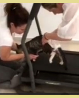
►Video 1. Paraplegic cat during physiotherapy on an escalator that developed spinal walking.
►Video 2. Positive cross extensor reflex during the testing of the hindlimb flexion reflex.
►Video 3. Paraplegic cat during spontaneous stepping without weight support.
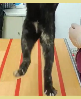
SW is clinically characterised by a lack of coordination of the hindlimbs in relation to the forelimbs, difficulty of the hindlimbs during the altering of the body direction, difficulty in moving backwards, and frequent falls. These falls mainly occur during turns and usually to one side, although some animals tend to try to avoid falling due to increased spasticity of the homolateral limb. In some cases, animals may be unable to perform locomotion on their own, but they do so if stimulation of the perineal region is induced by pinching of the area (Lovely et al. 1986, Rossignol et al. 1999). Nevertheless, several animals are able to walk for at least 10 steps unaided and without falling. During walking, several types of hindlimb movements can be observed. Specifically, some animals exhibit hypermetria due to spasticity of the flexor muscles, while in other animals, gait and support on the dorsal surface of the fingers (knuckling) of the hindlimbs is observed on the ground. Also, animals with SW usually rest their weight mainly on the forelimbs when attempting to come to an upright position. In addition, reflex disturbances are often observed a few days after the acute injury, taking for example the pathological reflex of hyperextension of the unilateral hind limb during the implementation of the withdrawal reflex (cross extensor reflex) (Lewis et al. 2020) (►Video 1) (►Video 2) (►Video 3).
Clinical presentation
Initially, upon presentation of these animals to the clinic (and of course, before SW develops), there is paralysis of the hindlimbs and loss of DPP in both hindlimbs (Fitzmaurice 2010, Gallucci et al. 2017).
During the neurological examination of an animal that has developed SW, abnormal gait with ataxia and paraparesis will initially be observed, as mentioned above. Subsequently, there is a disturbance of proprioceptive sensation which may manifest in various ways such as delayed response to the hopping test and non-response to the extensor postural thrust test of the hindlimbs. It should be noted that the paw replacement test may be normal. Spinal reflexes are usually increased, and spasticity of extensor and flexor muscles is often also increased. Reflex movements of the muscles corresponding to neuromeres of the SC that are posterior to the area of injury (mass reflex) are also observed at the same time: involuntary spasms of the flexor muscles of the hindlimbs, reflex tail raising, bladder or colonic emptying during tactile or other sensory stimulation (e.g., applying pressure to empty the bladder) (Lewis et al. 2020).
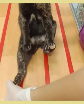
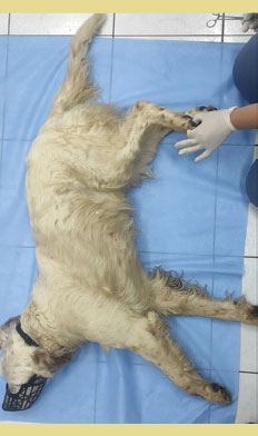
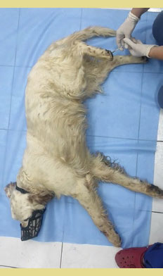
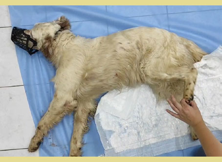
It should be noted that very often during neurological examination, the positive flexion reflex is mistakenly taken as DPP, so it is necessary to separate these two entities. The presence of a positive flexion reflex does not indicate that an animal has DPP (Fitzmaurice 2010) (►Video 4). Simple flexion of the limb is evidence that the lower motor neurons are intact, which allows the reflex to function without providing information about DPP, which requires communication with higher control centers and thus is not guaranteed by the integrity of the lower motor neurons (LMNs) alone (Fitzmau- rice 2010, De Lahunta et al. 2021). DPP is examined by applying a painful stimulus such as the pinching of an interdigital fold or a phalanx with hemostatic forceps (Garosi 2004) (►Video 5). The test is considered positive when the animal expresses a conscious - behavioural response, i.e., head turning towards the stimulus, attempting to bite or screaming (Garosi 2004) (►Video 6). It is important to test both the inner and outer fingers of all limbs, as depending on the findings, information about the location of the lesion can be extracted (Añor 2004). In addition, to ensure that an animal does not have DPP, sensation at the base of the tail should also be tested. The conscious response of the animal to any of the above stimuli implies the presence of DPP (Olby et al. 2020). Therefore, the confirmation that an animal has SW and not ataxia/ paresis of other etiology is the absence of DPP.
►Video 4. Positive hindlimb flexion reflex in a paraplegic dog without deep pain perception.
►Video 5. Deep hindlimb pain perception testing in a paraplegic dog. Test negative.
►Video 6. Positive hindlimb flexion reflex and negative deep pain perception control in the same limb in a paraplegic dog. Positive deep pain perception test in the normal forelimb.
Pathophysiology
Although the central nervous system is known to have little regenerative capacity, it displays impressive plasticity (Lewis et al. 2020). Plasticity is the reorganization of surviving neurons into a functional circuit that can continue to occur even weeks or months after injury (Jeffery & Blakemore 1999). Studies suggest that recovery of gait can occur through processes such as: a. Regeneration of neuro-axons at the site of injury, b. Restoration of function of intact neuro-axons of upper motor neurons (UMNs) crossing the site of injury, c. more autonomous role of central pattern generator (CPGs); d. changes in excitability of interneurons and LMNs posterior to the injury site; e. activation of «silent» synapses; f. changes in the «gravity» (or dynamics) of some synapses; g. changes in the perception of sensory stimuli at the levels of the SC posterior to the lesion (Lewis et al. 2020, De Lahunta et al. 2021).
Further explanation regarding CPGs is needed. Firstly, CPGs are self-contained neural circuits that produce repetitive patterns of motor behaviour independent of sensory stimulation or feedback (Bucher 2009). These circuits are also present in the SC and contribute to the generation of movement from the hindlimbs (Lewis et al. 2020). CPGs theoretically extend along the entire SC, but those located in the lumbar spine, involving the hindlimbs, have been studied primarily (Lewis et al. 2020). They have the ability to establish a basic walking pattern, as well as coordinate the right and left limbs (Lewis et al. 2020). This movement can be generated independently of brain communication and is achieved through the cooperation of interneurons (neurons that transmit impulses between other neurons) and LMNs that innervate skeletal muscles (Lewis et al. 2020). While in normal animals CPGs are subject to brain control to initiate their activity, they have been shown to gain greater autonomy, contributing to SW (Lewis et al. 2020) in animals with thoracolumbar injury. In conclusion, the activation of CPGs, appears to be assisted by the function of interstitial neurons in the SC, by changes in the excitability of motor neurons and alterations in the perception of sensory stimuli (Lewis et al. 2020).
DPP is the most important indirect predictor of recovery of voluntary ambulation in case of SC injury as in its absence the chances of full recovery of voluntary movement are extremely reduced (Olby et al. 2004). To understand the explanation of this phenomenon, it must first be mentioned that the large-sized myelinated nerve fibres that form the bundles that are involved in gait are more sensitive to injury compared to small-sized unmyelinated nerve fibres (Sharp & Wheeler 2005). So, when the damage involves up to the resistant unmyelinated fibers, which is perceived by the lack of DPP, it is considered that the neurological damage is very severe (Sharp & Wheeler 2005). Furthermore, the neuro-axons of the neurons responsible for DPP travel on the inner surface of the lateral white matter bundle of the SC, i.e., the innermost parts, forming the spinothalamic bundle (Schatzberg et al. 2012). Therefore, in case of heavy damage to these neuro-axons, it means that there is also heavy damage to the neurons of the more peripheral bundles (which are myelinated) and are responsible for walking (Schatzberg et al. 2012). Therefore, in such animals the chances for DPP recovery and gait are less likely (Schatzberg et al. 2012).
It is worth mentioning that in dogs with Hansen’s type I intervertebral disc prolapse (IVDP) and subsequent loss of DPP, DPP and walking ability was recovered in 58% of dogs (Olby et al. 2003) after decompression surgery. In contrast, dogs with Hansen’s type I IVDP, without loss of DPP, success rates were 72-100% port surgery (Kerwin et al. 2012).
Predisposing factors for spinal walking development
It should be noted that the majority of clinical information on dogs comes from cases of Hansen’s type I IVDP, as it is the most common pathological condition of the SC in this species (Granger & Carwardine 2014). In contrast, the most common aetiology of myelopathy in cats is traumatic (Granger & Carwardine 2014). Also, in cats there is a lack of literature regarding naturally induced injuries, as, apart from the study by Gallucci et al (2021), most studies are experimental (Gallucci et al. 2021).
1. Animals’ characteristics
In one study, the body weight of dogs appears to be important, as smaller dogs, specifically under 7.8 kg, are more likely to develop SW (Gallucci et al. 2017). It is speculated that it may be easier for smaller dogs to transfer extra weight to the forelimbs, thus ‘offloading’ the hind, weaker limbs (Lewis et al. 2020). Contrariwise, it may be more difficult for taller dogs with a higher centre of gravity to have good balance, preventing them from regaining gait ability (Lewis et al. 2020). Subsequently, based on research by Gallucci et al. (2017), it appears that younger dogs, under 60 months old, are more likely to develop SW, although other research has shown that age is irrelevant (Jeffery et al. 2016) or that younger age is only associated with more rapid SW development (Olby et al. 2003). It should be noted, however, that in Gallucci et al’s (2017) study, these dogs had undergone physiotherapy, which influenced the outcome. In relation to cats, an experimental study showed that 2-weekold paraplegic kittens were more likely to develop SW than 12-week-old paraplegic cats (Smith et al. 1982). Specifically, in this study all 2-week-old kittens developed SW, regardless of whether or not they underwent physiotherapy (Smith et al. 1982). In contrast, only 3 of the 5 12-week-old cats that underwent physiotherapy developed SW, while those that did not, had not developed SW (Smith et al. 1982).
2. Clinical presentation
The presence of the following clinical signs at presentation may provide information to the possibility of developing SW. Initially, according to the research of Lewis & Olby (2017) of particular importance appears to be the increased duration of flexor muscle spasm in the hindlimbs, as there is a positive correlation between this and the development of some form of gait. These spasms were elicited following pinpointing of the bottom of the foot (Lewis & Olby 2017). However, this experiment was performed in animals that had suffered an acute SC injury at least 3 months previously and having a stable clinical presentation, so it is possible that the increased duration of spasm of hind limb flexor muscles may not be used as a predictive marker in more recent cases (Lewis & Olby 2017).
Regarding cats, according to the study by Gallucci et al. (2021), the presence of the cross extensor reflex at the time of presentation was found to have a positive correlation with the development of SW (Gallucci et al. 2021).
It is worth noting that recovery of motor activity in dogs is seen less frequently in cases of spinal cord injury of traumatic origin (Olby et al. 2003). This may be since in such cases more extensive or multiple lesions are usually induced, as well as transection of the SC, or that in such cases euthanasia is more often chosen (Olby et al. 2003, Lewis et al. 2020). It is also worth mentioning that injuries posterior to the thoracolumbar SC do not result in SW, since damage to the CPG itself then occurs (De Lahunta et al. 2021).
In cats, no correlation has been found between the level of symptom severity and the development or not of SW (Gallucci et al. 2021).
3. Laboratory tests
Attempts have been made to identify blood and cerebrospinal fluid parameters that could be used as predictors of outcome in animals without DPP, but without success (Olby et al. 2020).
Frequency and timing of spinal walking
The percentages of dogs that were able to develop SW after naturally induced damage to the thoracolumbar SC and loss of DPP show a wide range, namely from 10% to 59%. This may be due to differences in population, type of injury and the more likely choice of euthanasia in trauma-induced injuries (Olby et al. 2003, Gallucci et al. 2017, Hu et al. 2018, Lewis et al. 2019, Lewis et al. 2020). It is worth mentioning at this point that the highest rate of SW development (59%) was observed in a study where dogs underwent intensive physiotherapy (Gallucci et al. 2017). Regarding Hansen type I IVDP, the percentage of dogs that managed to develop SW was 18% - 31% (Olby et al. 2003, Aikawa et al. 2012).
The median time of SW presentation in dogs shows great variation. The median presentation time in one study was 9 months (range: 4 to 18 months), while in a second study in which patients underwent physiotherapy, the median time was only 75 days (range: 16 to 350 days) (Olby et al. 2003, Gallucci et al. 2017). In Hansen’s type I IVDP cases, the median time of SW presentation was also 9 months, with a range of 2 to 28 months (Olby et al. 2003, Aikawa T et al. 2012).
In cats, in the available in the literature clinical study that is recording cases of physical injuries, none of the 7 cats managed to develop SW (Gallucci et al. 2021). In contrast, in 2 experimental studies, 83%-87.5% of cats were found to develop SW (Lovely et al. 1986, De Leon et al. 1998).
Myelomalacia
Dogs with DPP loss are at risk of developing myelo- malacia, for which there is no cure, and which usually leads the animal to death (Castel et al. 2019). It has been found that rates of myelomalacia due to Hansen’s type I IVDP in dogs without DPP range from 9 to 33% (Olby et al. 2020). Clinical signs of myelomalacia may be present at presentation or develop up to 5 days later (Castel et al. 2017). In caudal extension there is loss of spinal reflexes of the hindlimbs, reduced abdominal wall tone, reduced or absent perineal reflex and reduced or absent anal tone (Castel et al. 2017). As myelomalacia extends to the thoracic and cervical SC, there is respiratory distress due to paralysis of the intercostal muscles, loss of spinal reflexes of the forelimbs, bilateral Horner’s syndrome and finally death (Castel et al. 2019).
Supportive measures of the paraplegic animal with loss of deep pain perception
As already mentioned, the CNS is characterised by plasticity, and therefore can mitigate the effects of resulting damage through various mechanisms. Through plasticity, and with the help of repeated external stimulation, the remaining neurons essentially acquire new functions, resulting in the possibility of recovery even in severe spinal cord injuries (Sherman & Olby 2004).
As is well known, disuse of the limbs leads to loss of muscle mass, permanent muscle group contractions and deterioration of joints and associated structures (ankylosis). Physiotherapy is considered critical in arresting these issues and achieving maximum rehabilitation (Sherman & Olby 2004).
To date, there are limited clinical studies developing and demonstrating the efficacy of specific therapeutic interventions in the development of SW in dogs without DPP (Lewis et al. 2020). However, information from experimental models and human spinal cord injuries may be useful for application to dogs.
In order to choose the most appropriate rehabilitation programme for each animal, the following clinical signs should be taken into account: the ability to walk and the presence of other health problems. In any case, the plan must respect the following rules:
1. Existence of supportive care to prevent secondary problems and provide the right environmentt for rehabilitation.
2. Attempt to halt the progression of damage within the nervous system.
3. Maintain muscle mass and range of motion, maximise plasticity within the nervous system and stimulate proprioceptive neural pathways to relearn normal gait (Sherman & Olby 2004).
Urination
Injury to the thoracolumbar spine resulting in SW, will lead to subsequent bladder dysfunction due to damage to the sphincter UMNs, their continued increased tone and subsequent incontinence from retention (Granger et al. 2020). Because of this, the animal is exposed to the risk of lower urinary tract infections, urinary ulcers, bladder wall damage due to overstretching, uroperitoneum and, in more severe cases, damage to the upper urinary tract (Sherman & Olby 2004). It is therefore evident that bladder dysfunction can compromise the quality of life of the animal, so it is an additional challenge for the caretaker and must be addressed.
Animals with SW should therefore undergo mechanical bladder emptying 3 - 4 times a day (Sherman & Olby 2004). The resulting retained bladder is hard in palpation and emptying is expected to be difficult (Cook et al. 2019). Alternatively, intermittent aseptic urethral catheterisation can be performed, as permanent catheterisation can cause secondary complications such as urinary tract infections and urinary wall injury (Sherman & Olby 2004). This may be a temporary solution anyway, especially in male animals, since catheterisation of females requires advanced skills from the clinician (Cook et al. 2019).
To facilitate mechanical emptying of the spastic bladder, meditation is usually required in the acute phase of the SC injury. The following drugs are listed as examples:
Α) Diazepam 0.25 - 0.5 mg kg-1 orally 20 minutes before voiding causes relaxation of the external urethral sphincter (Sherman & Olby 2004) (Granger et al. 2020)
Β) Phenoxybenzamine 0.5 mg kg-1 orally every 8-12 hours causes relaxation of the internal urethral sphincter (Sherman & Olby 2004) (Granger et al. 2020)
C) Other alpha-adrenergic blockers can also be used (e.g., prazosin, alfuzosin) (Olby et al. 2022) (Granger et al. 2020)
There are no studies to support the combination of these drugs. It also appears that their use to facilitate mechanical bladder emptying is not required after a few weeks (Granger et al. 2020). Possible UTI should be treated with appropriate antibiotics following a culture and sensitivity testing (Sherman & Olby 2004). The veterinarian is required to differentiate UTI from subclinical bacteriuria, in which antibiotherapy is rarely required (Weese et al. 2019).
Defecation
As with urination, the normal function of defecation is also impaired in SC injury (Granger et al. 2020).
In thoracolumbar spine lesions, the resulting problem is the constant tone of the external anal sphincter and the accumulation of feces in the bowel until the increased pressure and distension of the bowel causes reflex involuntary emptying (Granger et al. 2020). The most practical and effective treatment for fecal incontinence is high nutrient and low residue diets (e.g., gastrointestinal diets, hypoallergenic diets), which reduces both the volume and moisture content of feces, so the problem becomes more manageable (Granger et al. 2020). There is, however, debate about the correct fiber content of the ration. In addition, establishing a daily schedule may also be helpful in management. Along with bladder emptying, defecation usually occurs, which can be assisted by gentle stimulation of the perineum (Granger et al. 2020). For some dogs, a meal or exercise is required to enable them to defecate (Granger et al. 2020). General cleaning and grooming of the perineal skin are essential to prevent infections and other adverse conditions (Granger et al. 2020). It should therefore be emphasised that practically no fecal incontinence occurs and that reflex movements of the anal sphincter aid defecation.
From the above, animals with SW are bound to have urination and defecation problems, but these can be managed, at least to some extent.
Physical rehabilitation
Various techniques have been proposed aiming to help an animal develop SW, but it is not the purpose of this article to discuss physiotherapy in detail.
Although not proven, there is evidence that an intensive rehabilitation program may benefit an animal with DPP loss more than a basic program that includes only passive range of motion and assisted walking (Olby et al. 2022, Lewis et al. 2020).
The programme should start within 1 or 2 weeks after the lesion and last at least 8 weeks (Gallucci et al. 2020). Early initiation of rehabilitation in animals without DPP appears to improve development of SW rates (Olby et al. 2022).
In the literature there are reports of physiotherapy duration ranging from a few days to months (Olby et al. 2022, Gallucci et al. 2020, Gallucci et al. 2017, Lovely et al. 1985, Martins et al. 2021). However, greater intensity (i.e. more days of the week, more physiotherapy sessions and more hydrotherapy sessions on a treadmill under water) is associated with more marked improvement in neurological outcome and shorter time to achieving weight bearing walking (Olby et al. 2022).
Specifically in animals paralysed after Hansen’s type I IVDP in the thoracolumbar spine, the reported frequencies of physiotherapy are 1 to 3 times per day for hospitalised patients, 1 to 3 times per week for patients who are going to visit the clinic specifically for the session, and 1 to 5 times per day for patients who do their physiotherapy at home (Olby et al. 2022).
It is important to note that protocols should be designed for each individual case (Olby et al. 2022). They should also be dynamic and modified over time and as improvement or non-response is noted (Olby et al. 2022).
The techniques that can be used are listed below, in descending order of practicality and convenience.
Assisted standing
The effect of adding weight on gait recovery is well known (Lovely et al. 1985, Tillakaratne et al. 2001, Edgerton et al. 2007, See & de Leon 2012). Thus, a key part of the rehabilitation programme should be to enabling the animal to support its own weight (Campbell & Huntingford 2016, Olby et al. 2022).
Exercise (Step training)
Research evaluating walking ability in cats that exercised on a treadmill and in cats not exercised after SC transection (De Leon et al. 1998), showed that the former walked at a higher rate and better than the latter.
Retraining the animal to walk can be done on an electric treadmill and/or through hydrotherapy.
Sensory stimuli applied to the paw play an important role in the physiology of gait (Lovely et al. 1986, Tillakaratne et al. 2002).
Hydrotherapy, which is advisable to precede the use of a land-based treadmill (Spinella et al. 2022), can be done in two forms: walking on a treadmill or swimming (Sherman & Olby 2004).
Range of motion (ROM)
This is the technique in which the joint is subjected to passive or active flexion and extension. This avoids the adverse effects that would result from immobilisation, which include osteoporosis, morphological and biochemical changes in articular cartilage, loss of muscle elasticity, etc. (Sherman & Olby 2004).
Medication
Research is ongoing to see if various drugs can be used to facilitate spinal cord stimulation and response to sensory stimuli. These act through serotonergic, noradrenergic or glutaminergic action or, constrastingly, through mobilisation of the in- hibitory glycine and GABA neurotransmitters (Edg- erton et al. 2008). However, they have not yet been used in daily clinical practice, so we will not expand further (Martins et al. 2022).
Practice of proprioceptive sensation
These are exercises designed to increase the proprioception and use of the limbs by the animal. A prerequisite for them to be performed is that the animal can carry enough of its own weight and can walk with some assistance (Sherman & Olby 2004).
Dorsal stimulation
Electrode placement and tonic stimulation of the dorsal surface of the lumbosacral spine can induce movements in animals with a complete SC dissection (Edgerton et al. 2008). Studies in rats have shown that combining this method with drugs has a synergistic effect in achieving movement (Edgerton et al. 2008). Other forms of electrical stimulation incorporated into the rehabilitation program also appear to aid in the recovery of gait, such as Interferential Electrical Stimulation (IES), Functional Electrical Stimulation (FES), and Transcutaneous Electrical Spinal Cord Stimulation (TESCS) (Martins et al. 2021).
Effectiveness of rehabilitation techniques
Indications of improvement and recovery of SW in the course of rehabilitation are the lack of need for stimulation of the perineal reflex in order for the animal to stand/move, the lack of need for external assistance for the initial flexion of the hindlimbs, achieving more weight-bearing steps, improved gait quality and duration, the ability to walk at higher treadmill speeds, no need for external weight support and more correct foot positioning (Lovely et al. 1985, de Leon et al. 1998, See & de Leon, 2012). As already mentioned, the time required to achieve SW varies (Lovely et al. 1985, Gallucci et al. 2017, Gallucci et al. 2020, Martins et al. 2021, Olby et al. 2022), so the perception of some of the clinical signs above should alert the clinician to a possible positive outcome and encourage them to continue the rehabilitation effort.
In a study by Gallucci et al. (2017) including 81 dogs without DPP who underwent a rehabilitation programme, 59% of them developed SW over an average of 75 days. In a later study by Gallucci et al. in 2021, 45% of cats were found to have developed SW with an average duration of rehabilitation of 46 days (and range 15-302 days). It therefore appears that about half of the animals that underwent physiotherapy developed SW.
Conclusions
In conclusion, dogs and cats that suffer an acute injury to the thoracolumbar SC that causes loss of DPP are likely to develop SW. While this is attributed to the plasticity of the nervous system, it can also be assisted by a physical rehabilitation programme designed according to the needs of each animal, which both prepares the animal’s musculoskeletal system and assists the development of the physiological changes responsible for SW. SW offers potential in terms of walkability of the animal, but restoration of normal urination should not be expected. However, urinary and fecal incontinence could be managed by a dedicated caretaker, determined to find the right programme to suit their animal.
Conflict of interest
The authors declare that there is no conflict of interest.
Corresponding author:
Aliki Groutidou
This email address is being protected from spambots. You need JavaScript enabled to view it.
References
- Aikawa T, Fujita H, Kanazono S, Shibata M & Yoshigae Y (2012) Longterm neurologic outcome of hemilaminectomy and disk fenestration for treatment of dogs with thoracolumbar intervertebral disk herniation: 831 cases (2000–2007). J Am Vet Med Assoc 241, 1617–1626.
- Añor S (2004) Monoparesis. In: BSAVA Manual of Canine and Feline Neurology. Platt SR & Olby NJ eds. 3rd ed. BSAVA, Gloucester, UK, pp. 265-279.
- Bucher D (2009) Central pattern generators. In: Encyclopedia of Neuroscience. Squire L ed. Elsevier, pp. 691-700.
- Castel A, Olby NJ, Ru H, Mariani CL & Muñana KR, Early PJ (2019) Risk factors associated with progressive myelomalacia in dogs with complete sensorimotor loss following intervertebral disc extrusion: a retrospective case-control study. BMC Vet Res 15, 1–9.
- Castel A, Olby NJ, Ru H, Mariani CL & Muñana KR, Early PJ (2017) Clinical Characteristics of Dogs with Progressive Myelomalacia Following Acute Intervertebral Disc Extrusion. J Vet Intern Med 31, 1782-1789.
- De Lahunta A, Glass E, Kent M (2021) Lower Motor Neuron: General Visceral Efferent System. In: de Lahunta’s Veterinary Neuroanatomy and Clinical Neurology, 5th ed, Saunders Elsevier, Philadelphia, pp. 203-229.
- De Lahunta A, Glass E, Kent M (2021) Upper Motor Neuron. In: de Lahunta’s Veterinary Neuroanatomy and Clinical Neurology, 5th ed, Saunders Elsevier, Philadelphia, pp. 230-245.
- De Leon RD, Hodgson JA, Roy RR, Edgerton VR (1998) Full weight-bearing hindlimb standing following stand training in the adult spinal cat. J Neurophysiol 80(1), 83-91.
- De Leon RD, Hodgson JA, Roy RR & Edgerton VR (1998) Locomotor Capacity Attributable to Step Training Versus Spontaneous Recovery After Spinalization in Adult Cats. J Neurophysiol 79, 1329–1340.
- Edgerton VR, Courtine G, Gerasimenko YP, Lavrov I, Ichiyama RM, Fong AJ, Cai LL, Otoshi CK, Tillakaratne NJ, Burdick JW & Roy RR (2008) Training locomotor networks. Brain Res Rev 57(1), 241254.
- Fitzmaurice S (2010) The neurological examination. In: Small Animal Neurology. Nind F ed. Saunders Elsevier, Edinburgh, pp. 6-34.
- Fitzmaurice S (2010) Gait - an introduction. In: Small Animal Neurology. Nind F ed. Saunders Elsevier, Edinburgh, pp.181-185.
- Gallucci A, Dragone L, Al Kafaji T, Menchetti M, Del Magno S & Gandini G(2021) Outcome in Cats with Acute Onset of Severe Thoracolumbar Spinal Cord Injury Following Physical Rehabilitation. Vet Sci 8(2), 1-11.
- Gallucci A, Dragone L, Menchetti M, Gagliardo T, Pietra M, Cardinali M & Gandini G (2017) Acquisition of Involuntary Spinal Locomotion (Spinal Walking) in Dogs with Irreversible Thoracolumbar Spinal Cord Lesion: 81 Dogs. J Vet Intern Med 31, 492-497.
- Garosi L (2004) The neurological examination. In: BSAVA Manual of Canine and Feline Neurology. Platt SR & Olby NJ eds. 3rd ed. BSAVA, Gloucester, UK, pp. 1-23.
- Granger N, Carwardine D (2014) Acute Spinal Cord Injury Tetraplegia and Paraplegia in Small Animals. Vet Clin Small Anim 44, 1131-1156.
- Granger N, Olby NJ, Nout-Lomas YS & the Canine Spinal Cord Injury Consortium (CANSORT-SCI) (2020) Bladder and Bowel Management in Dogs With Spinal Cord Injury. Front Vet Sci 7, 583342.
- Hu HZ, Jeffery ND & Granger N (2018) Somatosensory and motor evoked potentials in dogs with chronic severe thoracolumbar spinal cord injury. Vet J 237, 49–54.
- Jeffery ND, Barker AK, Hu HZ, Alcott CJ, Kraus KH, Scanlin EM, Granger N & Levine JM (2016) Factors associated with recovery from paraplegia in dogs with loss of pain perception in the pelvic limbs following intervertebral disk herniation. J Am Vet Med Assoc 248, 386–394.
- Jeffery ND, Blakemore WF (1999) Spinal cord injury in small animals 1. Mechanisms of spontaneous recovery. Veterinary Record 144, 407–413.
- Kerwin S, Levine M & Hicks D (2012) Thoracolumbar Spine. In: Veterinary Surgery Small Animal. Tobias K & Johnston S ed. Saunders Elsevier, Canada, pp. 449-475.
- Lewis MJ, Cohen EB & Olby NJ (2018) Magnetic resonance imaging features of dogs with incomplete recovery after acute, severe spinal cord injury. Spinal Cord 56,133–141.
- Lewis MJ, Jeffery ND, Olby NJ & the Canine Spinal Cord Injury Consortium (CANSORT-SCI) (2020) Ambulation in Dogs With Absent Pain Perception After Acute Thoracolumbar Spinal Cord Injury. FrontVet Sci 7, 560.
- Lewis MJ, Laber E & Olby NJ (2019) Predictors of response to 4-aminopyridine in chronic canine spinal cord injury. J Neurotrauma 36, 1428–1434.
- Lewis MJ & Olby NJ (2017) Development of a clinical spasticity scale for evaluation of dogs with chronic thoracolumbar spinal cord injury. Am J Vet Res 78, 854-861.
- Lovely RG, Gregor RJ, Roy RR & Edgerton VR (1986) Effects of training on the recovery of full-weight-bearing stepping in the adult spinal cat. Experimental Neurology 92, 421–435.
- Olby N, da Costa R, Levine J, Stein V & the Canine Spinal Cord Injury Consortium (CANSORT SCI) (2020) Prognostic Factors in Canine Acute Intervertebral Disc Disease. Front Vet Sci 7, 596059.
- Olby N, Harris T, Burr J, Muñana K, Sharp N & Keene B (2004) Recovery of Pelvic Limb Function in Dogs following Acute Intervertebral Disc Herniations. J Neurotrauma 21, 49-59.
- Olby N, Jeffery N (2012) Pathogenesis and Physiology of Central Nervous System Disease and Injury. In: Veterinary Surgery: Small Animal vol 1. Tobias K & Johnston S eds. Elsevier Saunders, Misouri, pp. 374-511.
- Olby NJ, Levine J, Harris T, Muñana K, Skeen T& Sharp N (2003) Long-term functional outcome of dogs with severe injuries of the thoracolumbar spinal cord: 87 cases (1996–2001). J Am Vet Med Assoc 222, 762–769.
- Olby N, Moore S, Brisson B, Fenn J, Flegel T, Kortz G, Lewis M & Tipold A (2022) ACVIM consensus statement on diagnosis and management of acute canine thoracolumbar intervertebral disc extrusion. J Vet Intern Med 36, 1570-1596.
- Rossignol S, Chau C, Brustein E, Belanger M, Barbeau H & Drew T (1996) Locomotor capacities after complete and partial lesions of the spinal cord. Acta Neurobiol Exp 56, 449-463.
- Rossignol S, Drew T, Brustein E & Jiang W (1999) Locomotor performance and adaptation after partial or complete spinal cord lesions in the cat. Progress in Brain Research 123, 349-365.
- Schatzberg S, Kent M & Platt S (2012) Neurological Examination and Neuroanatomin Diagnosis. In: Veterinary Surgery: Small Animal, vol 1.Tobias K & Johnston S eds. Elsevier Saunders, Misouri, pp. 325-340.
- See PA& de Leon RD (2013) Robotic loading during treadmill training enhances locomotor recovery in rats spinally transected as neonates. J Neurophysiol 110, 760-767.
- Sharp N, Wheeler S (2005) Functional Anatomy. In: Small Animal Spinal Disorders: Diagnosis and Surgery. Rodenhuis J ed. 2nd ed. Elsevier Mosby, n.p, pp.1-17.
- Sherman J, Olby NJ (2004) Nursing and rehabilitation of the neurological patient. In: BSAVA Manual of Canine and Feline Neurology. Platt SR & Olby NJ eds. 3rd ed. BSAVA, Gloucester, UK, pp. 394-407.
- Smith JL, Smith LA, Zernicke RF & Hoy M (1982) Locomotion in exercised and nonexercised cats cordotomized at two or twelve weeks of age. Experimental Neurology 76(2), 393-413.
- Tefend Campbell M & Huntingford J (2016) Nursing Care and Rehabilitation Therapy for Patients with Neurologic Disease. In: Practical Guide to Canine and Feline Neurology. Dewey & da Costaeds. 3rd ed, Wiley, Iowa, pp. 559-584.
- Tillakaratne NJ, de Leon RD, Hoang TX, Roy RR, Edgerton VR, Tobin A J (2002) Use-dependent modulation of inhibitory capacity in the feline lumbar spinal cord. J Neurosci 22, 3130-3143.
- Weese J.S, Blondeau J, Boothe D, Guardabassi L.G, Gumley N, Papich M, Jessen L.R, Lappin M, Rankin S, Westropp J.L, Sykes J (2019) International Society for Companion Animal Infectious Diseases (ISCAID) guidelines for the diagnosis and management of bacterial urinary tract infections in dogs and cats, The Veterinary Journal 247, 8-25.
