

Nictitating membrane prolapse and eye tumefaction in a dog.
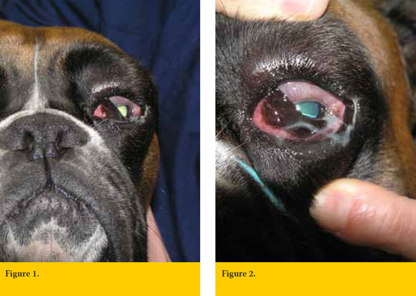
A five years old, male Boxer presented with severe tumefaction of the left eye and depression (Figure.1 and 2). A nictitating membrane prolapse was recorded, lasting four days. The last two days the dog was depressed and inappetent.
The general physical examination was unremarkable. The temperature of the dog was 40,3°C. Any attempt to open the the animal’s mouth, was painful.
- How would you describe ocular lesions? (Fig.1 and 2).
- What is the most likely diagnosis? What is your differential diagnosis and which clinical and laboratory tests would you recommend?
- What treatment would you suggest?
Answers
Nictitating membrane prolapse and eye tumefaction of a dog.
1. How would you describe the ocular lesions?
(Figures 1 and 2).
An intense nictitating membrane prolapse is noticed which is accompanied by edema and hyperemia. Extensive conjunctival edema (chemosis) is present, which covers part of the cornea. There is eyelid and periorbital swelling as well as mucopurulent discharge in the conjunctival sac and eyelids. The eye globe itself is normal.
2. What is the most likely diagnosis? From what other medical conditions would you make the differential diagnosis and what clinical examinations and laboratory tests would you recommend?
The sudden onset, the typical clinical manifestations and fever refer to retrobulbar/ peribulbar inflammation or abscess. Moreover, the intense pain, while opening the mouth, is a typical symptome of the disease1. Differential diagnosis includes zygomatic salivary retention cysts and mucoceles, eosinophilic myositis of mastication muscles and retrobulbar and periocular tumors 1,2,3,4.
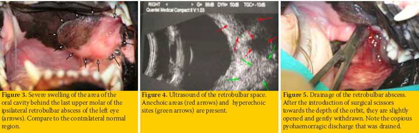
Oral cavity inspection is necessary for the diagnosis2. The inferior bony orbital wall is absent in the dog, so orbital structures lie adjacent to oral cavity and temporomandibular joint. In any case of suspected inflammation or abscess of the orbit, the area behind the ipsilateral last upper molar is swollen and therefore should be inspected. This was also true in our case (Figure 3). This examination is painful, so the procedure needs sedation. Laboratory investigation include total blood count, retrobulbar space cytology, orbital ultrasound and MRI, where available5,6,7. In this case, the blood leukocytes were 28.000/mm3. Orbital cytology revealed degenerative neutrophils with many free or phagocytized cocci. Finally, orbital ultrasound revealed, multiple hyperechoic areas and some anechoic sites were noticed, findings consistent with inflammation/abscess6 (Figure 4).
3. What is the treatment that you would recommend?
Treatment of orbital abscesses involves surgical drainage and systemic antibiotic therapy1,2. Access of retrobulbar space is performed through the oral cavity. Initially, with a scalpel blade, a small incision of the mucous membrane behind the last upper molar is made. Through this incision hemostatic forceps or closed blunt scissors are introduced towards the top of the orbit, behind the posterior pole of the eye. Then strands are opened slightly and forceps are withdrawn draining the abscess (Figure 5). In this case, amoxicillin/clavulanic acid (Synulox ® palatable tablets, Pfizer) at a dose of 20mg/kg every 12 hours for ten days was administered.
The exact cause of orbital abscesses often remains unidentified. Foreign bodies - that can be removed during drainage of abscess8,9, as well as hematogenous infection, injuries through the oral cavity and dental problems have been reffered 10.
> References
1. Van der Woerdt A. Orbital inflammatory disease and pseudotumor in dogs and cats. Vet Clin North Am Small Anim Pract 2008,38:389-401.
2. Spiess BM, Pot SA. Diseases and surgery of the canine orbit. In Veterinary Orhthalmology, .Gelatt KN (ed). 5th edn.Wiley-Blackwell Publishing: Ames Iowa,2013, pp 793- 831.
3. Attali-Soussay K,Jegou JP,Clerc B. Retrobulbar tumors in dogs and cats:25 cases. Vet Ophthalmol 2001,4:19-27.
4. Gilmour MA,Morgan RV, Moore FM. Masticatory myopathy in the dog: a retrospective study of 18 cases.J Am Anim Hosp Assoc 1992,28:300-306.
5. Boydell P. Fine needle aspiration biopsy in the diagnosis of exophthalmos. J Small Anim Pract 1991,32:542-546.
6. Mason DR,Lamb CR,McLellan GJ. Ultrasonographic findings in 50 dogs with retrobulbar disease. J Am Anim Hosp Assoc 2001,37:557-562.
7. Boroffka SA,Verbuggen AM,Grinwis GC et al. Assessment of ultrasonography and computed tomography for the evaluation of unilateral orbital disease in dogs. J Am Vet Med Assoc 2007,230:671-680.
8. Hartley C,McConnell JF,Doust R. Wooden orbital foreign body in a Weimaraner. Vet Ophthalmol 2007,10:390-393.
9. Tovar MC,Huguet E,Gomezi MA. Orbital cellulitis and intraocular abscess caused by migrating grass in a cat. Vet Ophthalmol 2005,8:353-356.
10. Ramsey D,Marreta S,Hamor R et al. Ophthalmic manifestations and complications of dental disease in dogs and cats. J Am Anim Hosp Assoc 1996,32:215-224.
