


> Abstract
The purpose of this study was the study of dogs with patellar luxation (PL) that were presented at the Companion Animal Clinic, School of Veterinary Medicine, Aristotle University of Thessaloniki, Greece during 2004 - 2010. Ninety-five dogs were included in the study. Among the information derived from the clinical examination records were breed, age, body weight, clinical signs upon presentation, site and grade of luxation, type of treatment and outcome of the dogs. Statistical analysis revealed that PL is observed more frequently in mixed breed (27.4%), small sized (61%) and female (52.6%) dogs. Regarding purebred dogs, PL has been more frequently diagnosed in Yorkshire Terriers, Poodles and Chihuahuas. PL was mostly medial (84.1%), bilateral (56.3%) and congenital/developmental (100%). In addition, it was noticed that diagnosis mainly concerned young dogs, aged less than two years (50.5%) and the major complaint upon presentation was lameness (69.5%). The progress of dogs which were surgically treated was considered excellent by the majority of the owners (75%), while post-operative complications were rare and mainly related to the presence of osteosynthesis materials. According to the owners, the majority of the dogs that were treated conservatively, lameness did not occur (35%) or remained stable (45%).
> Introduction
Patellar luxation (PL) is one of the most common orthopedic conditions concerning the hind limbs of dogs. It may present with pain and lameness and leads to secondary degenerative joint disease.1,2
Patellar luxation is mostly congenital/ developmental3 and less frequently acquired (traumatic). Congenital luxation is mainly observed in small sized breeds and rarely in medium and large breed dogs, or even in giant breed dogs.1,4,5 The highest percentage of congenital luxations is noticed in dogs younger than 3 years, 6-8 contrary, acquired luxation, is observed in all breeds of dogs regardless of age.9
Presentation of congenital PL in specific canine breeds, such as Miniature and Toy Poodles, Cavallier King Charles Spaniels, Yorkshire terriers, Chihuahuas and Griffons, reinforces the perception that it is a hereditary disease and, consequently, the use of these dogs for reproduction purposes is not recommended.5 The majority of congenital luxations is a result of musculoskeletal abnormalities, which concern the anatomical structures that participate in stifle extension and affect its alignment and consequently its function. Possible causes of improper alignment of this mechanism are considered to be the abnormal inclination and torsion angles of the femoral head and neck, although they have not been proved, yet.10
Upon presentation of dogs with low grade congenital PL, the owners usually report in various frequencies incidence of intermittent lameness, which does not seem to cause particular discomfort to the dog. Specifically, upon movement, the dog flexes the affected stifle abruptly and, shortly after, it brings it back to normal position, after a short cranial flick of the limb. Many of these animals are asymptomatic. In dogs with permanent PL, the stifle is held in partial flexion and if the luxation is bilateral, the dog presents “bunny hopping” walk type.1 Progressively, lameness is worsening due to secondary degenerative changes of the stifle joint and possible rupture of the cranial cruciate ligament. Lateral PL is frequently escorted with genu valgum distally to the stifle.5-8,11
In acquired PLs, lameness appears suddenly and can even lead to non-weight bearing one.12
In congenital PLs, the intensity of musculoskeletal abnormalities differs significantly according to the grade of luxation and this is the reason why several classification systems have been developed.1
Treatment choices can be either conservative or surgical. The former is indicated for asymptomatic patients or for those who rarely experience lameness episodes.1 In these cases, even the risk of development of degenerative lesions and cranial cruciate ligament rupture do not justify immediate surgical treatment,13 since dogs respond very well to later surgical reconstruction of the luxation, even if secondary rupture of the cranial cruciate ligament has already been established.5 The only exception are cases of very young, 3-4 months old, asymptomatic puppies,2 in which immediate surgical treatment is demanded, in order to avoid the aggravation of musculoskeletal malformations.5,9 Moreover, in medium and large breed asymptomatic dogs, it is suggested that surgical treatment must not be postponed, so as to prevent the destruction of the articular cartilage and the malformation of the trochlear groove.5 Conservative treatment constitutes of controlled exercise for the reinforcement of the quadriceps femoris muscle and demands frequent re-evaluations. Owners must inform the veterinarian regarding pain, lameness or reluctance to move.1
Generally speaking, surgical treatment is suitable for patients presenting with clinical symptoms due to PL14,15 and very young animals.16 Surgery aims to the restoration of normal biomechanics of the stifle joint and the elimination of constant injury of the articular surfaces,10 by reinstating the patella inside the trochlear groove permanently.17 Applied surgical techniques can be categorized into two groups: (a) those who concern soft tissues and (b) those who concern bony structures. The former includes reinforcement of the soft tissues on the other side than that of the luxation (articular capsule, fascia lata), release of the soft tissues on the luxation side, artificial retinaculum and release or transposition of the quadriceps femoris muscle. The latter mostly includes deepening of the trochlear groove (trochleoplasty), transposition of the tibial tuberosity and corrective osteotomy of the femur and/or the tibia. Trochleoplasty is achieved by trochlear sulcoplasty, chondroplasty and wedge or rectangular sulcoplasty.16 In most cases, more than one techniques are used and in various combinations.5,13,16,18,19
The outcome of surgical treatment of PL depends on the intensity of anatomical disorders existing pre-operatively.6 The percentage of complications and articular surfaces damage extent depend on the degree of luxation.20,21 Thus, prognosis concerning 1st, 2nd and 3rd grade luxations is very good, while for those of 4th grade is guarded.13,18 Animals treated conservatively have a good prognosis, although risk for developing degenerative lesions and cranial cruciate ligament rupture is present.22
The purpose of this paper was the retrospective study of the most important epidemiological and clinical aspects and, also, the therapeutical approaches and their results, from 95 dogs with patellar luxation.
> MATERIALS AND METHODS
From the clinical examination records of dogs with patellar luxation, presented in the Companion Animal Clinic (CAC), School of Veterinary Medicine, Aristitle University of Thessaloniki during the academic years 2004-05 to 2009-10, the following data were recorded and evaluated:
- breed,
- gender,
- age,
- body weight,
- cause of luxation
- clinical signs upon presentation,
- if the luxation was uni- or bilateral,
- direction of luxation,
- grade of luxation,
- treatment of choice,
- surgical technique and
- outcome.
Data concerning the progress of the patients (presence or not of lameness, type of lameness, use of analgesics) were derived from the clinical examination records and phone communication with the owners.
For data analysis, the dogs were divided according to body weight into (a) small sized breeds 1-9 kg, (b) medium sized breeds 10-23 kg, (c) large sized breeds 24-40 kg and (d) giant sized breeds >40 kg,23 while according to age, they were classified in groups of (a) 0-2 years old, (b) 2-8 years old and (c) >8 years old. Finally, for the classification of PL the well known four – graded scale was used.24,25
> RESULTS
Ninety five records were retrieved from the medical archives of the CAC, concerning dogs with PL. Based on the data registered in those records and due to lack of pre-existing trauma, all luxations were considered as congenital.
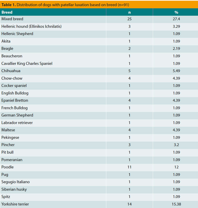
The majority of the dogs were listed as mixed breeds (n=25, 27.4%), while most of the purebreds were Yorkshire Terriers (n=14, 15.3%) and Poodles (n=11, 10.9%) (Table 1). For four dogs no data regarding breed was recorded.
Approximately 52.6% (n=50) of the dogs with PL were females, whereas the remaining 47.3% (n=45) were males. There was not adequate information whether those dogs were neutered or not.
Dogs’ age at the time of diagnosis of the PL varied from 0.5 to 15 years old, whereas, in two cases, age was omitted. Mean and median age was 3.97 and 2 years (n=93), respectively. Fifty percent (n=47) of the dogs examined were less than 2 years old, 35.4% (n=33) were 2.5-8 years old and only 13.9% (n=13) were more than 8 years old.
Body weight upon presentation ranged between 0.9 and 50 kg, whilst in three cases body weight was not registered in the medical records. Mean and median body weight was 10.7 and 7 kg, respectively (n=92). Almost 61% (n=57) of the dogs belonged to a small sized breed, 25% (n=23) were medium sized, 9.7% (n=9) were large sized and only 3.2% (n=3) belonged to a giant breed.
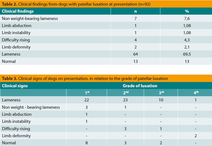
The main presenting complaint of dogs with PL was lameness concerning the affected limb (69.5%, n= 64) (Table 2). In Table 3, clinical appearance of dogs upon presentation related to the grade of luxation is recorded, with lameness dominating in the lowest grades. It should be noticed that in 14.1% (n=13) of the dogs, diagnosing PL was an accidental finding, since there was no mobility problem upon presentation in the CAC, whereas in three cases no reason for presentation was marked in the medical records.
Almost 43.6% (n=41) of the dogs included in the study appeared with unilateral PL and the other 56.3% (n=53) with bilateral. In one case this specific feature was not mentioned.
For 13 PL cases specification regarding luxation (medial or lateral) was lacking. From the remaining 82 dogs, 69 (84.1%) were presented with medial, 9 (10.9%) were presented with lateral, while 4 (4.8%) appeared with medial on one side and lateral on the other one.
From the 147 limbs with PL, grading of the luxation was absent in 10. In the rest 137 limbs, 54 (39.4%) of them were grade I, 44 (32.1%) were grade II, 32 (23.3%) were grade III and seven (5.1%) were grade IV. The distribution of the grade of luxation in relation to body weight of dogs appears in Table 4.
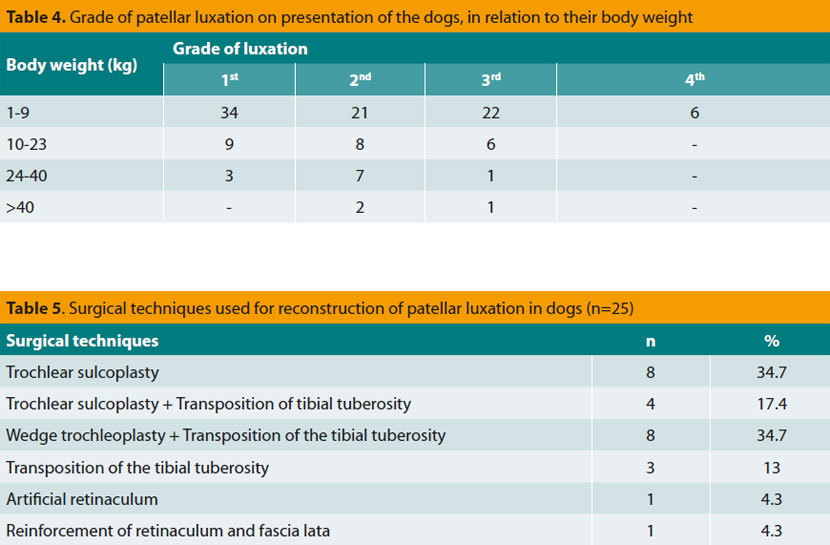
From the 95 dogs that were included in the study, in nine there is no treatment mentioned. From the remaining 86 animals, for which this specific feature is recorded, for 59 (68.6%) of them surgical treatment was not attempted. However it is not specified from the medical records if decision making for non surgical treatment of the PL was the result of medical issues, such as absence of intense symptoms, old age or concomitant animal health problems, or it was the result of owners’ denial. The rest 30% (n=25) of the dogs were submitted to surgical restoration of the PL (Table 5). In only two cases the method of choice was not mentioned.
The follow up of the cases was achieved through telephone communication with the owners and it was posssible in only 56 out of the 95. Time interval between initial presentation of the animals in the CAC and last communication with their owners varies from 3 until 58 months.
From the 40 animals that were not submitted to surgical treatment, 18 (45%) of them presented constant lameness to the limb of interest that did not seem to annoy the owners, two (5%) of them presented gradual worsening of the lameness, six (15%) of them presented at least an episode of lameness, while 14 (35%) of them, according to the owners, did not show any signs of lameness. None of these owners found the use of analgesic drugs necessary for the improvement of their dog’s mobility.
Among the dogs surgically treated (n=16), 12 (75%) did not show any signs of lameness, whereas the remaining four (25%) presented intermittent episodes of lameness. In two of them there were complications concerning the materials of osteosynthesis that were used for the surgical treatment of patellar luxation. In the first case, a seroma developed cranially to the tibial tuberosity due to the presence of pins underneath the skin. In the second case, a pin was migrated towards the caudal aspect of the tibia, which was finally removed spontaneously. Based on clinical presentation of the surgically treated animals, the use of analgesic drugs (e.g. non steroidal anti-inflammatory drugs - NSAIDs) was not considered necessary in almost 87% (n=14) cases according to their owners opinion. Nevertheless, in 12% (n=2) of the cases NSAIDs were used during lameness crisis only.
> DISCUSSION
Patellar luxation is one of the most common orthopedic conditions in dogs.8,12,26,27 Among purebred dogs, PL is observed more frequently in Yorkshire Terriers, Poodles and Chihuahuas, as noted from the majority of researchers.7,8,28-30 However, in our study, it was mixed breed dogs that were presented more frequently with PL (27.4%), probably due to the fact that large numbers of such dogs live in our country and consequently they are brought in the CAC.
Diagnosing PL more frequently in small sized breeds comes in total agreement with most of the authors,1,30 even though in recent studies the exact opposite is reported.11,31 The location of the CAC in an urban region, justifies the great population of small sized dogs presented to it, whereas the increasing frequency of PL presentation in large breeds is probably because of their rise in total dog population.11
The vast majority of PL is congenital,5 as shown in our study, in which there was no luxation caused by traumatic reasons.
Our results confirm the observations of other researchers7,8,11,21,32 that female dogs are predisposed in presenting PL (males/females: 1/1.13), even though it is reported that in large breeds the above ratio was 1.8/1.6 Bound et al. (2009)11 in an attempt to reconcile all the above results, claim that PL is more frequent in male large breed dogs and female small sized dogs. It is obvious that, in order to reinforce or contradict this opinion, the use of much higher numbers of dogs examined is required, that will be distributed in all the different sizes of breeds and the two genders.
Even though average age at which PL was diagnosed was 3.9 years, almost 50% of the dogs was less than 2 years old. These ages do not differ dramatically from the diagnosing age referred in other studies which varies from 2.1 until 3 years old.8,11
The greatest percentage (69.5%) of the animals brought in the CAC with PL mainly presented lameness and most of them had grade I or II PL. It is remarkable that two out of three dogs with grade IV PL had a significant malformation of the limb alignment. So, it seems that the greater the grade of PL, the more serious the musculoskeletal disorders of the affected limb.
In a significant percentage (14.1%) of the patients, the diagnosis of the PL was accidentally made and it mainly concerned grade I and II PLs. Vidoni et al. (2005)30 report that 40% of the dogs diagnosed with patellar luxation was asymptomatic and would not be otherwise diagnosed if it was not for an orthopedic examination conducted for another reason. Consequently, investigation for possible PL should be a part of the general clinical examination, regardless the reason for presentation.
The greatest percentage (84.1%) of dogs, that participated in the study, presented medial PL, as referred in most of the studies.6-8,11 It is worth noting that four of the dogs included in our study were diagnosed with bilateral luxation, with one limb presenting medial luxation and the other one lateral, a fact that has never been recorded in the bibliography available to us.
The aspect that lateral PL appears more frequently in large and giant breed dogs, while medial luxation appears more frequently in small breed dogs,5,7 contradicts the results of our study. In particular, all the lateral PLs were diagnosed in small and medium sized breeds, all but one, which concerned a large breed dog. Maybe this is due to the fact that not many dogs more than 24 kg were included in the study.
For the unilateral or bilateral nature of the PL, bibliographic data are controversial.7,8,33 However, in our study, the biggest percentage of animals (56.3%) presented bilateral luxation.
The choice of surgical method in which the animals with PL are submitted, depends on the grade of luxation, the animal’s pre-operative evaluation, the findings during the operation and the surgeon’s preference.8 Tibial tuberosity translocation combined with deepening of the femoral trochlea (trochleoplasty), usually is an effective techniques for the successful restoration of the luxated patella.34 Generally, the higher the grade, the more surgical corrective techniques required for the affected limb28,35 and the higher the incidence of post-operative complications, due to greater difficulties in restoring the skeletal abnormalities during the operation.12,20 The relevant small number of grade I luxations submitted in surgery in the CAC probably has to do with lack of serious clinical symptoms.8,14 Usually, in these animals conservative treatment is introduced, which consists of intense mobility restriction. From the case study, the result is that grade II and III luxations are the ones submitted more commonly in surgery. This is probably because of the fact that these animals present more intense symptomatology.8 Finally, the owners of animals with grade IV PL, regardless the guarded prognosis, seem to consent in surgical restoration of the luxated patella with high percentage (71%).
Post-operative progress was judged by the owners as excellent in 75% and satisfactory in 25% of the cases. In similar studies the progress of surgical reconstruction of PLs was judged as excellent in 63-88%, satisfactory in 5.5-30% and moderate or poor in 5.5-6%.6,8,28,31 The wide range of the results in various studies most likely reveals the fact that the distribution of the animals based on the grade of the luxation was not equal in all of them, because prognosis is worsening while grade of luxation and intensity of preoperative clinical symptoms are increasing.12
Many post-operative complications have been reported after surgically treatment the luxated patella.6,8,12,16,20,28,31,34,36 The ones recorded in our study (migration of the pin, seroma formation) were caused because of the presence of osteopaedic implants, that were used during the tibial tuberosity transposition and mainly regarded dogs weighing 23 and 17 kg, respectively. According to Gibbons et al. (2006),31 dogs of larger breeds present post-operative complications relevant to orthopaedic implants in higher frequency and this refers to the use of non appropriate materials of osteosynthesis and the great load of forces exercised to the joints of dogs with higher body weight.20 Unfortunately, there is no adequate data available in the medical records of the dogs, so as to ascertain if there are mistakes done during choosing and/or using the materials of osteosynthesis in these cases.
Movement of the higher percentage of the dogs that were not submitted to surgery, was generally good (80%), as they did not show signs of lameness or the one already present remained stable. But, even for the dogs that presented episodes of lameness or aggravation of it (20%), the owners did not consider the use of NSAIDs as necessary. Most likely, in these dogs there was worsening of the musculoskeletal abnormalities, due to improper alignment of the mechanism of stifle extension10 or due to degenerative changes’ formation in the joint, because of the repeated mechanical trauma of the articular surfaces of the stifle.21
In conclusion, PL is observed in higher frequency in young, mixed breed, small sized, female dogs and usually it was medial, bilateral and congenital. The main clinical sign during initial presentation was lameness, even though in many animals PL was asymptomatic. Post-operative progress of the dogs was noted as excellent from the majority of the owners, while post-operative complications were rare. The higher percentage of the dogs treated conservatively did not show lameness or the pre-existing one remained stable.
> Bibliography
1. Denny HR, Butterworth SJ. The stifle. In: A Guide to Canine and Feline Orthopedic Surgery. Denny HR, Butterworth SJ (eds). 4th edn. Blackwell: Oxford, 2000, pp. 517–525.
2. L’ Eplattenier H, Montavon P. Patellar luxation in dogs and cats: Pathogenesis and diagnosis. Compend Contin Educ Pract Vet 2002, 24: 234–239.
3. Singleton BW. Stifle joint surgery in the dog. Can Vet J 1963, 4: 142-150.
4. Hodgman SFJ. Abnormalities and defects in pedigree dogs–I. An investigation into the existence of abnormalities in pedigree dogs in the British isles. J Small Anim Pract 1963, 4: 447-456.
5. Piermattei DL, Flo GL. The stifle joint. In: Handbook of Small Animal Orthopedics and Fracture Repair. Piermattei DL, Flo GL, DeCamp CE (eds). 4th edn. Saunders: Philadelphia, 2006, pp. 562–632.
6. Remedios AM, Basher AWP, Runyon CL, Fries CL. Medial patellar luxation in 16 large dogs. A retrospective study. Vet Surg 1992, 21: 5-9.
7. Hayes AG, Boudrieau RJ, Hungerford LL. Frequency and distribution of medial and lateral patellar luxation in dogs: 124 cases (1982-1992). J Am Vet Med Assoc 1994, 205: 716-720.
8. Alam MR, Lee JI, Kang HS, Kim IS, Park SY, Lee KC, Kim NS. Frequency and distribution of patellar luxation in dogs. 134 cases (2000 to 2005). Vet Comp Orthop Traumatol 2007, 20: 59- 64.
9. Hulse DA. The stifle joint. In: Small Animal Orthopedics. Olmstead ML (ed). 1st edn. Mosby: St. Louis, 1995, pp. 395–443.
10. McKee WM, Cook JL. The stifle. In: Manual of Canine and Feline Musculoskeletal Disorders. Houlton JEF, Cook JL, Innes JF, Langley-Hobbs SL (eds). 1st edn. BSAVA: Waterwells, 2006, pp. 350-374.
11. Bound N, Zakai D, Butterworth SJ, Pead M. The prevalence of canine patellar luxation in three centres. Clinical features and radiographic evidence of limb deviation. Vet Comp Orthop Traumatol 2009, 22: 32-37.
12. Roush JK. Canine patellar luxation. Vet Clin North Am Small Anim Pract 1993, 23: 855-868.
13. Vasseur PB. Stifle joint. In: Textbook of Small Animal Surgery. Slatter DS (ed). 3rd edn. Elsevier: Philadelphia, 2003, pp. 2090-2133.
14. Ferguson J. Patellar luxation in the dog and cat. In Practice 1997, 19: 174-184.
15. Levine D, Taylor RA, Millis DR. Common orthopedic conditions and their physical rehabilitation. In: Canine Rehabilitation and Physical Therapy. Levine D, Taylor RA, Millis DR (eds). 1st edn. Elsevier: St. Louis, 2004, pp. 355-387.
16. Harasen G. Patellar luxation: pathogenesis and surgical correction. Can Vet J 2006, 47: 1037-1039.
17. Duedland RT, Palmisano M. Orthopedic disorders of the stifle. In: Saunder’s Manual of Small Animal Practice. 3rd edn. Saunders: St. Louis, 2006, pp. 1030-1040.
18. Schulz K. Diseases of the joints. In: Small Animal Surgery. Fossum TW (ed). 3rd edn. Mosby: St. Louis, 2007, pp. 1143–1356.
19. Wangdee C, Kalpravidh M. Tube realignment for patellar luxation repair in dogs. Thai J Vet Med 2008, 38: 39-44.
20. Arthurs GI, Langley-Hobbs SJ. Complications associated with corrective surgery for patellar luxation in 109 dogs. Vet Surg 2006, 35: 559-566.
21. Daems R, Janssens LA, Beosier YM. Grossly apparent cartilage erosion of the patellar articular surface in dogs with congenital medial patellar luxation. Vet Comp Orthop Traumatol 2009, 22: 222-224.
22. Arnoczky SP, Tarvin GB. Surgical repair of patellar luxations and fractures. In: Current Techniques in Small Animal Surgery. Bojrab MJ (ed). 3rd edn. Lea and Febiger: Philadelphia, 1990, pp. 714-721.
23. Goldstone RT. Preface to geriatrics and gerontology. Vet Clin North Am Small Anim Pract 1989, 19: 9-10.
24. Putnam G. Patella luxation in the dog. MSc thesis. University of Guelph: Ontario, Canada, 1968.
25. Singleton WB. The surgical correction of stifle deformities in the dog. J Small Anim Pract 1969, 10: 59-69.
26. Johnson AL, Probst CW, Decamp CE, Rosenstein DS, Hauptman JG, Weaver BT, Kern TL. Comparison of trochlear block recession and trochlear wedge recession for canine patellar luxation using a cadaver model. Vet Surg 2001, 30: 140- 150.
27. Filho JGP, Neto FAD, Dórea HC. Treatment of the lateral patellar luxation in toy poodles. Ciência Rural 2005, 35: 843-7.
28. Willauer CC, Vasseur PB. Clinical results of surgical correction of medial luxation of the patella in dogs. Vet Surg 1987, 16: 31-36.
29. LaFond E, Breur GJ, Austin CC. Breed susceptibility for developmental orthopedic diseases in dogs. J Am Anim Hosp Assoc 2002, 38: 467-477.
30. Vidoni B, Sommerfeld-Stur I, Eisenmenger E. Diagnostic and genetic aspects of patellar luxation in small and miniature breed dogs in Austria. Wien Tierarztl Mschr 2005, 92: 170-81.
31. Gibbons SE, Macias C, Tonzing MA, Pinchbeck GL, McKee WM. Patellar luxation in 70 large breed dogs. J Small Anim Pract 2006, 47: 3-9.
32. Denny HR, Minter HM. The long term results of surgery of canine stifle disorders. J Small Anim Pract 1973, 14: 695-713.
33. Mostafa AA, Griffon DJ, Thomas MW, Constable PD. Proximodistal alignment of the canine patella: radiographic evaluation and association with medial and lateral patellar luxation. Vet Surg 2008, 37: 201-211.
34. Towle HA, Griffon DJ, Thomas MW, Siegel AM, Dunning D, Johnson A. Pre- and postoperative radiographic and computed tomographic evaluation of dogs with medial patellar luxation. Vet Surg 2005, 34: 265-272.
35. Campbell JR, Pond MJ. The canine stifle joint. II. Medical luxation of the patella. An assessment of lateral capsular overlap and more radical surgery. J Small Anim Pract 1972, 13: 11-8.
36. Roy RG, Wallace LJ, Johnston GR, Wickstrom SL. A retrospective evaluation of stifle osteoarthritis in dogs with bilateral medial patellar luxation and unilateral surgical repair. Vet Surg 1992, 21: 475-479.
