

Remember how ...
Schirmer tear and fluorescein tests
> Introduction
The Schirmer tear test and the fluorescein test are two simple diagnostic procedures applied to establish a diagnosis of ocular diseases in domestic animals. Accurate performance of these tests minimizes misinterpretation of their results and leads to a safe diagnosis.
> Schirmer tear test
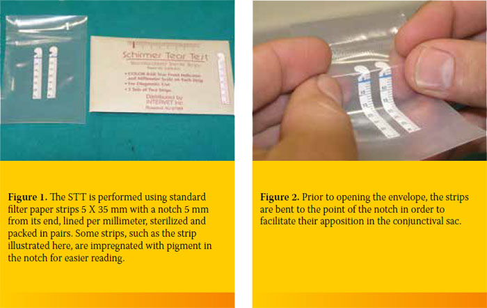
The Schirmer tear test (STT) estimates the aqueous phase of tears. Hence, it is used for diagnosing keratoconjunctivitis sicca (KCS) and quantitative KCS, in particular. Diagnosis of qualitative KCS is established using other tests. In clinical practice, STT I is used commonly as it does not require local anesthesia. STT I measures both basic and reflex tear production. Normal values in the STT I range between 18.64 ± 4.47 mm/min and 23.90 ± 5.12 mm/min in the adult dog and 14.3 ± 4.7 mm/min and 16.92 ± 5.73 mm/min in the adult cat, although feline KCS is uncommon. In dogs, values lower than 5 mm indicate severe KCS. Values 5-10 mm indicate KCS while 10-15mm raises suspicion of the disease and should be evaluated in association with other clinical symptoms.
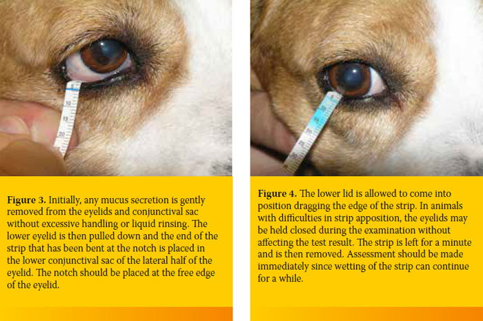
The STT precedes the ophthalmic examination to avoid the induction of reflex tears due to handling, and before the topical use of other diagnostic solutions. Prior to re-examinations, topical application of ophthalmic solutions should be avoided. Before the test, any mucus discharge that may be present on the eyelids and conjunctival sac is gently removed without excessive handling or liquid rinsing.
> Fluorescein test
The most common indication for topical use of fluorescein, which will also be mentioned here, is the detection of corneal ulcers. Fluorescein is additionally used to detect aqueous humour leakage from the anterior chamber (Seidel test), for qualitative KCS diagnosis (break up time), and to examine lacrimal apparatus patency.
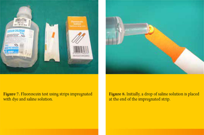
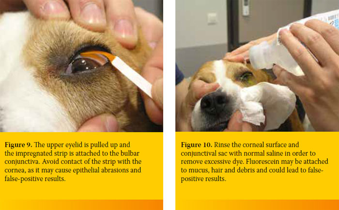
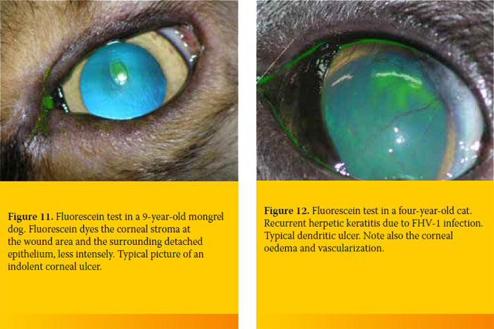
Fluorescein has the maximum spectral absorption of light at 490 nm (blue light) and has the capacity to convert 100% of the absorbed light at 520 nm (green light). For this reason, its visualization is easier when illuminated with blue light. Fluorescein is highly lipophobic and hydrophilic and thus not normally absorbed by the corneal epithelium because of fat contained in the cell membranes. With the occurrence of epithelial damage, fluorescein is absorbed by the corneal stroma giving the characteristic green colour. Fluorescein is not absorbed by Descemet’s membrane. Consequently, only the periphery of a pre- Descemet ulcer is stained rather than its bottom.
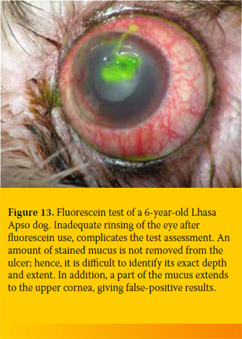
Fluorescein is available on the market either in single dose vials of 2% solution or in disposable sterile paper strips. The second form is easier to find in Greece. Multi-dose solutions should be avoided as they are considered to be contaminated. A fluorescein stain may be seen directly. For a safer evaluation of the test, a blue colour light in a dark environment can be used with the aid of magnification.
> Suggested Reading
1. Featherstone HJ, Heinrich CL. Ophthalmic examination and diagnostics Part 1. In : Veterinary Ophthalmology. Gelatt KN, Gilger BC, Kern TJ (ed). 5th edn. John Wiley & Sons, Inc: 2013, pp. 533-613
