

Remember how...
Jugular vein catheter placement
Indications & technique
Indications
Jugular vein catheter (JVC) placement in the dog is a relatively quick and easy to be performed procedure that does not demand sedation of the animal, although the presence of a veterinary nurse is deemed necessary. Various catheter types that are appropriate for jugular vein catheterization are available in the market. Catheters designed for humans can also be used. Jugular vein catheters are categorized according to the number of channels that they have (signlechanneled, double-channeled, triple-channeled), their length (6 to 18 cm) and their diameter (14 to 20 gauge). The length and diameter of the JVC that will be used are analogous to the dog’s body size, while the number of channels is chosen according to the purpose that the catheter is going to be used. For example, if JVC is placed for caustic drug administration (intravenous administration of doxycyclin) a signle-channeled type is chosen, while if JVC placement is decided for long-term fluid administration a multi-channeled type is preferred. If a JVC is not available at the veterinary clinic a simple over the needle vein catheter (16 or 18 gauge) can be used. The most common indications for JVC placement in the dog are:
A. Long-term fluid administration (e.g. chronic renal failure)
B. Parenteral nutrition (high diameter veins are demanded)
C. Measurement of central venous pressureD. Multiple blood samples in short term (e.g. diabetes mellitus) or multiple blood samples or drug administration to fractious animals
Ε. Blood transfusion
The most serious contradictions for JVC placement are:
Α. Non cooperative animal to whom sedation is forbidden for JVC placement (sedation is not a requirement for the placement of JVC in cooperative animals)
B. Severe coagulopathies
Complications related to JVC placement, even though they are rare, can be seen both upon placement and during long-term presence of the catheter in the jugular vein. When rules regarding safe placement are followed as well as hygiene rules, complications can be prevented. Some of the most common are:
A. Severe blood loss upon procedure B. Vein rupture upon catheter placement
C. Arrhythmias
D. Phlebitis
E. Bacterial endocarditis
F. Septicemia
G. Air embolism
H. Skin infection at the insertion site
I. Head and neck edema due to tight bandaging
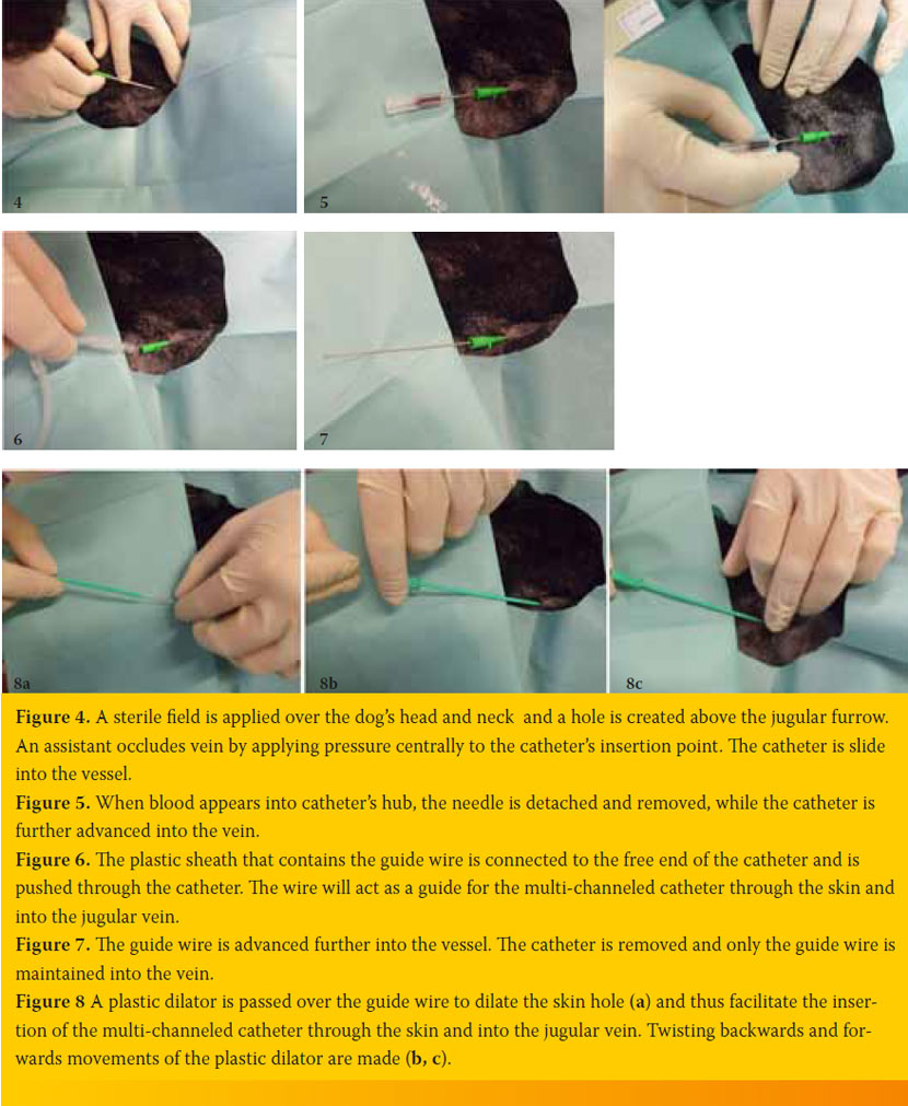
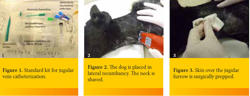
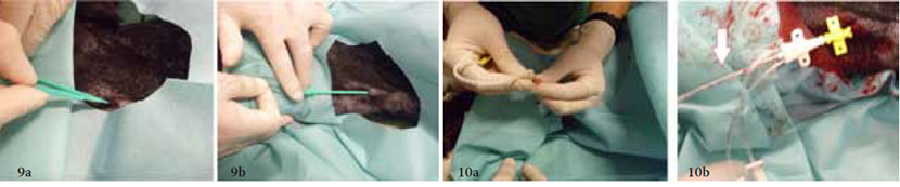
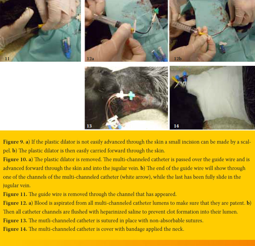
Procedure of jugular vein catheter placement
In figures 1-14 a step by step, placement of a triple-channeled jugular vein catheter in a nine year old, male, German shorthair pointer dog
suffering for chronic renal failure. The procedure was performed by one veterinarian and its nurse, and without sedation.
> Suggested reading
1. Abrams-Ogg ACG, Kruth SA, Carter RF, Valli VEO, Kamel-Reid S, Dube JD. The use of an implantable central venous (Hickman) catheter for long term venous access in dogs undergoing bone marrow transplatation. Can J Vet Res 1992, 56(4): 382-386.
2. Bexfield N, Lee K. Intravenous catheter placement – (b) jugular vein (modified Seldinger technique). In: BSAVA guide to procedures in small animal practice. BSAVA: Glousester, 2011, pp. 128-131.
3. Mesfin GM, Higgins MJ, Brown WP, Rosnick D. Cardiovascular complications of chronic catheterization of the jugular vein in the dog. Vet Pathol 1988, 25 (6): 492-502.
