Choose the Volume to see a summary of Abstracts
-
Abstracts Volume 7
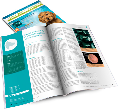
Canine thyroid tumours: diagnosis and treatment
Τhyroid neoplasia accounts for 1-3,8% of all canine tumours, and the most common types include adenoma and carcinoma, the latter being identified in 90% of cases. Exposure to radiation, iodine deficiency, hypothyroidism, or even genetic mutations, are included among the triggering factors. Benign adenomas are usually small and movable, without infiltration of surrounding tissues and the presence of metastasis. In contrast, carcinomas are larger, with extensive infiltrating tendencies, primarily in the lungs and regional lymph nodes. Diagnosis is based on clinical signs emerging due to compression of adjacent organs (oesophagus, trachea, larynx, pharynx), endocrine testing and diagnostic imaging. To reach a definitive diagnosis biopsies for histopathological examination need to be obtained, which will confirm the origin of the mass and will differentiate benign from malignant neoplastic tissue. In cases of thyroid adenoma, surgical excision of the tumour is the treatment of choice and leads to clinical cure. The treatment of choice and the prognosis for thyroid carcinoma depend on tumour size, the extent of infiltration of surrounding tissues, the presence of metastases and the possibility of using alternative treatments. Surgical management is indicated in cases of small and movable carcinomas or with superficial infiltration of surrounding tissues, but it is not recommended in cases of extensively infiltrative and fixed carcinomas.
Transpelvic urethrostomy in three cats
This study describes the application of transpelvic urethrostomy in three male, domestic, shorthair cats. Two male cats, aged one and three years old, presented with micturition disorders, due to stoma stenosis, following perineal urethrostomy, indicated to treat recurrent obstruction of the lower urinary tract. The first cat had undergone a second procedure to restore the stoma stricture. The third cat, aged three, presented with urethral obstruction attributed to lithiasis at the level of the bulbourethral glands, and was admitted with micturition inability, following a failed urinary catheterisation. Transpelvic urethrostomy was selected in all three cases, as this surgical technique allows urethrostomy to be performed approximately 1 centimetre anterior to the bulbourethral glands. Post-operatively, cat No 1 occasionally presented with urinary tract infections, treated with appropriate antimicrobial medication. Cat No 2 revealed pyuria and suppuration of the surgical wound, treated with the placement of a Foley catheter and appropriate antimicrobial medication. Cat No 3 was free of symptoms post-operatively. Stoma stenosis did not occur in any of the cases.
Exposure of a cat to human-edible mushrooms: were they toxic?
The owners of a 3-month-old female DSH cat witnessed her eating raw mushrooms of the species Boletus edulis, Boletus aereus and Amanita caesarea. These mushrooms are edible for humans and highly prized in various cuisines. Vomiting, hypersalivation, horizontal head oscillation and limb muscle tremor were developed within 6 hours. Two days later the cat was admitted due to depression and anorexia, while the neurologic signs had subsided. Dehydration, depression, lymphopenia, increased serum urea nitrogen concentration, proteinuria and bilirubinuria were detected. During the 5-day-hospitalisation period, treatment comprised of intravenous fluids, and per os vitamin E and hepatoprotectants (SAMe – vitamin Ε – vitamin C – silibinin complex). Due to mucohaemorrhagic diarrhoea present on the first day of hospitalisation, ampicillin and sucralfate were subsequently added. The kitten recovered completely a week later and was still healthy 8 months later. Mushrooms in general, are classified as edible or poisonous; the latter could be hepatotoxic, neurotoxic, nephrotoxic, gastroenterotoxic, muscarinic or coprinoid. This basic classification based on human experience may not apply to other species, and consequently “edible” mushroom species may be potentially toxic for animals. In addition, in many cases of mushroom ingestion in animals, the species involved remained unidentified. Thus, this case report describes presumed poisoning from three identified mushrooms, Boletus edulis, Boletus aereus and/or Amanita caesarea, which are considered edible for humans, but caused gastrointestinal, hepatic and neurologic signs in a cat. Prognosis in these cases may be favourable, if early supportive care is instituted.
Anaesthesia in pregnant dogs and cats
Anaesthesia during pregnancy in companion animals is necessary in cases of programmed or emergency Caesarean section or in cases when surgical intervention is required. During pregnancy, there are physiological changes in pregnant animals that should be taken into consideration during anaesthesia. These changes mainly affect the cardiovascular and respiratory system and influence to a lesser extent other systems, such as the digestive tract. In case of a Caesarean section, the preanaesthetic agents are usually either avoided or opioids are selected, although recent data support the use of alpha2-adrenergic agonists. Preparation for surgery occurs prior to the induction of anaesthesia, while oxygen is already being inhaled. The standard intravenous anaesthetics can be used for the induction of anaesthesia. In dogs, propofol is superior to thiopental, whereas etomidate is a very good choice in severely debilitated patients. For cats, the use of an α2-adrenergic agonist with ketamine is an adequate choice in order to ensure anaesthetic maintenance. The latter can be achieved in dogs with inhaled anaesthetic agents such as isoflurane, or with injectable anaesthetics like propofol. Epidural anaesthesia is a viable option in some cases. Postsurgical analgesia is based on opioids and/or nonsteroidal anti-inflammatory drugs and it is necessary so that the mother can take care of the neonates. In cases where the pregnant animal undergoes anaesthesia for a non-obstetric surgical procedure, the risk of embryotoxicity must be prevented, e.g. by avoiding benzodiazepines during the first stages of pregnancy, and accurately assessing anaesthetic depth.
Protein-losing enteropathy in the dog: a report of two clinical cases
Protein-losing Enteropathy (PLE) stems from multiple causes and it manifests as malabsorption syndrome. In the present report two canine cases of PLE are described, due to eosinophilic, lymphoplasmacytic enteritis and intestinal lymphangiectasia. History and physical examination findings included chronic intermittent diarrhoea, weight loss, subcutaneous oedema and ascites. Biochemistry revealed hypoalbuminaemia and hypoproteinaemia. Both cases underwent exploratory laparotomy in order for multiple histopathology samples from the small intestine to be collected. Treatment was administered in both cases for the primary cause. Response to treatment was satisfactory, despite complications due to long-term administration of medications. These specific cases are of particular interest due to comparatively long-term survival times and satisfactory clinical responses following initiation of treatment, despite the poor prognosis usually reported in the literature.
Study of the bacterial population of the duodenum and presence of bacteria in the bile of cats with chronic inflammatory bowel disease, cholangitis, pancreatitis, triaditis and small intestinal lymphoma, in comparison to healthy cats
The etiopathogenetic relationship between the intestinal flora and the presence of bacteria in bile in feline gastrointestinal disorders has not been studied previously. The aim of the study was the bacteriological analysis of duodenal juice and bile in cats with chronic inflammatory bowel disease (IBD), cholangitis, pancreatitis, and their combinations (triaditis), as well as in cats with intestinal lymphoma. In this prospective study 49 sick cats were included, 45 (25 symptomatic, 20 asymptomatic) with histopathological evidence of IBD, and/or cholangitis, and/or pancreatitis and four with intestinal lymphoma, as well as eight healthy cats. Samples of duodenal juice and bile were collected during exploratory laparotomy and cultured under aerobic, anaerobic and microaerobic conditions in order to isolate, enumerate and identify bacteria following standard microbiological guidelines. Comparisons of the bacterial populations of the duodenum among the groups of cats of the study regarding the growth of aerobic (P=0,831), anaerobic (P=0,406) and the total population of bacteria (P=0,752) did not outline any statistically significant differences. A statistically significant difference was noted in cats with triaditis regarding the growth of anaerobic Clostridium spp. (P=0,055). Τhe bile samples of the normal and most (48/49, 98%) of the sick cats were bacteriologically negative. However, growth of a strain of Enterobacter cloacae was noted in a bile sample of a cat with IBD and pancreatitis. Inflammatory disorders of the small intestine, the liver, and the pancreas are not related to bacterial growth in the bile. In order to confirm the possibility of triaditis being correlated with an overgrowth of anaerobic intestinal, such as Clostridium spp., further research using sensitive molecular diagnostics will be necessary.
Diagnostic dilemma: neurological or orthopaedic case?
A diagnostic dilemma commonly encountered by the clinician is whether the abnormal gait of a dog can be attributed to a neurological or an orthopaedic origin. In general, lameness is considered to result from orthopaedic disorders, whereas ataxia is expected to be neurological in origin. However, it is not uncommon to encounter cases with uncoordinated gait due to orthopaedic disorders (e.g. hip dysplasia) or lameness due to neurological disorders (e.g. nerve root signature). In order to solve this diagnostic riddle, the careful collection of data from the physical examination and, especially, from the orthopaedic and neurological examination, as well as from several diagnostic tests including imaging and electrophysiology testing are necessary.
Canine anal furunculosis: is there a place for surgery?
Anal furunculosis is a chronic progressive canine inflammatory disorder affecting the anus and the perianal region, resulting in ulcers and blind fistula in the skin and subcutaneous tissues of the perianal region. German shepherds are predisposed to this condition. The origins of the disorder are unknown but it can be immune- mediated. Treatment can be medical or surgical, but complete cure is uncommon. Medical treatment with immunomodulatory drugs, mostly cyclosporine or cyclosporine and ketoconazole usually provides satisfactory results. Surgical treatment is employed in cases with no response to medical management, due to increased cost or prolonged duration of the latter and includes complete anoplasty and simultaneous anal sacculectomy.
-
Abstracts Volume 6
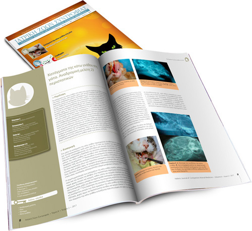
Fractures of the mandible in cats. Retrospective study of 23 cases
Fractures of the mandible in cats often occur due to falls from heights, car accidents or human abuse. The current retrospective study describes 23 cases of feline mandibular fractures, which were treated at the Companion Animal Clinic, at the School of Veterinary Medicine of the Aristotle University of Thessaloniki, during 2008-2015. Young male animals were mostly affected. Eight fractures were caused by car accidents, one after falling from great height, one by human abuse and thirteen were of unknown cause. However, thirty-three fractures are discussed in total, as four cats exhibited multiple fractures. In particular, eleven fractures were located in the mandibular symphysis, seventeen in the rammus and five in the body of the mandible. Mandibular symphyseal separation was treated either with metal wire introduced circumferentially behind the canines or with a figure-of-eight wire placed intraorally between the canines, bonded with acrylic resin. Fractures of the body were repaired with wire, placed either interdentally reinforced with acrylic resin or intrabony combined with screws. Eighteen animals were hospitalised postoperatively. Finally, repetitive surgery was required in one case, while recovery was uncomplicated or minimally complicated for the rest.
Hypothermia in companion animals
Companion animals maintain their body temperature through thermoregulatory mechanisms located in the hypothalamus that stimulate the production or reduction of heat accordingly in the event of a sudden fall or increase in environmental temperature. Hypothermia occurs when the body temperature falls below 37.5οC. Young, aged, malnourished and small animals are more susceptible. Hypothermia can be caused by exposure to low ambient temperatures or through the disruption of thermoregulatory mechanisms due to various factors including surgical procedures, trauma, systemic disorders, drug administration etc. A drop in temperature results in a lower basal metabolic rate and abnormal function of the cardiovascular, respiratory, urinary and central nervous systems, as well as acid-base balance and haemostasis. Bradycardia, hypotension, bradypnoea, pulmonary hypoventilation and deterioration of consciousness, shivering, hypovolaemia, electrolyte disorders, hyperglycaemia, as well as respiratory and metabolic acidosis are exhibited. Treatment includes rewarming therapy and stabilization via oxygen and fluid administration. The rewarming techniques may be external or internal. External passive techniques include wrapping the animal in blankets and heating its environment. Active external rewarming entails placing a heat source around the animal’s body, such as warm iv solution bags and heating pads, whereas active internal rewarming involves intravenous therapy of warm fluids and more invasive techniques such as abdominal or pleural lavage with warmed solutions.
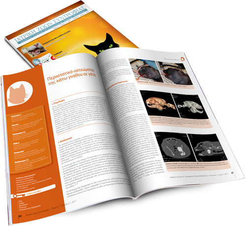
Osteoma of the mandible in a cat
A seven-year-old cat was admitted due to dysphagia caused by a mandibular tumor. Upon examination, a round, non-painful, firm mass which was attached to the underlying bone was revealed. Radiography and computed tomography of the mandible were performed. The mass involved the area between the last premolar tooth and the angular process, up to the neck of the condyloid process. Based on the findings of the computed tomography, a tentative diagnosis of a peripheral osteoma was made. Caudal mandibulectomy was performed and the mass was excised en bloc. Histopathological examination confirmed the diagnosis of an osteoma. Recovery was uneventful; the animal was able to eat quite normally a week after surgery and is symptom-free one year later.
Ovarian teratoma with inflammatory components in a bitch
A 2-year-old female mixed breed dog was presented with a history of abdominal distention of 2 month duration. Physical examination revealed a non-painful firm abdominal mass. Diagnostic imaging demonstrated a mid-abdominal mass with numerous mineralized densities. An exploratory coeliotomy revealed a mass associated with the left ovary, which was removed through a complete ovariohysterectomy. Histopathological examination of the mass was compatible with a benign ovarian teratoma with inflammatory components. Three years after surgery the dog was reported to be well.
Congenital esophageal diverticulum in a dog. A case report
A rare case of congenital esophageal diverticulum in a male, intact, 2-month-old Yorkshire terrier is described in the present report. This is a congenital malformation of the esophageal wall that occurred during fetal development, resulting in incomplete formation of the lamina muscularis and, consequently, herniation of the esophageal wall and distension of the esophageal lumen. The main causes of admission included vomiting and regurgitation. This congenital defect was diagnosed based on esophageal endoscopy findings. Treatment was conservative and included the administration of metoclopramide and dietary guidelines.
Elbow hygroma in the dog. Which treatment works better
Elbow hygroma is a serous fluid accumulation over the olecranon caused by repetitive trauma in dogs lying on hard surfaces during housing. Diagnosis is made by physical examination and fi ne needle aspiration of the hygroma cavity. Treatment may be conservative or surgical. Padded bandages and soft bedding are provided for small hygromas until the formation of a callus. Surgical intervention in reserved for recurrent, large or complicated hygromas and includes drainage or surgical excision.
Pleural effusion in the cat: a focus on laboratory diagnosis
Pleural effusions constitute a common entity in feline medicine. Laboratory evaluation of pleural fluids remains the cornerstone of a proper diagnosis. Total nucleated cell count, total protein concentration and haematocrit values are the most important indices, which, along with cytological findings, are used for the classification of an effusion (transudate, modified transudate, exudate, haemorrhagic effusion). Occasionally, the biochemical examination of effusions is necessary in order to determine the presence of fluid of a more specific aetiology (chyle, feline infectious peritonitis effusions, septic exudates), while microbiological examination remains a standard procedure in suspected septic effusions. After having considered the obtained information, clinicians are usually able to understand the aetiology behind cavitary fluid accumulation and thus make the best therapeutic decision for the cat patient.
Full-thickness mesh skin grafts in dogs and cats. Indications, pathophysiology of graft taking, surgical techniques and complications
Skin grafts are comprised of the epidermis and dermis. They are harvested from the donor site and transported to the recipient site of the same animal where they undergo adhesion, osmotic fl ow of plasma into the graft (plasmatic imbibition), vascular anastomosis and revascularization in order to be accepted by the recipient site. Full- thickness mesh skin grafts are most commonly used in the veterinary clinical setting in both dogs and cats for the reconstruction of skin defects located mainly on the limbs, where other reconstruction methods are not available. Grafts are placed on skin defects with healthy granulation tissue or on surgical wounds with adequate blood perfusion. Survival of feline grafts is far superior to that of canine grafts.
-
Abstracts Volume 5
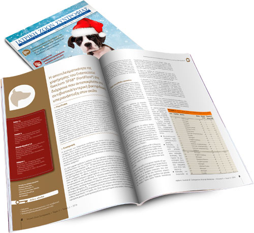
Effect of Enterococcus faecium SF68® (FortiFlora®) administration in dogs with antibiotic responsive or small intestinal bacterial overgrowth diarrhοea
The objective of this study was to assess whether dogs suff ering from small intestinal diarrhoea due to small intestinal bacterial overgrowth or antibiotic responsive diarrhoea would benefi t from a combination of Purina Veterinary Diets® EN Gastroenteric® Canine Formula and Purina Veterinary Diets® FortiFlora® Canine Probiotic Nutritional Supplement (Enterococcus faecium SF68®). The study involved twenty-six adult dogs presenting with symptoms compatible with chronic small intestinal diarrhoea that could not be attributed to any specifi c cause. The dogs were randomly divided into two groups: A (EN Gastroenteric plus FortiFlora sachet) and B (EN Gastroenteric plus placebo sachet). Reassessment of symptoms was scheduled every seven days over a 20-day period. Faeces were characterised based on their consistency. Biochemical and haematological parameters of all dogs participating in the study were within normal limits throughout the entire study period. During the fi rst week of the study, no statistically signifi cant diff erences were found between groups A and B in the characteristics of the diarrhoea. After a 14-day period, comparisons between groups showed that there was a statistically signifi cant diff erence (p = 0.0002) regarding resolution of diarrhoea in group A. During the third week of administration, group A maintained the positive outcome (p = 0.0001).
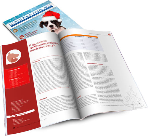
The interpretation of erythrogram in dog and cat
Εrythrogram is part of the complete blood count and includes the number of erythrocytes, hemoglobin concentration, hematocrit, erythrocyte indices (mean corpuscular volume, mean corpuscular hemoglobin, mean corpuscular hemoglobin concentration), red cell distribution width, the number and percentage of reticulocytes and erythrocyte morphology. Appropriate blood sampling and handling are essential for the validity of the erythrogram interpretation. Erythrogram major abnormalities are anemia, erythrocytosis/polycythemia, erythrocyte morphologic abnormalities and erythrocyte inclusions. Anemia can be either regenerative or nonregenerative. Regenerative anemia is divided into blood loss anemia and hemolytic anemia. Common causes of blood loss anemia are traumatic injuries and hemostatic disorders, while hemolytic anemia can be immune-mediated, microangiopathic, associated with Heinzbody formation or due to genetic defects of red blood cells. Nonregenerative anemia includes anemia of chronic disease, anemia of chronic renal failure, aplastic anemia and nutritional anemia. Erythrocytosis/polycythemia can be relative (hemoconcentration) or absolute, which is further divided into primary (polycythemia vera) and secondary. The appearance of abnormal erythrocytes can be either an artifact or associated with certain disorders. The erythrocyte inclusions are either of non-infectious origin, such as Howell-Jolly and Heinz bodies or related to infections, such as Babesia spp. and hemotropic Mycoplasma spp., the observation of which into the erythrocytes sets the defi nitive diagnosis of the relevant diseases.
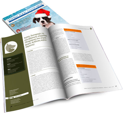
Simple acid-base disorders (management at the clinic without blood gas analysis)
Acid-base balance is an important homeostatic mechanism that focuses on maintaining a constant concentration of hydrogen ions in body fluids and it is expressed by the negative base-10 logarithm of hydrogen ion concentration (pH). The normal ranges of pH are between 7.35 and 7.45. However, when the concentration of H+ rises, a decrease of pH is observed and this condition is called acidosis. Conversely, a decrease in concentration of H+ increases pH, a condition termed alkalosis. When the above disturbances are caused by a change in the partial pressure of carbon dioxide (CO2), they are defi ned as respiratory disturbances (respiratory acidosis and respiratory alkalosis), whereas if caused by a change in the concentration of bicarbonate ions (HCO3), they are described as metabolic disturbances (metabolic acidosis and metabolic alkalosis). It is often possible that a combination of two or more disorders may occur (mixed disorders). Typically, the diagnosis of acid-base disorders is based on the measurement of pH, partial pressure of CO2 and the concentration of HCO3 in an arterial blood sample. In cases where the above control is not possible, the diagnosis or suspicion of occurrence of an acid-base disorder is established on the fi ndings of the clinical examination and electronic monitoring of vital functions. Knowledge of the mechanism by which a disease can cause an acid-base disorder is of signifi cant assistance to the veterinarian. The detection of any acid-base disturbance leads to early and intensive treatment which aims to eliminate the cause of primary disease.
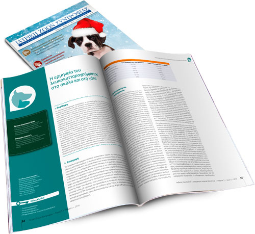
The interpretation of leukogram in dog and cat
Leukogram includes total and differential white blood cell counts, as well as leukocyte morphology. Appropriate blood sampling and handling are essential for the validity of the leukogram interpretation. Leukocytosis and leukopenia are associated with physiologic or pathologic conditions. From a clinical point of view, neutrophilia, neutropenia, lymphocytosis, lymphopenia, monocytosis and eosinophilia are considered the most important alterations in leukocyte numbers. Neutrophilia and monocytosis usually accompany infl ammatory diseases, but they are also observed in excess of steroids. Additionally, neutrophilia can be physiologic, as well as, lymphocytosis, which is further associated with chronic infl ammatory diseases. Eosinophilia typically occurs in hypersensitivity reactions and parasitisms. Neutropenia is mainly related to infectious diseases, while lymphopenia is usually steroid-induced or related to acute infectious diseases. Monocytopenia, eosinopenia, basophilia and basopenia have limited diagnostic signifi cance.Common morphologic changes of the leukocytes include the presence of immature, hypersegmented or toxic neutrophils, as well as reactive lymphocytes and various leukocyte inclusions. These may be of infectious origin, such as morulae of Ehrlichia canis and Ehrlichia ewingii or of non-infectious origin, such as siderotic inclusions.
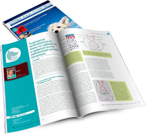
Chromatic pupillary light reflex and its application in small animal ophthalmology
The recent discovery of the existence in the retina of a third group of photosensitive cells, other than cones and rods, which have the capacity of stimulating the pupillary light reflex, has changed our knowledge of how the iris reacts to different wavelengths of light and introduced the concept of chromatic pupillary light reflex in eye examination. This is a test whereby the pupillary response is stimulated not by monochromatic white light, but successively by red and blue light, allowing the selective stimulation of photoreceptors. The chromatic pupillary light reflex is particularly useful in the diagnosis of sudden acquired retinal degeneration, progressive retinal atrophy, chorioretinopathies, retinal detachment, glaucoma, disorders of the optic nerve and optic chiasm and certain brain diseases that cause blindness.
Canine hip dysplasia. Part I: Aetiopathogenesis & diagnostic approach
Hip dysplasia in dogs is a multifactorial disease caused by hereditary and environmental factors. Most commonly seen among very young giant and chondrodystrophic breeds, it manifests as an abnormal development of the round ligament of the femoral head that leads to coxofemoral joint instability and (sub)luxation. The disease presents as hind limb lameness. In the young animal, this is due to pain caused by hyperextension of the soft tissues of the hip joint, whereas in adults it is caused by osteoarthritis resulting from the degenerative development of the disease. In addition to radiological examination, diagnosis is based on clinical signs and specific clinical trials in both the awake animal and the animal under deep sedation or/and general anaesthesia. Radiologic imaging requires several views, and findings are evaluated according to the animal’s age, stage of disease and clinical signs. The need for early assessment of hip dysplasia resulted in Prassinos N.N. the establishment of different classification systems according to radiological findings.
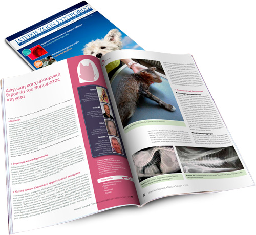
Diagnosis and surgical treatment of thymoma in the cat
Thymoma is a rare neoplasm seen in aged cats that derives from thymic epithelial cells and is usually located in the cranial mediastinum. Clinical signs include dyspnoea, coughing, anorexia, lethargy or regurgitation. Feline thymoma may also be associated with myasthenia gravis or exfoliative dermatitis. Diagnosis of the tumour is based on diagnostic imaging. Ultrasonography provides information concerning the consistency of the mass. The extent and invasiveness of the mass can only be determined by computed tomography. Cytologic examination can also facilitate diagnosis where thymic epithelial cells and small lymphocytes dominate. Surgery is the treatment of choice for feline thymomas with favourable results. Thymomas are approached through a median sternotomy or intercostal thoracotomy. Definite diagnosis of thymoma can only be confirmed by histopathologic examination. The prognosis of feline thymoma is favourable, providing it is not associated with paraneoplastic syndromes or metastatic disease.
-
Abstracts Volume 4
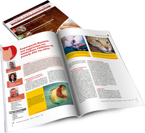
Surgical treatment of squamous cell carcinoma of the nasal planum in cats
Squamous cell carcinoma (SCC) of the nasal planum is a malignant neoplasm commonly seen in older cats. Long-term ultraviolet light exposure and light pigmentation or depigmentation and sparsely-haired skin are considered main risk factors for SCC. This tumour can be slowly progressive and locally invasive with a low rate of metastasis to the regional lymph nodes or lungs. Skin lesions are characterized by erythema, crusts, erosions or deep ulcers. Diagnosis is based on history and physical examination and can be confirmed by histopathological examination of the skin. Nosectomy is the recommended treatment for the invasive SCC. Prognosis is usually good to excellent with good cosmetic results in completely resected tumours.
Management of an extensive partial and full thickness skin burn in a dog with the aid of medical honey
A 9-year-old male mixed breed dog was admitted for the management of a partial and full thickness thermal burn that was covering 45% of his body including the dorsal, lateral and ventral thorax and abdomen, medial thigh and caudal elbow. Medical honey (L-Mesitran® soft) was applied to the burn area on a daily basis and dressed with absorbent pads resulting in complete healing by contraction and epithelialisation 35 days after initial admission. Medical honey may be used as an effective alternative for the treatment of extensive skin burns.
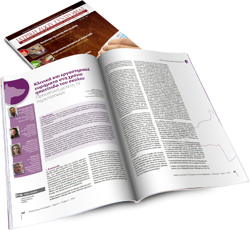
Clinical and laboratory findings in canine chronic hepatitis
Prospective study of 19 cases Canine chronic hepatitis (CH) is a group of liver diseases, the causes of which are different and little known, with similar clinical presentation and laboratory findings but with differences in the histological findings, prognosis and treatment.
The current prospective study concerns 19 cases of CH that were admitted, examined, diagnosed, treated and followed up at the Companion Animal Clinic, School of Veterinary Medicine, A.U.Th. between October 2012 and October 2014. During that period, 35 animals were admitted with clinical symptoms suggestive of liver disease. However, only 19 animals with histologically confirmed CH were included in the study.
The majority of dogs were of mixed-breed with a mean age of 6.5 years. The most common symptoms included depression, anorexia and vomiting. Though observed in certain cases, disorders in the blood count were uncommon. The main laboratory finding was that of increased liver enzyme activity: alanine aminotransferase (ALT) and alkaline phosphatase (ALP). Histopathology revealed a varying degree of inflammation, mostly involving lymphocytes and plasma cells in every case, fibrosis in several cases and apoptosis and/or necrosis and regeneration in few cases. Finally, one case was characterized as copper-associated CH.
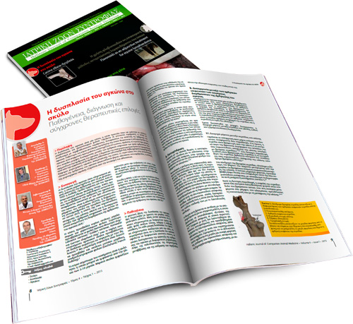
Canine elbow dysplasia Aetiopathogenesis, diagnosis and current treatment recommendations
Elbow dysplasia is a condition that causes pain and lameness in large and giant breed dogs. Its origins are genetic and when combined with environmental factors, development of the elbow joint becomes abnormal. Originally, elbow osteochondrosis was considered to be the main cause of this condition. Modern studies claim that the condition, in most cases, is caused by various forms of incongruity between articular surfaces of the three joints forming the elbow. Treatment is surgical and should be performed before the development of osteoarthritic lesions in the joint. Multiple surgical techniques to correct this condition are described in the literature. In cases when radiological examination of the elbow joint reveals severe osteoarthritic lesions, the selection of surgical technique depends on lesion localization.
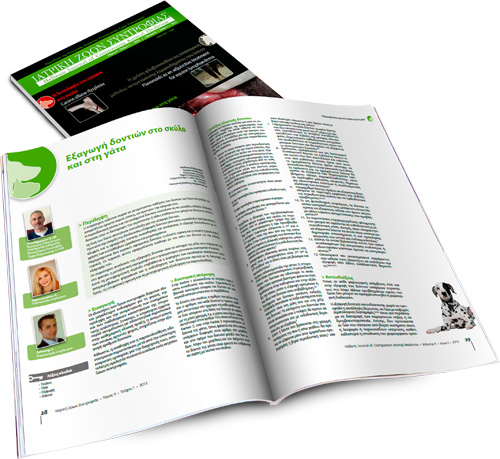
Tooth extractions in dogs and cats
Tooth problems are very common among small animals. The veterinarian encounters them frequently, and quite often has to suggest tooth extraction. Periodontal disease and tooth fractures are the most common indication for extraction, while clotting defects or other pathological conditions which may endanger the animal’s life are contraindications. Appropriate pre-anaesthetic control and regional anaesthesia and analgesia should be considered before the extractions. The veterinarian should use proper instruments and pieces of equipment for such procedures. There are two basic extraction techniques: namely, the open and the closed technique. The choice of either technique will depend on several factors. The most usual complications after extraction are root fractures, massive post operative bleeding and soft tissue trauma. Postoperatively, the animal should receive the appropriate analgesics, antimicrobials, recommendations concerning the animal’s diet and home dental care.
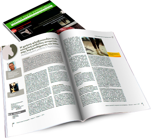
Flavonoids as an adjunctive treatment for equine lymphoedema: Retrospective study of five cases
Equine lymphoedema, a common pathologic condition characterized by lymph stasis, requires immediate treatment to ensure a positive outcome. Ascertaining the specific causative agent is not always possible. Despite typical signs of the syndrome, the complex pathogenesis impedes the establishment of an aetiologic treatment protocol. The use of flavonoids in human lymphoedema in recent years has provided the basis for their experimental use in equine lymphoedema. In this study, a treatment protocol for the management of lymphoedema was applied to five horses. The treatment, which included conservative measures, anti-inflammatory and antimicrobial therapy as well as flavoinoids, provided encouraging results, indicating that flavonoids could be a promising therapeutic option for the treatment of equine lymphoedema.
-
Abstracts Volume 3
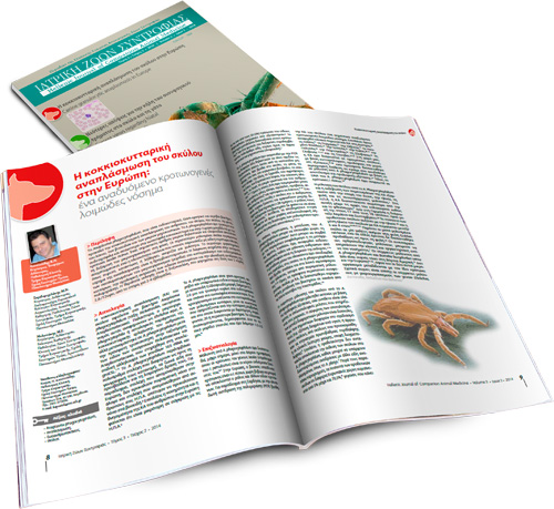
Canine granulocytic anaplasmosis in Europe: an emerging tick-borne infectious disease
Anaplasma phagocytophilum, an intracellular, gram-negative aerobic bacterium, is the cause for granulocytic anaplasmosis in people, horses, dogs, cats, wolves, cattle and small ruminants. A. phagocytophilum mainly targets neutrophils and rarely eosinophilic granulocytes; in Europe it is transmitted by the tick Ixodes ricinus. A significant number of small mammals and deer comprise the reservoir for the microorganism in the wild. The majority of infected dogs remain asymptomatic, whereas those that develop clinical signs mostly present with non-specific signs like fever, depression or lethargy, anorexia and lameness. The most common laboratory finding of anaplasmosis is thrombocytopenia. Diagnosis is based on finding aggregates of the organism (morulae) in the cytoplasm of neutrophils, serological detection of specific antibodies and polymerase chain reaction. The treatment of choice is Doxycycline, administered per os at a dose of 5 mg/kg Β.W./12 hours for 2-4 weeks.
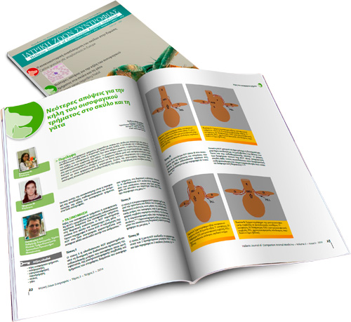
Current views regarding hiatal hernia in dogs and cats
Τhe cause and pathophysiology of hiatal hernia are not yet fully understood, but hiatal hernia seldom appears in dogs and is rarer in cats. Sliding hiatal hernia is the most common type to present in clinical practice. Chinese Shar-Pei dogs are predisposed to the development of this type of hernia. The majority of clinical signs are due to gastroesophageal reflux. Diagnosis is based on diagnostic imaging, and esophagoscopy may provide useful information. It can be managed medically and surgically. The aim of surgical treatment is the anatomical repair and stabilization of the hernia. Prognosis for patients with hiatal hernia is usually favorable.
The aim of this review is to describe the etiology, pathophysiology and treatment of hiatal hernia in companion animals.Update on the diagnosis and therapy of canine cruciate ligament rupture
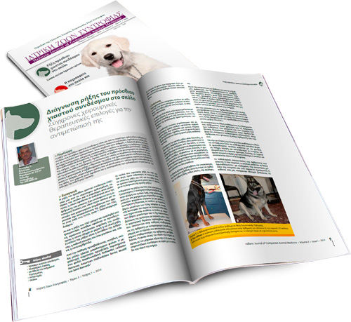
Rupture of the cranial cruciate ligament is one of the most common canine orthopedic disorders, usually caused by progressive degeneration of unknown etiology. Rarely, it can be caused by trauma, similar to that which occurs in people. Diagnosing this condition requires a detailed history, observation of the dog and physical examination of the stifle joint. Two orthopedic examinations are mandatory: the cranial “drawer” sign and the tibial compression test. Radiographic evaluation of the stifle joint must always be included to differentiate it from other pathological conditions and, also, to receive information regarding the acute or chronic nature of the rupture. Treatment for cranial cruciate ligament rupture can be either conservative or surgical. A number of surgical techniques have been previously described in the literature; however, no study has yet conclusively proven that a single technique is better than any other. Because of this fact, technique selection has been a matter of debate among orthopedic veterinary surgeons.
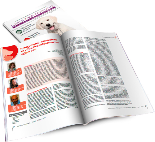
Obesity in dogs and cats: Discovering the enemy
Obesity ranks as the most common nutritional disorder reaching epidemic proportions among dogs and cats, at least in the developed world. Caused by energy intake exceeding energy loss, it results in adipose tissue accumulation in the organism negatively affecting an individual’s health. The list of hormones, neurotransmitters and substances secreted by adipose tissue itself, proven to have an active part in body weight modulation, is extensive. Predisposing factors contributing to the appearance of this disease include age, gender, breed and living conditions. The most common conditions to be associated with obesity are osteoarthritis, diseases of the cardiovascular and respiratory system, hypertension, hepatic lipidosis and type II diabetes mellitus. The evaluation and grading of obesity is based on the findings of observation and palpation of the animal, taking into consideration the predefined guidelines. The aim of any treatment plan applied in obesity management is the reduction of energy input and increase in output. This is mainly accomplished by reducing daily consumed calories and increasing physical activity. A fundamental requirement for successful management is that the owner understands the animal’s problem and is willing to cooperate with their veterinarian on a long-term basis. Finally, re-evaluation of body weight should occur at monthly intervals so that the dietary plan can be adjusted accordingly.
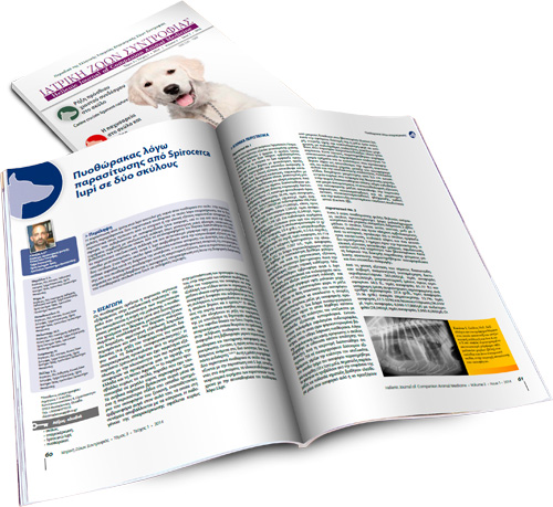
Spirocerca lupi-associated pyothorax in two dogs
Spirocerca lupi infection is an uncommon cause of pyothorax in the dog. In the present report, two cases of canine spirocercosis-associated pyothorax are described. Both cases demonstrated historical or clinical evidence of esophageal dysphagia, manifested as odynophagia and/or regurgitation, and one showed clinical findings suggestive of pleural effusion such as weakness, depression and muffled heart sounds. Thoracic radiography in the first case revealed pleural effusion and soft tissue opacity located at the caudodorsal aspect of the mediastinum, while S. lupi ova were found in fecal examination of both dogs. The first dog was euthanized at his owner’s request, while the second died suddenly during hospitalization. The definitive association between spirocercosis and pyothorax was established post mortem. These cases emphasize the importance of considering esophageal spirocercosis as a cause of canine pyothorax in highly endemic areas.
-
Abstracts Volume 2
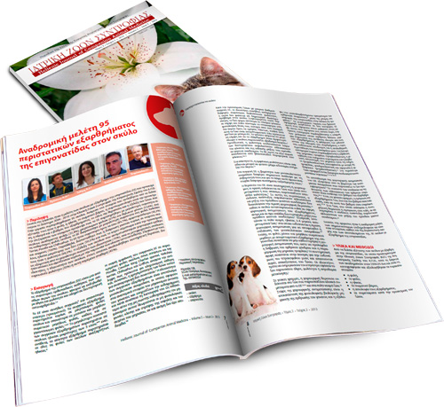
Retrospective study of 95 dogs with patellar luxation
The purpose of this study was the study of dogs with patellar luxation (PL) that were presented at the Companion Animal Clinic, School of Veterinary Medicine, Aristotle University of Thessaloniki, Greece during 2004 - 2010. Ninety-five dogs were included in the study. Among the information derived from the clinical examination records were breed, age, body weight, clinical signs upon presentation, site and grade of luxation, type of treatment and outcome of the dogs. Statistical analysis revealed that PL is observed more frequently in mixed breed (27.4%), small sized (61%) and female (52.6%) dogs. Regarding purebred dogs, PL has been more frequently diagnosed in Yorkshire Terriers, Poodles and Chihuahuas. PL was mostly medial (84.1%), bilateral (56.3%) and congenital/developmental (100%). In addition, it was noticed that diagnosis mainly concerned young dogs, aged less than two years (50.5%) and the major complaint upon presentation was lameness (69.5%). The progress of dogs which were surgically treated was considered excellent by the majority of the owners (75%), while post-operative complications were rare and mainly related to the presence of osteosynthesis materials. According to the owners, the majority of the dogs that were treated conservatively, lameness did not occur (35%) or remained stable (45%).
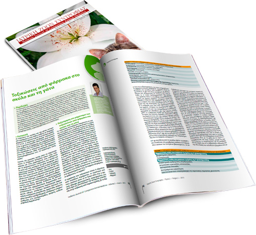
Drug Toxicities in Dogs and Cats
Drug toxicities are relatively common in dogs and cats and they can be classified into type Α or predictable which are caused by the pharmacological or the intrinsic toxic effects of the responsible drug and into type Β or non-predictable that are unrelated to the above. The appearance of type A drug toxicities depends on multiple factors that are related to the affected animal, the dosage regimen and the simultaneous administration of other drugs. Clinical manifestations most commonly originate from organ systems where the responsible drug accumulates or those that are characterized by an increased metabolic rate. In contrast, type B drug toxicities commonly affect organs presenting suitable proteins that after coupling with the drug or its metabolites (haptens) form complete antigens or organs that trap circulating immune complexes. Drugs most commonly responsible for toxicities in dogs and cats include aminoglycosides, macrocyclic lactones (avermectins and milbemycins), pyrethroids, non-steroidal anti-inflammatories, phenobarbital and diazepam.
White Line Disease: Retrospective study of 56 cases
White Line Disease (W.L.D.) refers to hoof wall separation at the junction between the stratum medium and stratum internum of the epidermis that subsequently forms a cavity. This study included 56 horses with W.L.D. that were admitted to the Equine Unit, Companion Animal Clinic, Faculty of Veterinary Medicine, Aristotle University of Thessaloniki over the last 3 years. The cause of W.L.D. has been attributed to incorrect hot shoeing (overheated-dried out hoof) in 18 horses, overhydration of the hoof in 10 horses, dehydration of the hoof due to environmental factors in 6 horses, improper shoeing (“nail bind”- small or inappropriate horseshoe, contamination of nail holes) in 8 horses and combination of the above in 6 horses. Stall hygiene (stall bedding) and training ground were inappropriate in 48 cases. Disease affected the forelimbs, unilaterally or bilaterally in 39 (69.64%) horses and hindlimbs, unilaterally or bilaterally in 10 (17.85%) horses. In the remaining 7 (12.5%) horses forelimbs and hindlimbs were randomly affected. Therapeutically, debridement of the cavity, daily rinsing with aqueous solution eosin 2%, heart-bar shoe, biotin and rest were recommended. The majority of horses (91%) responded positively in the treatment protocol described above.
This retrospective study reveals the relatively high prevalence of W.L.D. in the region of Thessaloniki considering the fact that 15-20 horses were affected per year (4% of the total population), while a 20% present with secondary disease with guarded or poor prognosis.Dermatologic emergencies in the dog and the cat
Dermatologic emergencies are rare in clinical practice and most commonly have an acute onset. A canine or feline skin disease can become life-threatening because of sepsis and toxemia, loss of fluids, protein, and electrolytes and the simultaneous involvement of vital internal organs. Emergency skin diseases include bacterial cellulitis in dogs, necrotizing fasciitis in dogs and cats, toxic shock syndrome in dogs, subcutaneous and systemic fungal infections in dogs and cats, angioedema in dogs and cats, autoimmune skin diseases with extensive ulceration in dogs and cats, Stevens-Johnson syndrome and toxic epidermal necrolysis in dogs and cats, vasculitis in dogs and cats, sterile postural erythroderma (superficial suppurative necrolytic dermatitis) of miniature Schnauzers, sulfonamide hypersensitivity syndrome in dogs, and sterile neutrophilic dermatosis (subcorneal and follicular neutrophilic pustular dermatitis or Sweet’s syndrome) in dogs. The diagnosis of these diseases is based on history, clinical signs and the results of various laboratory examinations (cytologic, microbiologic, histopathologic etc.). However, in addition to the definitive diagnosis of the skin disease itself, clinical and laboratory (complete blood count, serum biochemistry, urinalysis, coagulation profile etc.) examinations for potential systemic complications are also important. Treatment should begin as soon as possible and includes the general supportive measures applicable to all emergency cases and the specific treatment of the skin disease.
-
Abstracts Volume 1
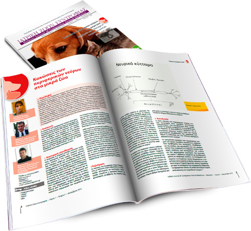
Peripheral nerve damage in companion animals
Peripheral nerve damage can occur as a consequence of accidental or iatrogenic injury caused by sharp or blunt trauma. Damage to peripheral nerves often accompanies orthopedic injuries (e.g. fracture, dislocation) and of particular clinical importance is considered the damage to nerves of the limbs. The mechanism of nerve degeneration and regeneration after nerve injury is complex. Failure of nerve restoration of normal function and the emergence of complications may both lead to permanent disability. Knowledge of pathophysiology and regeneration process is considered fundamental for the clinician, because clinical symptoms can be interpreted more accurately. Serial physical examinations and electrophysiology studies can be used to evaluate the prognosis and the suitable method of treatment. In many cases surgical exploration of the affected area can provide accurate prognostic information. Regardless of the treatment method chosen, recovery progresses relatively slow. The owner should be informed about the increased nursing care requirements involved in those cases, because his collaboration is considered essential in their management.
Traumatic brain injury in the dog and cat
Traumatic brain injury (TBI) is a frequent occurrence in dogs and cats and it is mainly caused by motor vehicle accident, fall, human violent acts and attacks from other animals. Damages in TBI are divided in primary and secondary. Primary damages take place immediately as a result of the direct mechanical destruction of the neural tissue at the time of trauma, while secondary brain damages occur within a few minutes or days following the traumatic event and they are caused by systemic extracranial injuries and intracranial biochemical alterations.
Initial assessment of an animal with TBI is focused on the life-threatening injuries and it is followed by the performance of neurologic examination.
It is difficult for the clinician to control primary brain damage. Treatment efforts must start immediately and their aim is to stabilize the animal, prevent and treat the secondary brain damages. At first general measures are taken to restore and maintain brain oxygenation. This is achieved by supporting the circulatory system with fluids and by oxygen supplementation. The aim of instituting specific measures is to minimize the brain injury.
TBI is associated with high mortality rates in both humans and animals. However, dogs and cats exhibit remarkable rehabilitation ability, provided that extended follow up is granted after a severe brain trauma. For this reason, it is strongly recommended to not infer hasty conclusions about prognosis based on the initial status of an animal presented with TBI.Aetiopathogenesis and consequences of chronic feline kidney disease: Improving by understanding
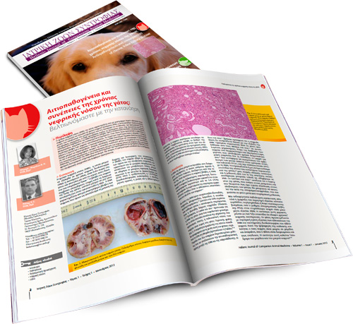
Feline chronic kidney disease (CKD) is characterized by irreversible structural lesions of the kidneys and may lead to chronic renal failure (CRF), which eventually results in accumulation of metabolic toxins and dysregulation of fluid, electrolyte, and acid-base balance. CKD mainly affects geriatric cats. In the majority of animals the initiating factor of CKD remains unclear. Idiopathic, familial, congenital, inflammatory, infectious, and neoplastic causes have been suggested. Once lesions have adequately progressed, the condition is generally self-perpetuated. The main clinical signs are anorexia, weight loss, vomiting, and diarrhoea. Early diagnosis of CKD is crucial. Anaemia, azotaemia, hyperphosphataemia, and hypokalaemia may be detected by laboratory examination. Radiology and ultrasonography of the abdominal cavity may contribute to identification of the initiating factor. Renal histopathology may aid in diagnosing the primary cause. Consequences of CKD are multisystemic and include arterial hypertension, renal secondary hyperparathyroidism, anaemia, gastrointestinal complications, and acid-base and diverse electrolyte disturbances.
Clinical signs of corneal lesions in dog and cat
Corneal damage in dog and cat can alter its clarity and transparency in light, which are essential for its function. Corneal lesions include edema, neovascularisation, pigmentation, microcrystal depositions, ulceration, inflammation or regenerative tissue formation, scar formation and finally those involving its size and curvature. This lesion may occur solely or in conjunction and may be caused by corneal, other ophthalmic or systemic diseases. In this study the above mentioned corneal lesions of the dog and cat are extensively reported and described.
 PDF Download