Scientific Journal

Scientific Journal of the Hellenic Companion Animal Veterinary Society (HCAVS)


Hellenic Journal of Companion Animal Medicine - Volume 7 - Issue 2 - 2018

Table of Contents

  • Bullet71 1

    Editorial

  • Bullet72 2

    Canine thyroid tumours: diagnosis and treatment

  • Bullet72 3

    Transpelvic urethrostomy in three cats

  • Bullet72 4

    Exposure of a cat to human-edible mushrooms: were they toxic?

  • Bullet72 5

    Anaesthesia in pregnant dogs and cats

  • Bullet72 8

    Instructions for authors


Editorial

Hellenic Journal of Companion Animal Medicine - Volume 7 - Issue 2 - 2018The present editorial was drawn up with regard to the publication of guidelines for veterinary dentistry, issued by the World Small Animal Veterinary Association (WSAVA). It is a document of major importance, covering 165 pages. The Hellenic Companion Animal Veterinary Society took the initiative to translate the guidelines into the Greek language, with a view to distributing the document to its members in the 10th anniversary Forum in March 2019.

Historically, dentistry in veterinary medicine seems to have been applied in equidae several centuries B. C. There has also been some reference to the treatment of dental problems and the attempt to determine the age of equidae since the sixth century B.C. in China. It seems that the ancient Greeks conquered the estimation of a horse’s age based on tooth growth. In the fifth century B.C., Simon of Athens, in his book “The Veterinary art, inspection of horses” referred to the age when teeth tend to erupt in horses, as well as the way to estimate their age based on their dentition. Subsequently, in 333 B.C., Aristotle in his book “History of Animals”, gave an account on periodontal disease in horses. The baton was taken by the Romans, who managed to develop significantly the dental medicine in equidae.

In the modern age, the key figure of Companion Animal Dentistry was Joseph Bodingbauer who distinguished himself in the 1930s in Vienna, while in the USA this particular field of science started to develop relatively recently, especially, after 1970. Specifically, Companion Animal Dentistry (CAD) was recognised as a separate specialty of Veterinary Medicine in the USA in 1988, after the establishment of the American Veterinary Dentistry College (AVDC) and in Europe in 1998 when the European Veterinary Dentistry College (EVDC) was founded. The first time that veterinary dentistry was established as a separate department was at the School of Veterinary Medicine at the University of Pennsylvania (U-Penn) in Philadelphia, USA in 1970s, with Dr. Colin Harvey, Professor of Surgery, being the leading figure in this endeavour. The development of Companion Animal Dentistry has been rapid over the last two decades and, according to many authors, dental, oral and maxillofacial diseases are among the most common medical conditions confronted in companion animal veterinary medicine. In North America and Europe, a significant part of the revenues of a veterinary office comes from the Companion Animal Dentistry.

Within the framework of the CAD, veterinarians combat cases of periodontal disease, which is the most common medical condition in dogs and cats, dental fractures (that are possible to be treated not only by means of extraction, but also with restoration, root canal therapy and metal crowns), inflammatory diseases of the oral cavity, orthodontic problems leading to traumatic occlusion, fractures of the mandible, neoplasms of the oral cavity, oro-nasal fistula, etc.

As stressed in the WSAVA global guidelines, dental and oral diseases cause great pain, as well as local and systemic infections. For the aforementioned reasons, it is considered that, in case dental diseases cannot be cured or they are undertreated, the animal’s welfare is compromised. The guidelines also include an extensive reference to the disastrous effects caused by the application of dental procedures without anaesthesia (anaesthesia-free, AFD or non-anaesthetic dentistry, NAD), which is definitely ineffective or even harmful. Another point of interest is the negative effect of undiagnosed or/and uncured dental diseases on the animal’s health, as well as the way they are associated with the well-being of animals.

As also stated in the guidelines, that dentistry is a specialty of veterinary medicine that has received insufficient attention so far and many inaccuracies and misunderstandings have been related to it. Teaching of veterinary dentistry in the Schools of veterinary medicine is a way to help this specialty to develop and flourish, with a view to providing the sick animals with high level medical care, as well as improving their general health condition and welfare.

Transpelvic urethrostomy in three cats


> Abstract

This study describes the application of transpelvic urethrostomy in three male, domestic, shorthair cats. Two male cats, aged one and three years old, presented with micturition disorders, due to stoma stenosis, following perineal urethrostomy, indicated to treat recurrent obstruction of the lower urinary tract. The first cat had undergone a second procedure to restore the stoma stricture. The third cat, aged three, presented with urethral obstruction attributed to lithiasis at the level of the bulbourethral glands, and was admitted with micturition inability, following a failed urinary catheterisation. Transpelvic urethrostomy was selected in all three cases, as this surgical technique allows urethrostomy to be performed approximately 1 centimetre anterior to the bulbourethral glands. Post-operatively, cat No 1 occasionally presented with urinary tract infections, treated with appropriate antimicrobial medication. Cat No 2 revealed pyuria and suppuration of the surgical wound, treated with the placement of a Foley catheter and appropriate antimicrobial medication. Cat No 3 was free of symptoms post-operatively. Stoma stenosis did not occur in any of the cases.

> Introduction

Feline idiopathic lower urinary tract disease, also referred to as feline urological syndrome or feline interstitial cystitis, resembles interstitial cystitis in humans.1 The disease is frequent in cats and may cause partial or total urethral obstruction, which is treated by catheterisation. In cases where urinary catheterisation is inapplicable, as well as in cases of recurrent urethral obstruction, unresponsive to conservative treatment, perineal urethrostomy (PU) is implemented.2-5 However, in cases of urethral obstruction, where PU cannot be applied or has failed, other urethrostomy techniques have been proposed, such as prepubic urethrostomy, subpubic urethrostomy and transpelvic urethrostomy (TPU). PU and prepubic urethrostomy have been extensively described,2-13 as opposed to subpubic urethrostomy and TPU, which are poorly cited in the relevant literature.14-16

The purpose of this study is to describe the technique, the progression, the outcome and the complications of TPU in three male cats.

> Interesting cases

Case 1

A one-year-old Domestic Shorthair (DSH) male cat was admitted due to micturition disorders. The cat lived indoors along with other cats and dogs and was fully vaccinated and dewormed. Two months prior to its admission, it had undergone PU, due to recurrent lower urinary tract obstruction. However, one month post-operatively, it exhibited stranguria and dysuria, due to stricture of the urethrostomy, and underwent a corrective PU. Clinical examination identified severe stenosis of the urethrostomy. The cat’s urethra was catheterised with a 1 mmdiameter catheter, following a small incision of the fibrotic ring formed at the site of the stoma, and a TPU was performed.

Case 2

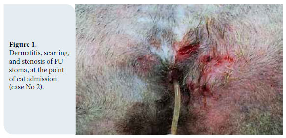
A three- year- old DSH male cat was admitted due to micturition disorders. The cat lived indoors, as well as outdoors along with other cats, and was fully vaccinated and dewormed. Four months prior to its admission it had undergone PU, due to recurrent lower urinary tract obstruction. However, two months post-operatively it presented with gradually aggravating stranguria and dysuria. When admitted, the cat had been administered enrofloxacin (5 mg/ kg/24h, po) to treat a diagnosed lower urinary tract infection. Clinical examination revealed scarring and stenosis of the urethrostomy. There was inflammation and thickening of the skin in the area surrounding the stoma (Figure 1). The urethra was catheterised, with difficulty, using a 1mm diameter catheter, and a TPU was performed.

Transpelvic urethrostomy in three cats

Case 3

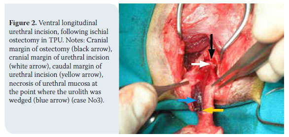
An intact DSH three-year-old cat was admitted with micturition inability, due to lower urinary tract obstruction and urolithiasis. This was the third obstruction episode in a period of two months. A few hours prior to the admission, there had been several unsuccessful attempts to catheterise the urethra. Clinical examination revealed inflammation injuries of the prepuce and penis. Subsequently, a 1mm diameter catheter was introduced into the urethra, up to the level of the bulbourethral glands, where the obstruction had been localised. The calculi were pushed back to the bladder mechanically via hydropropulsion, and the bladder was catheterised. A TPU was performed, considering the localisation of the obstruction and the expected injury of the urethra at that segment (Figure 2).

Transpelvic urethrostomy in three cats

> Management

In all cases, CBC and biochemical results were within normal limits. Urinalysis revealed crystalluria, without concomitant urinary tract infection in cats No 1 and 3, and they were prophylactically administered amoxicillin-clavulanic acid (20 mg/ kg/12h, sc). Cat No 2 continued to be treated with enrofloxacin.

In all cases, medetomidine (20-40 μg/kg, im) was used for sedation, 30 minutes before anaesthesia was induced with propofol (2-4 mg/kg, iv). Following tracheal intubation, anaesthesia was maintained with isoflurane. A purse string suture was temporarily placed in the anus. The surgical procedure was performed according to Bernande and Viguier’s description (2006) in six stages.15,16

Transpelvic urethrostomy in three cats1. Cat positioning and antisepsis

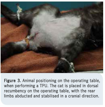
The cat is positioned in dorsal recumbency, with the rear legs secured to the surgical table in a cranial position (Figure 3). This position allows the ischiopubic symphysis to be exposed during the surgery. Subsequently, the field, which includes the base of the tail, the perineum, the posterior abdominal area, and the inner surface of the thighs, is surgically prepared (shaving, antisepsis). If a catheter has not been already introduced to the urethra, it is done at this stage.

2. Skin incision and exposure of the adductor muscles

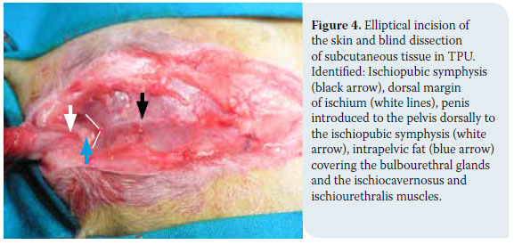
An elliptical incision is made around the scrotum and prepuce, exactly as in PU, and the incision extends cranially up to the cranial margin of the pubis. or even more anterior, in the case of an obese animal. In entire males, the testicles are then exposed and castration is performed (case No 3). Subsequently, a blind dissection of subcutaneous tissue is performed, along with the removal of a portion of subcutaneous fat in obese animals, and the ischiopubic symphysis is identified. The penis is introduced to the pelvis dorsally to the ischiopubic symphysis, which is posteriorly defined by the “V” formed by the adductor muscle fibers. The bulbourethral glands, as well as the ischiocavernosus and ischiourethralis muscles are identified and left intact (Figure 4).

Transpelvic urethrostomy in three cats

3. Detachment of adductor muscles and ischial ostectomy

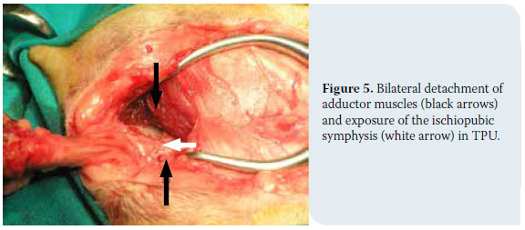
With the use of a periosteal elevator, the adductor muscles’ insertions are retracted from the ischiopubic symphysis, and its ventral part is exposed (Figure 5). Using a bone Ronguer, an approximately 12mm long and 10mm wide bilateral ischial ostectomy is performed. Attention is required so as not to include soft tissue in the excision, and so that no bone splinters remain at the edge of the osteotomy. Subsequently, the pelvic urethra is exposed and palpated, with the catheter within its canal.

Transpelvic urethrostomy in three cats

4. Urethrotomy

In cases where it is impossible to catheterise the urethra pre-operatively, the urethral canal is catheterised following a partial transverse incision of the penis at the level of the bulbourethral glands. This was not necessary in any of the three cases described here, as they had been successfully catheterised pre-operatively. Subsequently, a ventral longitudinal urethral incision is made with a No 15 blade, or, preferably, with iris scissors, from the level of the bulbourethral glands, to a point 2-3 mm caudal to the cranial margin of the ostectomy. The urethrotomy should be 10-12mm long (Figure 2).

5. Urethrostomy

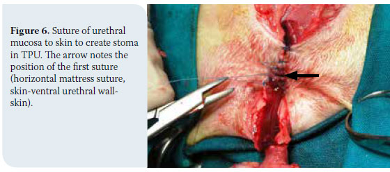
The urethral wall is manipulated with a nontraumatic DeBakey forceps. Using a nonabsorbable, monofilament, taper-point needle, 4-0 suture (e.g. Nylon, polypropylene), the urethral mucosa is sutured to the skin in a simple interrupted pattern. The suture extends from the anterior end of the stoma, with the first two sutures placed at a 45º degree angle to the mid line in a cranial direction, or with the first suture done in a horizontal mattress fashion passing twice through the ventral wall of the urethra -the latter being the suture that was applied in the three cases described here (Figure 6). The suture continues caudally on both sides of the urethra. In obese cats, it is probably required to remove fatty tissue around the stoma (case No 1), or even anteriorly (case No 3), in order to reduce suture tension. The penis distal to the bulbourethral glands is amputated, having had priorly placed a horizontal mattress suture with an absorbable, 3/0 monofilament suture at the stump of the penis for haemostasis.

Transpelvic urethrostomy in three cats

6. Skin closure

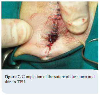
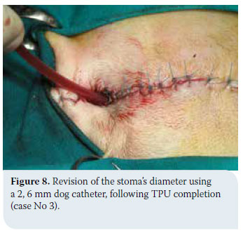
The suture of the remaining incision is done in a simple interrupted pattern (Figure 7). The urinary catheter is removed and the diameter of the stoma is checked so as to ensure that a 2-2.6 mm dog catheter or a curved Mosquito haemostat (Figure 8) can be passed through it. The anal suture is removed, and, with the cat placed in a lateral position, the bladder is emptied via ventral wall pressure. Via this manipulation, in cases No 1 and 2, haemorrhagic urine was expelled, containing mucus and blood clots, and in case No 3 urine was expelled, with small, up to 3mm in diameter, calculi.

Transpelvic urethrostomy in three catsTranspelvic urethrostomy in three cats

Post-operatively, in all cases an Elizabethan collar was placed to prevent self-induced injury. Enrofloxacin (5 mg/kg/24h, po) was administered for 10 days (Case No 2), and amoxicillin-clavulanic acid (20 mg/kg/12h, po) was administered for 4 days (Cases No 1 and 3). All animals were administered meloxicam (0,15 mg/kg/24h, sc) for three days. Fucidic acid ointment was applied daily on the site of the trauma, for 10 days. The sand in the litter box was replaced by newspaper, until the sutures were removed (12 days post operatively), to avoid sand covering to the urethrostomy. Chemical analysis of uroliths of Cat No 3 revealed calcium oxalate uroliths. Dietary measures were recommended for all cats, as they suffered from idiopathic lower urinary tract disease. Following suture removal, the cats were reexamined at one, three, and six months, and every time the diameter of the stoma was examined by the aforementioned methods.

> Outcome

Post-operatively, no cat presented with incontinence and all cats urinated normally within the first 24 hours. Haematuria was observed during the first three days. Cats No 1 and 3 were hospitalised for four days, and cat No 2, for ten days.

Cat No 1 has occasionally presented with haematuria and polyuria within a period of twelve years since the procedure. Each time, urine culture confirms a urinary tract infection, which is treated with the targeted antimicrobial therapy, while the psychogenic causes of cystitis recurrence are also addressed.

Cat No 2, which suffered from urinary tract infection when admitted, revealed pyuria and suppuration of the surgical wound on the third post-operative day. A Foley catheter was placed in the urethra, and replaced every 24 hours. The urine culture revealed Corynebacterium spp, which was resistant to most antimicrobial medication. Therefore, vancomycin hydrochloride (15mg/kg/8h, iv) was administered for four days, at which point the symptoms subsided and the Foley catheter was removed. Antibiotic therapy continued with the administration of linezolid (10mg/kg/8h, po) for another ten days. Two months post-operatively he presented urinary tract infection symptoms. Clinical examination did not reveal stoma stenosis. The urine culture was positive and the isolated micro-organism (Escherichia coli) was sensitive to the combination of amoxicillin and clavulanic acid, which was administered for two weeks (20 mg/kg/12h po). One week after the end of the treatment, urine culture was negative and the cat was asymptomatic for one year.

Cat No 3 was free of symptoms post-operatively.

Cats No 1, No 2, and No 3 are now aged 13, 4, and 5 respectively, and they remain asymptomatic with regard to stenosis/urethral stricture recurrence.

> Discussion

For the majority of surgeons, including ourselves, PU, as described by Wilson & Harrison (1971), is the preferred surgical technique to treat the obstructive type of feline idiopathic lower urinary tract disease.2-7 One of the most serious complications of PU is stoma stenosis, which results to similar clinical presentation as of the urethral stricture.3,17,18 It is usually suggested to perform a corrective PU to treat post-operative stenosis, yet this procedure is not always technically feasible.3,19 In this case, as well as in cases where PU is not eligible, due to a deficiency of healthy urethral tissue, prepubic urethrostomy,11 subpubic urethrostomy,14 and finally TPU15 have all been suggested.

In cases No 1 and 2, we applied TPU to restore stenosis caused by PU. Stenosis is the most serious complication of PU, and it usually occurs at the mucocutaneous margin.20 Post-operative stenosis is usually due to surgical errors, such as inadequate dissection of the urethra anterior to the bulbourethral glands, inability to mobilise the penis due to defective loosening of its junctions to the pelvis, resulting to a heightened tension upon anastomosis, urethral rupture during suturing, poor apposition of mucosa to the skin of the perineum, and post-operative rupture and/or suppuration of the surgical wound.3,17-19 In these cases, the anastomosis of the urethra to a more anterior position via TPU was preferred in lieu of performing a corrective PU, because it was impossible to identify the exact causes of the stenosis, in order to adequately treat them. Moreover, in case No 1, the cat had already undergone a corrective PU, and in case No 2, a concomitant urinary tract infection and the deteriorated state of the tissue in the area, rendered PU precarious. In case No 3, TPU was selected on grounds of insufficient healthy urethral area to perform a PU. This was due to the site and the cause of the stricture (at the level of the bulbourethral glands by a solid urolith), the visible injury of the penis, and the expected injury of the urethra at the site of the stricture, caused by the urolith and previous manipulations (Figure 6).

TPU was preferred in lieu of prepubic urethrostomy and subpubic urethrostomy in all 3 cases, as prepubic urethrostomy, which has been mostly cited in the relevant literature,11-13 is accompanied by serious, and usually irreversible complications, such as skin abrasions and necrosis, incontinence, and chronic urinary tract infections. In a study of 32 cats, which had undergone prepubic urethrostomy, one third of the cats died or were euthanised due to post-operative complications. Moreover, the owners of the cats that survived were dissatisfied, because of chronic irritant contact dermatitis at the region of the stoma.12 In a more recent study, 11 out of 16 cats who underwent prepubic urethrostomy presented with complications, and six of them were euthaniszed 1 to 23 months post-operatively.13 Subpubic urethrostomy probably presents fewer complications compared to prepubic urethrostomy, however the literature is poor.14 Additionally, it is an intensely traumatic technique, as it entails an osteotomy of the ischiopubic symphysis and a transpelvic mobilisation of the urethra.

TPU is considered a modified, less traumatic technique of subpubic urethrostomy.15 TPU has only been described in two studies by the same authors (Bernarde & Viguier 2004, 2006). In the second study, 19 cats participated, of which only 3 suffered from PU stenosis. 8 out of 19 cats presented post-operative complications. These were: rupture of surgical wound (1/19), transient incontinence (1/19), urinary tract infection (1/19), permanent or transient hair discolouration by urine (3/19), and feline idiopathic lower urinary tract disease (2/19).16

Two of our cases, for which TPU was a second or third treatment choice due to a failed PU, presented post-operative urinary tract infection, while such complication was not present in the third case, where TPU was applied as a first choice technique. Although the sample is small, this is in accord with Bernarde & Viguier study, in which, no urinary tract infection presented in any of the 16 cases where TPU was implemented as a first choice technique.16 This is possibly related to the ventral access to the urethra and the avoidance of injury of pelvic and pudendal plexus. Predisposing factors that have been identified for the presentation of urinary tract infection include the pre-operative use or overuse of catheters to establish urethral passage, idiopathic obstructive uropathy, due to feline idiopathic lower urinary tract disease, and the alterations of anatomical and functional barriers of the urethra.3,5,17,19 In one of our cases, occasional urinary tract infections were attributed to recurrent lower urinary tract disease and an ascending infection due to the loss of anatomical and functional urethral barriers. In the second case, that of the cat suffering from a urinary tract infection when operated, the suppuration of the surgical wound was attributed to the development of resistant strains. In order to prevent nosocomial infections, immediately after the infection was controlled, the catheter was removed and the cat was discharged from the clinic, continuing the appropriate antimicrobial treatment by oral administration. Despite this complication, there was no stenosis of the stoma, and this is consistent with Bernarde & Viguier’s research, according to which the rupture of the suture in one case (1/19) did not result in stenosis. In our case, two months later there was a recurrence of urinary tract infection, caused by a different microbial strain, and since then, the cat has been symptom-free. This urinary tract infection falls within the range of expected random urinary tract infections often occurring in animals that suffer from feline idiopathic urinary tract disease and that have undergone urethrostomy.6

In our cases, the skin/urethral suture was done with a synthetic (nylon), non-absorbable, 4/0 suture in an interrupted pattern. It is reported in the literature that, in PU, monofilament or multifilament absorbable sutures (polydioxanone, polyglactin 910) may also be used in simple interrupted or continuous suture patterns.21 This could be applied to TPU as well, reducing animal irritation, as well as trauma of the stoma, during suture removal.3

In case No 3, due to the severe obesity of the cat, extensive subcutaneous fat removal was required throughout the incision. While in the description of the technique16 it is generally reported that during surgical access fatty tissue is being removed from the area, limited removal of fat around the urethrostomy does not appear to adequately reduce suture tension in obese animals. In our opinion, in obese animals, the surgical incision should be longer cranially, and a sufficient amount of subcutaneous fat should be removed. This would ensure the anatomical symmetry of the area following suture of the incision, and reduce suture tension and stoma retraction. In this case, as in the other two, no stenosis of the stoma was observed, and this is in line with the literature so far. In TPU, the stoma is created approximately 15-18 mm anterior to the bulbourethral glands, where the diameter of the urethra is larger, thus contributing to prevent stenosis.

> Conclusions

TPU was performed to repair a failed PU in the case of two cats, and as a first choice surgical treatment in the case of one cat. Post-operative stenosis of the stoma did not occur in any animal, and mild, manageable complications only occurred in the two cats, which had already undergone a failed PU. In our opinion, this technique can be used in case of PU failure, as an alternative to subpubic urethrostomy and prepubic urethrostomy. Compared to PU, TPU could be the preferred technique, as, although it requires ostectomy, it allows ventral access to the urethra, reducing nerve injury in the area, which results in better functioning of the urinary system postoperatively, and in fewer complications. However, the bibliographic references that would support its suitability as an alternative to PU, to treat the obstructive form of feline idiopathic lower urinary tract disease, are still few, and its application as a first choice technique mainly concerns cases in which urethrostomy is required to be located cranially to the bulbourethral glands.


> References

Gieg JA, Chew DJ, McLoughlin MA. Παθήσεις της ουροδόχου κύ- στης. In: Saunders Εγχειρίδιο κτηνιατρικής των μικρών ζώων Birchard SJ, Sherding RG (ed) 3rd edn, Saunders: St Louis, 2006, pp. 895-914. Για την Ελληνική γλώσσα: MENDOR Editions SA 2008.

2. Smith CW Perineal urethrostomy. Vet Clin Small Anim Pract 2002, 32: 917-925.

3. Papazoglou LG, Basdani E. Perineal urethrostomy in the cat. Technique and complications. J Hellenic Vet Med Soc 2011, 62(2): 150-160.

4. Wilson GP, Harrison JW. Perineal urethrostomy in cats. J AmVet Med Assoc 1971, 159: 1789-1793.

5. Wilson GP, Kusba JK. Urethra. In: Current Techniques in Small Animal Surgery Bojrab MJ (ed) 2nd edn, Lea and Febiger, Philadelphia, 1983, pp. 325-333.

6. Flanders JA, Harvey HJ. Surgery of the urinary tract. In: The Cat Diseases and Clinical Management Sherding RG (ed) 2nd edn, Churchill Living stone, New York, 1994, pp. 1825-1845.

7. Caywood DD, Raffe MR. Perspectives on surgical management of feline urethral obstruction. Vet Clin North Am Small Anim Pract 1984, 14: 677-690.

8. Goldman AC, Beckman SL. Traumatic urethral avulsion at the preputial fornix in a cat. J Am Vet Med Assoc 1989, 194: 88-90.

9. Fox SM. Surgical repair of a traumatic perineal laceration with urethral transection: a case report. J Am Anim Hosp Assoc 1990, 26: 301-304.

10. Holt PE Non-prostatic dysuria. In: Urological Disorders in the Dog and Cat. 2nd ed, Manson Publishing, London, 2008, pp. 59-90.

11. Brandley RL. Prebubic urethrostomy. An acceptable urinary diversion technique. Prob Vet Med 1989, 1: 120-127.

12. Mendham (JH). - A description and evaluation of antepubic urethrostomy in the male cat. J Small Anim Pract 1970, 11: 709- 721.

13. Baines SJ, Rennie S, White RAS Prepubic urehrostomy: a longterm study in 16 cats. Vet Surg 2001, 30: 107-113.

14. Ellison GW, Lewis DD, Boren FC. Subpubic urethrostomy to salvage a failed perineal urethrostomy in a cat. Comp Cont Educ Pract Vet 1989, 11: 946-951.

15. Bernarde A, Viguier E. Transpelvic urethrostomy in 11 cats using an ischial oeteotomy. Vet Surg 2004, 33: 246-252.

16. Bernarde A, Viguier E. Transpelvic urethrostomy (TPU) in the cat: a new technique. Prospective survey: 19 cases. The European J Comp Anim Pract. 2006, 16: 41-49.

17. Smith CW, Schiller AG. Perineal urethrostomy in the cat: a retrospective study of complications. J Am Anim Hosp Assoc 1978, 14: 225-228.

18. Bass M, Howard J, Gerber B, Messmer M. Retrospective study of indications for and outcome of perineal urethrostomy in cats. J Small Anim Pract 2005, 46: 227-231.

19. Kusba JK, Lipowitz AJ. Repair of strictures following perineal urethrostomy in the cat. J Am Anim Hosp Assoc 1982, 18: 308- 310.

20. Phillips H, Holt DE. Surgical removal of the urethral stoma following perineal urethrostomy in 11 cats: (1998-2004). J Am Anim Hosp Assoc 2006, 42: 218-222.

21. Agrodnia MD, Hauptman JG, Stanley BJ, Walshaw R. A simple continuous pattern using absorbable suture material for perineal urethrostomy in the cat:18 cases (2000-2002). J Am Anim Hosp Assoc 2004, 40: 479-483.


Anaesthesia in pregnant dogs and cats


> Abstract

Anaesthesia during pregnancy in companion animals is necessary in cases of programmed or emergency Caesarean section or in cases when surgical intervention is required. During pregnancy, there are physiological changes in pregnant animals that should be taken into consideration during anaesthesia. These changes mainly affect the cardiovascular and respiratory system and influence to a lesser extent other systems, such as the digestive tract. In case of a Caesarean section, the preanaesthetic agents are usually either avoided or opioids are selected, although recent data support the use of alpha2-adrenergic agonists. Preparation for surgery occurs prior to the induction of anaesthesia, while oxygen is already being inhaled. The standard intravenous anaesthetics can be used for the induction of anaesthesia. In dogs, propofol is superior to thiopental, whereas etomidate is a very good choice in severely debilitated patients. For cats, the use of an α2-adrenergic agonist with ketamine is an adequate choice in order to ensure anaesthetic maintenance. The latter can be achieved in dogs with inhaled anaesthetic agents such as isoflurane, or with injectable anaesthetics like propofol. Epidural anaesthesia is a viable option in some cases. Postsurgical analgesia is based on opioids and/or nonsteroidal anti-inflammatory drugs and it is necessary so that the mother can take care of the neonates. In cases where the pregnant animal undergoes anaesthesia for a non-obstetric surgical procedure, the risk of embryotoxicity must be prevented, e.g. by avoiding benzodiazepines during the first stages of pregnancy, and accurately assessing anaesthetic depth.

> Introduction

Anaesthesia of the pregnant animal must provide sufficient sedation, while ensuring the health of the dam and viability of the litter.

The pregnant animal usually undergoes anaesthesia for a Caesarean section which is either programmed or an emergency due to dystocia. Dystocia is expected in 5% of deliveries in dogs and in 3-8% of deliveries in cats.3 Furthermore, a pregnant animal may undergo anaesthesia when its life is at risk, e.g. after a motor vehicle accident. In such cases, anaesthesia should be safe for the mother and the foetuses and not contribute to the initiation of premature labour, which may occur due to trauma, surgical manipulation of tissues and the effect of general anaesthetics.3,5 In cases of termination of unwanted pregnancy, the primary focus of anaesthetic care is the dam.

It is worthy of note that there are no active substances that can be used with absolute proven safety in all patients, nor any anaesthetic protocol that can “release” the veterinarian from his/her concern about all possible scenarios. Therefore, the clinician is required to evaluate each case separately and select the anaesthetic protocol based on the animal’s condition. In order to make the correct decisions, the clinician should originally be aware of the physiological changes in the dam’s body during pregnancy and labour, which will be mentioned in this paper, as well as the basic pharmacology of anaesthetic medications.2

The aim of the present article is to inform the clinician about the general anaesthetic options that are safe for pregnant companion animals, according to the latest published literature.

> Physiology of pregnant companion animals

During pregnancy, the dam’s metabolic needs increase, triggering a series of changes in various organ systems.1 Most of the relevant data originate from humans and sheep.1,16 Nevertheless, presuming that the same study results apply also in companion animals is considered acceptable, because hormonal changes during pregnancy are similar between humans and animals.16 Progesterone serum levels increase during the first stages of pregnancy, remain increased until the midterm and then begin to decrease reaching baseline values (< 2 ng/ml in serum) 24 hours prior to the beginning of labour. Further significant hormonal changes occur in the final weeks of pregnancy, such as an increase in serum levels of prolactine, relaxine and oestrogen. Moreover, as the day of delivery approaches, there is an increase in cortisol, prostaglandins and oxytocin, which may lead to the initiation of parturition. Other changes from an anaesthesiological perspective, which are expected in the second half of pregnancy are attributed to mechanical causes due to pressure exerted on the abdomen by the gravid uterus.2 It has not been determined if the exerted pressure is more severe in companion animals than it is in humans or other animal species and whether the weight of the fetal and uteroplacental tissues contribute to this. It is worthy of note that the mean weight of neonate kittens and puppies on the day of delivery is equal to 13.2% and 16.1% of the mother’s body weight, respectively, whereas the corresponding percentage for people and sheep are 5.7% and 11.4% respectively.1

The most important physiological changes during pregnancy mainly affect the cardiovascular and respiratory system of the dam and can directly influence the course of anaesthesia. However, changes occur in other body systems as well, such as the digestive and the urinary tract. Understanding these changes is a prerequisite in order to plan a safe anaesthetic protocol for both dam and fetuses.16

Cardiovascular system

Pregnancy affects the cardiovascular system through various pathophysiological mechanisms.16 During pregnancy the circulating blood volume gradually increases by about 40%.1 More specifically, it is plasma volume that increases the most and not so much the total red blood cell numbers, resulting in a reduction in haematocrit and blood haemoglobin concentration (this reduction is proportional to the size of the litter).1,2,4,33 Therefore, the presence of normal haematocrit values in advanced pregnancy can be suggestive of haemoconcentration or a small litter. Also, according to the increase in circulating blood volume, the cardiac output also increases by 30-50%, and there is a simultaneous increase in cardiac frequency.1 Nevertheless, mean arterial pressure (MAP) is preserved in the normal range.1,2 The increased intra-abdominal pressure due to the enlarged uterus during advanced stages of pregnancy, combined with dorsal recumbency, can decrease venous return due to compression of the caudal vena cava. Due to compression of the aorta there is a decrease in cardiac output, bradycardia, hypotension and consequent decrease in uterine and renal perfusion. However, in companion animals it seems that this phenomenon does not have the same clinical impact as it does in humans.1,2 In fact, Abitbol (1978)7 caused an artificial temporary compression of the caudal vena cava by ligating both the latter and the renal arteries in pregnant and non-pregnant dogs, and noticed a reduction in arterial pressure in pregnant dogs but none of the severe clinical consequences that develop in humans, such as bradycardia and hypovolemic shock. Therefore, even though the dorsal recumbency position is not forbidden, it is preferred for pregnant animals not to remain in this position for an extended time period prior to initiation of surgery.6 Finally, during pregnancy the cardiac load is increased, leading to relapse in patients with cardiological disorders that were previously under control or subclinical patients, and sometimes leading to temporary congestive heart failure.1,2 Α similar feline case has been reported, in which clinical signs of congestive heart failure developed in a cat in the final stages of pregnancy. The cat underwent treatment for pulmonary oedema and then ovariohysterectomy was performed. Signs of congestive heart failure receded after the procedure. Four months later, treatment for heart failure was discontinued because the cat was healthy and remained asymptomatic for nine years without treatment.30 A case of congestive heart failure has also been described in a dog six months post labour, which ended in sudden death. Post mortem examination and histopathology of cardiac tissue concurred with peripartum or postpartum cardiomyopathy in people.29

Respiratory tract

The volume of inhaled air per minute increases during pregnancy, including the respiratory frequency and lung volume, resulting in a gradual decrease of partial pressure of carbon dioxide in arterial blood (PaCO2), around 28-32 mmHg, in contrast to normal animals in which it ranges between 35-45 mmHg.4,16 The resulting chronic respiratory alkalosis does not affect circulating blood pH because there is time for adequate renal regulation by reducing the reabsorption of bicarbonates (HCO3 -). Furthermore, during pregnancy oxygen consumption increased by 20%.1 Αlso, due to the enlarged uterus, the abdomen is distended, the intraabdominal organs are driven dorsally and frontally, the diaphragm is compressed and intraabdominal pressure increases. This leads to a reduction of the lung functional residual capacity (FRC), which can predispose to pulmonary atelectasis, resulting in hypoventilation of the dam.1,2,6 Based on the aforementioned, in pregnant animals hypoxemia and hypercapnia occur faster than in non-pregnant ones. For this reason, it is recommended that prior to induction of anaesthesia, pure oxygen or oxygen mixed with environmental air should be provided for at least three minutes. The induction of anaesthesia with inhaled anaesthetics occurs faster in pregnant animals due to the increased respiratory volume, decreased lung FRC and possibly due to increased progesterone levels which act as a natural sedative. Moreover, during surgery, support with positive ventilation is necessary in some cases.1,2,4,6 Finally, in pregnant animals a reduced concentration of inhaled anaesthetic agents is sufficient. In particular, the minimal alveolar concentration of isoflurane or sevoflurane in pregnant animals is reduced by 40%. This pathophysiologic mechanism has not been fully elucidated but it seems to be caused by high levels of progesterone-endorphins in the CNS.1,6,16

Other body systems

The increased intra-abdominal pressure, the relaxation of the cardiac sphincter and the reduction in gastrointestinal motility lead to an increased risk of gastroesophageal reflux and possibly vomiting, a phenomenon which has also been described by Αnagnostou et al. (2010).8 Therefore, pregnant animals which undergo general anaesthesia should always be intubated swiftly and the cuff of the endotracheal tube should be filled adequately, so that the risk of aspiration of refluxing gastric contents can be avoided.1,6,16 It was once suggested by Paddleford (1992)23 to administer metoclopramide and cimetidine in order to prevent gastroesophageal reflux and aspiration pneumonia, but this treatment was effective only in much higher doses than standard metoclopramide dosage regimen. However this was not proven in the canine species during pregnancy.34 Nowadays, more antiemetic agents are available such as maropitant, ondansetron, and cisapride, although a research-proven preventative result has not been substantiated in the pregnant dam.1,36

Liver function is not extensively affected by pregnancy.2 It is worthy of note that the levels of plasma proteins are reduced. Most inhaled anaesthetic agents are extensively ionised and bind strongly with plasma proteins. Reduction of such protein levels leads to a prolonged effect of anaesthetic agents due to an increased amount of unbound active component.1,16

Renal function is also slightly affected. Creatinine and blood urea nitrogen levels are slightly decreased. Normal range levels in pregnant animals can be caused by kidney disorders, or physiological compensation of the dam’s body.1

> Anaesthesia in the pregnant for caesarean section

Τhe anaesthetic protocol that is used in companion animals undergoing Caesarean section should achieve swift induction of anaesthesia, preserve uterine perfusion and provide the veterinarian with an option of reversing the anaesthetic effect in the mother as well as the neonates, after delivery. Furthermore, the recovery of the dam should occur in a brief time period after obtaining the neonates so that the dam can provide care to the litter.31 However, in emergency cases it is considered safer to select an anaesthetic protocol the clinician is familiar with, even if it is not considered an ideal solution in cases of Caesarean section.24

Preparation of the pregnant, such as clipping, scrubbing and sterilising the surgical site on the abdomen as well as placement on the surgical table should ideally be performed prior to anaesthetic induction, in order to reduce the total duration of anaesthesia.16 Also, the animal must be pre-oxygenated, as previously mentioned, and hypovolemia, electrolyte disorders and mostly hypocalcaemia and hypoglycaemia should be corrected.4,16

All the anaesthetic agents, minus muscle relaxants, cross the placenta to an extensive degree, enter the foetal systemic circulation and have an impact depending on the administered dose and the duration of action of each agent.1,16 Sedation of the parturient animal should ideally be avoided, or if applied, depending on the case, mild opioids should be used, such as butorphanol or short-acting opioids, like fentanyl. The use of naloxone when available (0.02 mg/kg sublingually, intramuscularly or through the umbilical cord), on the neonates can counteract the effect of opioids.3,16 Acepromazine, which is classified as a phenothiazine, is usually avoided due to its prolonged duration of action.1 On the other hand, it is true that phenothiazines have not been implicated for higher dam or neonate mortality. Therefore, they can be used on occasion, such as in the study of Luna et al. (2004)18, in which chlorpromazine was administered 0,5 mg/ kg iv 15 minutes prior to induction of anaesthesia in bitches that underwent Caesarean section. Nevertheless, due to the negative effect on the cardiovascular system (resulting in hypotension caused by their effect on a1-adrenergic receptors), their prolonged duration of action and the fact that they are metabolised in the liver (the neonate liver is slower in metabolising compared to the dam), they should be avoided.3,24 Furthermore, benzodiazepines should also be avoided because they have been implicated for lethargy, depression, hypothermia and neonatal apnea directly postdelivery. Naturally, in cases when benzodiazepines are administered and prolonged lethargy is noted in the neonates or the dam, their effect can be countered by flumazenil, although the latter seems to have no effect on cats.1,3 Moreover, some of the α2-adrenergic agonists should be avoided because they lead to suppression of cardiovascular and respiratory function in the dam and neonates.1 Also xylazine has been implicated for higher rates of neonate puppy mortality.21 There are no relevant studies in companion animals regarding the use of dexmedetomidine prior to Caesarean section.1 In cases that it is used, its effect can be reversed through atipamezole in neonates that are lethargic after delivery. Recently a retrospective study has been conducted in pregnant dogs that underwent Caesarean section, and medetomidine (7 μg/kg im) was part of the preanaesthetic plan. Τhe results were particularly encouraging regarding dam and neonate survival, a fact that reassures and assists the clinician to a certain extent, when the latter is accustomed to using α2-adrenergic agonists.35

Induction of anaesthesia can occur with any of the standard intravenous anaesthetic agents. Traditionally propofol (6-8 mg/kg iv without sedation or 2-5 mg/kg iv if sedation has been administered) is preferred to thiopental due to higher puppy survival rates.14,28 Moreover, propofol is superior to thiopental and the combination of midazolam/ ketamine, because the percentage of neonate puppy lethargy is lower, as proven by Luna et.al. (2004).18 Also, in a different study by Moon-Massat et al. (2002)22 the activity level of puppies immediately post Caesarean section was evaluated and was found reduced in cases where ketamine and/or thiopental were injected for anaesthetic induction compared to puppies in which propofol had been used. It is noteworthy that viability of the foetuses does not only rely on the anaesthetic protocol, but also on the duration of delivery and the physical condition of the foetuses and the dam, however there is no mention in the published reports of any of the latter.3 Obtaining the neonates should occur 15-20 minutes after induction with propofol, so that the anaesthetic agents have been metabolised and redistributed and the neonatal respiratory system can function as naturally as possible. There are no published clinical studies about etomidate in companion animals.16 Etomidate (1-2 mg/kg iv) is considered a viable option for patients with preexisting cardiomyopathies or patients in critical condition.27 The combination of ketamine (4-6 mg/ kg) – midazolam (0.1-0.3 mg/kg) in dogs causes suppression of the respiratory system in neonates because of both the ketamine and midazolam. Therefore the neonates may not breathe on their own and may need respiratory support.28

Regarding the injectable anaesthetic steroid alfaxalone (not yet released in Greece), Doebeli et al. (2013)11 compared the survival rates of puppies 5, 15 and 60 minutes post Caesarean section, using the Apgar score (which has been developed to assess the effect of anaesthesia in neonates) after injecting the dams with alfaxalone (1-2 mg/kg iv) or propofol (2-6 mg/kg iv). They observed that neonatal survival rates were higher after delivery in the group that received alfaxalone, but the survival rates three months later were essentially the same. The survival rates of puppies 24 hours after delivery were the same in the study by Metcalfe et al. (2014),20 where the effect of alfaxalone was compared to propofol in bitches that underwent Caesarean section.

At this point it should be stressed that the selection of agents is based on the clinical condition of the dam and whether it is already debilitated by an already existing major body system failure or at a high risk of the latter, such as the cardiovascular or the central nervous system in cases of cranial trauma.16 Also, the intravenous anaesthetics should be injected slowly in patients in critical condition, so as to prevent any sudden or major decrease in mean arterial pressure (ΜΑP) and uterine perfusion. Injecting a small dose of fentanyl prior to their administration or a simultaneous injection of lidocaine (0.25-1.0 mg/kg iv) with a low dose of propofol (1-2mg/kg) or thiopental (2-5 mg/kg), may facilitate intubation, so that a high dose of the previous injectable anaesthetic agents is no longer necessary.3

The induction of anaesthesia through inhalation, either by mask or in an anaesthetic chamber, may increase stress resulting in the release of catecholamines, which results in vasoconstriction and foetal hypoxia and finally in acidosis in the parturient animal. Furthermore, because its duration is longer than intravenous induction and as there is a constant risk of regurgitation and aspiration, it is best avoided in pregnant bitches.28 However, as mentioned in the retrospective study of Moon et al. (2000),21 in 34% of the animals that were included in the study, isoflurane was used during induction, as well as maintenance of anaesthesia, with excellent neonate survival rates. Isoflurane, sevoflurane and desflurane are the preferred inhaled anaesthetics because induction and recovery are faster compared to other inhaled anaesthetic agents.1

The maintenance of general anaesthesia can occur either with inhaled or injectable agents.2,16 The former is considered safer for the foetuses because, according to a study involving the administration of propofol in pregnant sheep, the plasma protein levels of the foetuses are reduced compared to the dam. Therefore the concentration of the active form of the drug is higher than in the dam, and the concentration of propofol in fetal blood was maintained for prolonged periods of time until it was metabolised.9 Moreover, in a study that focused on anaesthetic maintenance during Caesarean section in dogs, the use of alfaxalone was compared to isoflurane. It was also noted that in the group which received alfaxalone, the duration of recovery was prolonged and the neonates presented with more pronounced lethargy compared to the group that received isoflurane.10 The levels of inhaled anaesthetic agents should be reduced by 30-60% compared to what is commonly used in nonpregnant animals for the aforementioned reasons (please consult the Physiology section). Regarding cats, it is preferable to use inhaled anaesthesia after intubation.2 From the inhaled anaesthetic agents that are available, isoflurane is superior to alothane and methoxyflurane because it has been correlated with higher neonatal survival rates.21

Not many studies are available as regards the administration of anaesthesia in pregnant cats. In one of these studies Elovsson et al., (1996)13 mention that the use of xylazine-ketamine or medetomidineketamine are marginally superior to propofolisoflurane, because newborn kittens are more active when the former were selected. In the study of Robbins and Mullen (1994)26 in which 26 cats underwent Caesarean section due to dystocia, and induction of anaesthesia was achieved by isoflurane, neonate survival rates reached barely 41%. Due to the insufficient scientific data regarding anaesthesia in cats that undergo Caesarean section and their differences with dogs, selecting an anaesthetic protocol is currently based on anaesthetic drug availability, and the ability of the clinician to use them correctly having comprehended the physiological and pharmacological changes that occur during pregnancy is of particular importance.3

The use of epidural anaesthesia appears to be very effective for neonate survival, because local anaesthetics do not cross into the systemic circulation of the fetus.13,14,18,28,31 Nevertheless, it is not an ideal solution in all cases because it is time-consuming and the clinician needs to be acquainted with the procedure.16 Τhe most crucial disadvantage of epidural anaesthesia, however, is that in an attempt to prevent the animal regaining consciousness during the surgical procedure, stronger sedation or light generalised anaesthesia is required, negating, as a result, both the advantage of avoiding cardiovascular suppression by the general anaesthetics, and the advantage of epidural injection in neonate survival. Furthermore, oxygen supply can be accomplished by mask or the flowby method but artificial ventilation is excluded because the animal is not intubated.28 For the aforementioned reasons, epidural anaesthesia is not usually applied in cases of Caesarean section in companion animals.28 Αn undesirable side effect is restlessness-nervousness displayed by the mother during recovery due to reduced sensation and motility in the hind limbs (depending on the drugs that were used, for a duration of half an hour up to two hours) after epidural anaesthesia, because it has a negative effect on the dam’s interaction with the neonates. If epidural anaesthesia is desired nonetheless, a mixture of local anaesthetics, such as lidocaine and bupivacaine 1:1 is used, in which an opioid can be added for more effective analgesia.1 Pascoe and Moon (2001)24 prefer the use of lidocaine only, due to the shorter duration of action (about 60 minutes) so that the mother can be ready to take care of the litter immediately after the surgery. To the same effect, Traas (2008)31suggested that only opioids should be used when the cardiac frequency of the litter can be measured and is normal (200 b.p./min), because it provides adequate postsurgical analgesia and the hind limbs maintain their motility. In any case, the total volume of the drug that is injected is reduced by 25-35% due to the fact that the epidural space is reduced during pregnancy because of venous dilation in the particular anatomic region. The injected volume of the solution should not exceed 6 ml.28

Local anaesthesia is particularly useful during preoperative preparation of the dam in or around the surgical site. Local anaesthesia reduces the dose of intravenous and inhaled general anaesthetic agents and provides surgical and postsurgical analgesia.24,31 The option of splash-block is also available, meaning the instillation of local anaesthetic in the surgical resection site, after suturing the muscular wall and prior to skin closure. This specific technique of local anaesthesia offers excellent post-surgical analgesia and facilitates dam acceptance of the neonates for the first suckling episode.6

Postsurgical analgesia is of particular importance because, as it has been proven, postsurgical pain reduces milk production.31 If opioids have not been administered prior to surgery, then pethidine, methadone, buprenorphine or butorphanol can be given post neonate delivery and before the end of the procedure, to provide an excellent analgesic effect.6,19 Both opioids and non-steroidal anti-inflammatory drugs (NSAID) cross the bloodmammary barrier and can be traced in the milk. The use of NSAIDs is an adequate option for managing postoperative pain, because it provides the mother with analgesia without affecting the level of consciousness.19 Even though the preoperative administration of NSAIDs provides a better postoperative analgesic effect,12 their use prior to obtaining the neonates is avoided because they have been implicated for higher neonate mortality rates.21 Cyclooxygenase-2 inhibitors (COX- 2 inhibitors), such as karprofen and meloxicam, are recommended (based on obstetrician experience, without confirmation from published studies) to be offered only once in lactating animals, because they are excreted by the kidneys of the neonates, which do not attain full function until the age of 6-8 weeks and thus more frequent use can lead to severe kidney disorders. In the same manner, meaning a single use, paracetamol can also be prescribed in lactating bitches, but never in cats.19 Recently cimicoxib (an NSAID that has not been released in Greece) was prescribed on day 0 and day 28 post labour, after obtaining the litter in six lactating bitches and it was noted that the exposure of the newborns to the drug was insignificant and the risk of undesirable side effects was minimal.37 Finally, tramadol can be used safely, though there have been no clinical research studies in pregnant animals.3

The selection of a proper anaesthetic protocol is only one of the factors that are involved in neonate survival. The clinician should take into consideration factors including the time lapse from the initiation of labour, the degree of placental detachment and accordingly, the degree of hypoxia or anoxia, which each foetus has suffered prior to the administration of anaesthesia, the time lapse until the definitive management - resolution of dystocia, as well as the overuse or misuse of ecbolic drugs, that lead to unproductive uterine contractions.

> Anaesthesia in pregnant animals for non - obstetric procedures

In pregnant animals that undergo anaesthesia for non-obstetric procedures, the anaesthetic protocol should be adapted to the needs of the dam and the survival of the litter should be ensured, meaning that adequate perfusion and oxygenation should be accomplished and teratogenesis should be avoided.16 The pregnant animal should initially be relieved from stress, which can lead to resorption, abortion or premature labour depending on the stage of pregnancy. Ηypoxia, hypotension and anaemia should be managed if they are already present, and if not they should be prevented. In such patients, blood transfusion is administered in haematocrit values higher than a nonpregnant animal e.g. HCT 25%.24 Regarding the preanaesthetic treatment, mild short-acting opioids can be selected, such as butorphanol, which offers adequate analgesia, moderate sedation and has minimal effect on the respiratory function of the pregnant animal.1,31 The a2-adrenergic agonists should be avoided because it has been proven that they are connected to increased uterine contractility which can lead to miscarriage, usually in the initial or the final stages of pregnancy.2 From the latter, xylazine seems to result in more severe uterine contractility and higher risk of abortion than newer type a2-adrenergic agonists.2 Jedruch et al. (1989)15 observed in canine cases in the final stages of pregnancy that a single dose of 60 μg/ kg medetomidine causes a greater increase in electrical activity in the uterine wall than a 20 μg/kg dose, however no abortions were noted. Acepromazine is not considered a good choice, because other than vasodilation, hypotension and hypothermia that this drug leads to in the pregnant animal, the duration of its effect is also prolonged.6 Epidural or other types of topical anaesthesia are desirable, because they decrease the requirements for anaesthetic medications, stress, and postsurgical pain.24

Induction of anaesthesia can be achieved by intravenous injection of propofol or etomidate.16,31 The latter is an ideal choice for patients in critical condition as previously mentioned. Moreover, ketamine is also considered to be an acceptable choice for induction of anaesthesia in ill or debilitated pregnant animals, because it preserves cardiovascular function in the desirable levels.3 For maintenance of anaesthesia the standard inhaled anaesthetics are recommended. Postsurgical analgesia is mostly based on the administration of opioids19, even though their long - term use in pregnant animals has not been previously studied. Regarding NSAIDs, they should be avoided during pregnancy because studies in people have shown that they are implicated in cleft palate malformations and renal dysfunction in the newborn.19

The risk of embryotoxicity and/or teratogenesis is higher in the beginning of pregnancy (6-45 days), prior to and during organogenesis and especially in the first twenty days of pregnancy, even before the blastocyst implantation.25 However, when the life of the pregnant animal is at risk, depending on the owner or the clinician’s preference and, due to necessity and irrespective of the risk, the possibility of teratogenesis or miscarriage can be disregarded. In the last stages of pregnancy there is no such risk.24 Most anaesthetics and analgesics can cause teratogenesis at high doses, but the doses used in the clinical setting are considered to be safe. Only nitrous oxide seems to have teratogenic properties in companion animals after repeated administration on multiple days.3,17 Also, it would be preferable to avoid benzodiazepines, because, according to studies in people, they have been implicated in causing cleft palate malformation in neonates. In companion animals there are no such reports.3

> Anaesthesia in pregnant animals that undergo ovariohysterectomy

In cases of ovariohysterectomy, survival of the litter is not a priority. Τhe anaesthetic protocol does not have to adapt to the limitations that were previously mentioned in order to preserve the survival of the neonates.24 After the removal of the uterus from the abdominal cavity, foetal death will occur swiftly, without the sensation of mortal stress, if the uterus remains unopened and the respiratory center of the foetuses is not activated due to contact with the environmental air. If the resection of the uterus is necessary, then it should be performed one hour following uterine removal from the abdominal cavity. Foetal movements observed in advanced stages of pregnancy are believed to be involuntary and they occur spontaneously or by stimulation of the amniotic sac, e.g. due to manipulation.32


> References:

1. Grimm K, Lamont L, Tranquilli W, Greene S and Robertson S Veterinary Anesthesia and Analgesia The Fifth Edition of Lumb and Jones In: Anesthetic Considerations During Pregnancy and for the Newborn Raffe M. 5th edn., John Wiley & Sons, USA,UK, 2015, pp. 708-722.

2. Clarke K, Trim C, Hall L, Adams J, Borer-Weir K, Divers S, Hernandez S Veterinary Anaesthesia In: Anaesthesia for Obstetrics. 11th edn. Elsevier, China, 2014, pp. 585-598.

3. Duke-Novakovski T, Vries de M, Seymour C. BSAVA Manual of Canine and Feline Anaesthesia and Analgesia. In: Anaesthesia for Caesarian Section and for the Pregnant patient Claude A, Meyer R. 3rd edn., John Wiley & Sons, 2016, pp. 366-376.

4. Shelby A, McKune C Small Animal Anesthesia Techniques. In: Anesthetic Protocols for Specific Procedures and Other Conditions that Influence Anesthesia. 1st edn., John Wiley & Sons, 2014, pp. 116-122 and 186-7.

5. Snyder L, Johnson R Canine and Feline Anesthesia and Co- Existing Disease. In: Cesarean section and pregnancy, Aarnes T and Bednarski R, John Wiley & Sons, 2014, pp. 299-309.

6. Eberspacher E. Anasthesie Skills Perioperatives Management bei Klein-, Heim-, und Grosstieren. In: Medikamente und Gravider Patient, Kaiserschnitt, Schattauer, Stuttgart, 2016, pp. 47-49 and 243-247.

7. Abitbol M. Inferior vena cava compression in the pregnant dog. Am J Obstet Gynecol 1978, 130(2): 194-8.

8. Anagnostou T, Savvas I, Kazakos G, Ververidis H, Psalla D, Kostakis C, Skepastianos P, Raptopoulos D. The effect of the stage of the ovarian cycle (anoestrus or dioestrus) and of pregnancy on the incidence of gastro-oesophageal reflux in dogs undergoing ovariohysterectomy. Vet Anaesth Analg 2015, 42: 455-558.

9. Andaluz A, Tusell J, Trasserres O, Cristofol C, Capece B, Arboix M, Garcia F. Transplacental transfer of propofol in pregnant ewes. Vet J 2003, 166: 198–204.

10. Conde Ruiz C, Del Carro A, Rosset E, Guyot E, Maroiller L, Buff S & Portier K. Alfaxalone for total intravenous anaesthesia in bitches undergoing elective caesarean section and its effects on puppies: a randomized clinical trial. Vet Anaesth Analg 2016, 43: 281–290.

11. Doebeli A, Michel E, Bettschart R, Hartnack S, Reichler I. Apgar score after induction of anesthesia for canine cesarean section with alfaxalone versus propofol. Theriogenology 2013, 80: 850–854.

12. Dunkan B, Lascelles X, Cripps P, Jones A, Waterman-Pearson A. Efficacy and Kinetics of Carprofen, Administered Preoperatively or Postoperatively, for the Prevention of Pain in Dogs Undergoing Ovariohysterectomy. Vet Surg 1998, 27: 568-582.

13. Elovsson L, Funkquist P, Nyman G. Retrospective evaluation of anaesthetic techniques for Caesarian section in the cat. J Vet Anaesth 1996, 23: 80.

14. Funkquist P, Nyman G, Löfgren A, Fahlbrink E. Use of propofol-isoflurane as an anesthetic regimen for cesarean section in dogs. J Am Vet Med Assoc 1997, 211: 313-317.

15. Jedruch J, Gajewski Z, Ratajska-Michalczak K. Uterine motor responses to an alpha 2-adrenergic agonist medetomidine hydrochloride in the bitches during the end of gestation and the post-partum period. Acta Vet Scand Suppl 1989, 85: 129–134.

16. Kushnir Y and Epstein A. Anesthesia for the Pregnant Cat and Dog. Israel Journal of Veterinary Medicine 2012, 67: 19-23.

17. Lane G, Nahrwold M, Tait A, Taylor-Busch M, Cohen P and Beaudoin A Anesthetics as Teratogens: Nitrous Oxide Is Fetotoxic, Xenon is not. Science 1980, 210: 899-901.

18. Luna S, Cassu R, Castro G, Teixeria Neto F, Silva Junior J, Lopes M. Effects of four anaesthetic protocols on the neurological and cardiorespiratory variables of puppies born by caesarean section. Vet Rec 2004, 154: 387–389.

19. Mathwes K Pain. Management for the Pregnant, Lactating, and Neonatal to Pediatric Cat and Dog. Vet Clin Small Anim 2008, 38: 1291-1308.

20. Metcalfe S, Hulands-Nave A, Bell M, Kidd C, Pasloske K, O’Hagan B, Perkins N and Whittem T. Multicentre, randomised clinical trial evaluating the efficacy and safety of alfaxalone administered to bitches for induction of anaesthesia prior to caesarean section. Aust Vet J 2014, 92: 333–338.

21. Moon P, Erb H, Ludders J, Gleed R, Pascoe P. Perioperative risk factors for puppies delivered by cesarean section in the United States and Canada. J Am Anim Hosp Assoc 2000, 36: 359–368.

22. Moon-Massat P, Erb H. Perioperative factors associated with puppy vigor after delivery by cesarean section. J Am Anim Hosp Assoc 2002, 38: 90–96.

23. Paddleford R. Anesthesia for Cesarian Section in the Dog. Vet Clin North Am Small Anim Pract 1992, 22(2): 481-4.

24. Pascoe P, Moon P Periparturient and neonatal anesthesia. Vet Clin N Am Small Anim Pract 2001, 31: 315–341.

25. Rebuelto M, Loza M. Antibiotic Treatment of Dogs and Cats during Pregnancy. Vet Med Intern 2010, 14: Article ID 385640, 8 pages.

26. Robbins M, Mullen H En Bloc. Ovariohysterectomy as a Treatment for Dystocia in Dogs and Cats. Vet Surg 1994, 23: 48-52.

27. Robertson S. Advantages of Etomidate Use as an Anesthetic Agent. Vet Clin North Am Small Anim Pract 1992, 22(2): 277-80.

28. Ryan S, Wagner A. Cesarean Section in Dogs: Anesthetic Management. Compend Contin Educ Vet 2006, 28: 44-54.

29. Sandusky G, Cho D. Congestive Cardiomyopathy in a Dog associated with Pregnancy. Cornell Vet 1984, 74: 60-64.

30. Stoneham A, Graham J, Rozanski E, Rush J. Pregnancy- Associated Congestive Heart Failure in a Cat. J Am Anim Hosp Assoc 2006, 42: 457-461.

31. Traas AM. Surgical management of canine and feline dystocia. Theriogenology 2008, 70: 337–342.

32. White S. Prevention of fetal suffering during ovariohysterectomy of pregnant animals. JAVMA 2012, 240 (10): 1160-3.

33. Kaneko M1, Nakayama H, Igarashi N, Hirose H. Relationship between the number of fetuses and the blood constituents of beagles in late pregnancy. J Vet Med Sci 1993 Aug, 55(4): 681-2.

34. Wilson D, Evans A, Mauer W. Influence of metoclopramide on gastroesophageal reflux in anesthetized dogs. Am J Vet Res 2006 Jan, 67(1):26-31.

35. De Cramer K, Joubert K Nothling J. Puppy survival and vigor associated with the use of low dose medetomidine premedication, propofol induction and maintenance of anesthesia using sevoflurane gas-inhalation for cesarean section in the bitch. Theriogenology, 96 (2017): 10-15.

36. Zacuto A, Marks S, Osborn J, Douthitt K, Hollingshead K, Hayashi K, Kapatkin A, Pypendop B, Belafsky P. The influence of esomeprazole and cisapride on gastroesophageal reflux during anesthesia in dogs J Vet Intern Med 2012 May-Jun, 26(3): 518-25.

37. Schneider M, Kuchta A, Dron F, Woehrle F. Disposition of cimicoxib in plasma and milk of whelping bitches and in their puppies BMC Vet Res 2015, 11: 178.


Canine thyroid tumours: diagnosis and treatment


> Abstract

Τhyroid neoplasia accounts for 1-3,8% of all canine tumours, and the most common types include adenoma and carcinoma, the latter being identified in 90% of cases. Exposure to radiation, iodine deficiency, hypothyroidism, or even genetic mutations, are included among the triggering factors. Benign adenomas are usually small and movable, without infiltration of surrounding tissues and the presence of metastasis. In contrast, carcinomas are larger, with extensive infiltrating tendencies, primarily in the lungs and regional lymph nodes. Diagnosis is based on clinical signs emerging due to compression of adjacent organs (oesophagus, trachea, larynx, pharynx), endocrine testing and diagnostic imaging. To reach a definitive diagnosis biopsies for histopathological examination need to be obtained, which will confirm the origin of the mass and will differentiate benign from malignant neoplastic tissue. In cases of thyroid adenoma, surgical excision of the tumour is the treatment of choice and leads to clinical cure. The treatment of choice and the prognosis for thyroid carcinoma depend on tumour size, the extent of infiltration of surrounding tissues, the presence of metastases and the possibility of using alternative treatments. Surgical management is indicated in cases of small and movable carcinomas or with superficial infiltration of surrounding tissues, but it is not recommended in cases of extensively infiltrative and fixed carcinomas.

> Introduction

Τhyroid tumours represent 1-3,8 % of all canine tumours while 10% and 15% of neoplasms affecting the head and cervical region, respectively.1-5 Thyroid tumour types include adenoma and carcinoma, the latter being identified more often in 90 % of cases, and in dogs of median age ranging between 9 and 11 years.1,3,6-10 Beagles, Siberian Huskies, Boxers and Golden Retrievers are predisposed to thyroid neoplasia,1,3,4,11,12 but gender predisposition has not been noted.1-3,9-14

> Initiating causes and predisposing factors

Previously published studies in people as well as dogs have proven that thyroid exposure to radiation and iodine deficiency have been implicated in neoplastic growth in the gland.15 Iodine deficiency causes over secretion of thyroid-stimulating hormone (TSH) from the pituitary gland, which leads to thyroid epithelial cell hyperplasia and possibly to metaplasia.16,17 Furthermore, it has been proven that hypothyroidism caused by lymphocytic thyroiditis is related to thyroid neoplastic transformation in dogs.17 Studies from human medicine confirm that various mutations in the ras oncogenes and tumour suppressor genes can trigger thyroid neoplastic transformation and benign neoplasms may undergo malignant transformation respectively.18,19 Finally, aneuploidy is a common characteristic of canine thyroid carcinoma and it is identified at a rate higher than 50% of cases with primary tumours.20

> Biological behaviour

Thyroid tumours are usually located in the area surrounding the gland, including the ventral or ventrolateral surface of the cervical region, caudal to the larynx.3,10 The right and left lobe are affected with the same frequency, whereas 36-40% of carcinomas affect both lobes.1,7,8,11 Rarely neoplasms can be observed in ectopic sites, such as the base of the tongue, or they can be sublingual, and found subcutaneously dorsal to the trachea,21 in the cranial mediastinum, at the heart base, and in other endocardial sites.22-26 Benign adenomas develop at a slow rate, they are usually small, ranging from microscopical to several centimeters in diameter, soft, compact, well encapsulated and movable along their craniocaudal axis in the subcutaneous cervical region. They do not infiltrate surrounding tissues, they do not metastasise and they are usually discovered as an incidental finding during necropsy.1,6,11

Malignant carcinomas usually originate from follicular (adenocarcinoma) and rarely from parafollicular or C cells (myeloid carcinoma).5,27,28 They grow fast and are usually nodular, larger than adenomas, ranging in size from microscopical to a diameter over 10 centimeters.1,3,29 Their biological behaviour is extensively infiltrative affecting adjacent tissue structures such as the larynx, the trachea, the jugular veins, and the carotid sheath and they have a predilection for regional or distal metastasis.1,3,6 Τhyroid carcinomas usually metastasise in the lungs and regional lymph nodes, and rarely in the jugular vein, the heart, the kidneys, the adrenal glands, the liver, the spleen, the intestine, the omentum, and the brain.3,11 The possibility of diagnosing metastatic lesions at the time of diagnosis ranges from 16 to 38% and is directly related to tumour size.3,6,7,10 In a large retrospective study it was noted that all dogs in which the size of the carcinoma exceeded 100 cm3, would have distal metastatic sites in 100%, whereas in cases of carcinoma ranging from 21 to 100 cm3 and less than 20 cm3 metastatic lesions would be identified in 74% and 14% of cases, respectively.11

> Diagnosis and Staging

Clinical signs

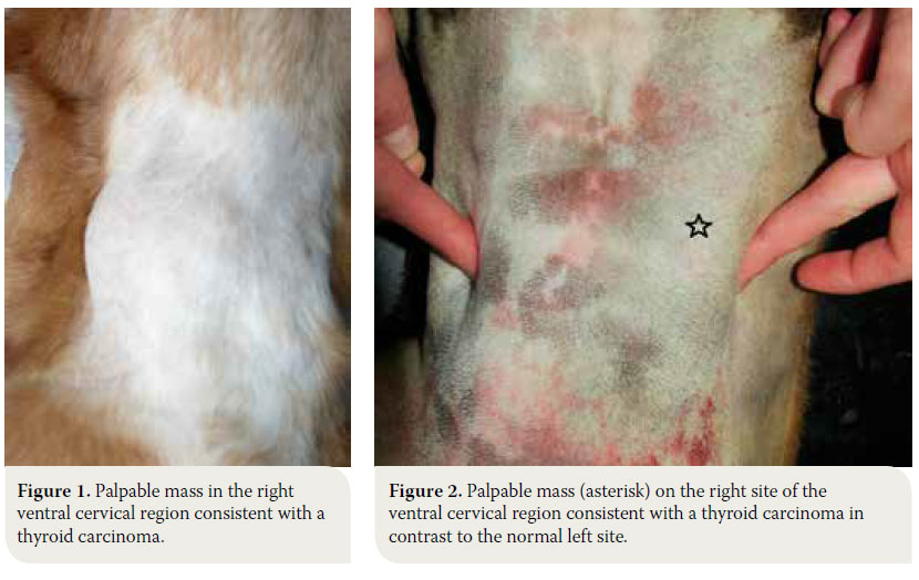
The dog is usually admitted after the owner has noted a mass in the cervical region, which rarely extends toward the thoracic inlet (Figures 1, 2).1-3,7,10,11 This mass is usually noted ventral to the trachea (65- 70 of cases %), is related to the larynx and can be well defined or invasive.11 Information from the history and physical examination can identify clinical sings from the respiratory and gastrointestinal tract. Τhe large size of the neoplasm, metastatic lesions in the lungs and lymph nodes and compression of adjacent tissues (oesophagus, trachea, larynx, pharynx) can manifest with signs including respiratory distress, coughing, wheezing, cyanosis, sneezing, reverse sneezing, laryngeal paralysis, dysphonia, regurgitation, vomiting, lethargy, peripheral lymphadenopathy, fever and Horner’s syndrome.1-3,11,30
 Canine thyroid tumours: diagnosis and treatment

It is worthy of note that 60% of dogs will remain clinically euthyroid, whereas 30% present with signs of hypothyroidism due to obliteration of the normal gland parenchyma.1-3,11 Cases of functional neoplasm have been reported in 10-22% of cases, when dogs developed signs of hyperthyroidism similar to hyperthyroid cats, including polyuria, polydipsia, polyphagia with simultaneous weight loss, generalised muscle weakness, restlessness, exercise intolerance, tachycardia and continuous searching for cold areas in the home environment due to a low tolerance to heat.3,29,31

Endocrine testing

Considering that most dogs are admitted in a euthyroid state, there are very few clinical studies evaluating thyroid function via estimation of hormone levels including triiodothyronine (Τ3), thyroxine (Τ4) and anterior pituitary lobe TSH.3,11 From a study in 36 dogs with thyroid tumours, 39% had reduced levels of Τ3 and Τ4, none of them presenting with clinical signs of hypothyroidism, whereas 31% had increased levels of Τ4, with only 2 cases presenting with clinical signs of hyperthyroidism.9 Irrespective of clinical signs of hypothyroidism or hyperthyroidism, it is recommended that all cases have total Τ4, free Τ4 and TSH levels measured to evaluate thyroid function.32

Diagnostic imaging

Radiography

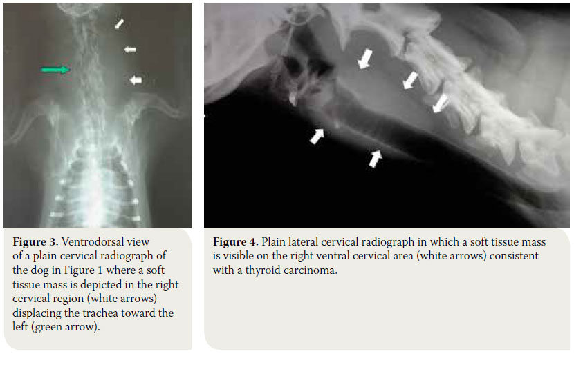
Radiographs of the cervical region will reveal, almost in every case, the presence of a soft tissue mass that may or may not displace the trachea or larynx, however, it does not provide information regarding vascular support and infiltration of surrounding tissues (Figures 3, 4).6 Thoracic radiography reveals the presence of pulmonary metastatic lesions in 33-77% of cases.1-3,11 If mediastinal findings compatible with a soft tissue mass that may related to an ectopic thyroid tumor are noted, ultrasonographic evaluation of the thorax is indicated, because the presence of fluid or fat in the thoracic cavity or the mediastinum may resemble a mass in plain radiographs.30,33
 Canine thyroid tumours: diagnosis and treatment

Ultrasonography

This is a valuable, relatively inexpensive non-invasive imaging modality, for which anaesthesia is rarely required. For all the previous reasons, it is often used as the method of choice to evaluate thyroid morphology. Ultrasonography can discern masses >2mm and can differentiate between solid and cystic nodules. Consequently, ultrasonographic evaluation is recommended for lesions <2 cm.34 The information that ultrasonography provides aids in differentiating thyroid tumours from other mass lesions that can be found in the ventral aspect of the cervical region and can determine if the gland is unilaterally or bilaterally affected.35 However, it can sometimes be challenging to substantiate if the mass originated from the thyroid gland if the normal anatomy of the cervical region has been altered.33,36 Moreover, it provides information about the degree of tumour vascularization and surrounding tissue infiltration, however without differentiating between benign and malignant neoplasms, unless peripheral lymphadenopathy and local infiltration are present.33 In case of tumours with an extensive vascular supply, ultrasound-guided needle aspiration biopsy is recommended to avoid any haemorrhage.30,33
 Canine thyroid tumours: diagnosis and treatment Computed tomography (CT) and magnetic resonance imaging (MRI)

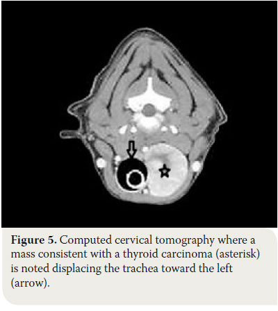
These imaging modalities are indicated for masses >3cm and can provide useful information regarding the origin and the degree of vascularisation, as well as the extent of surrounding tissue infiltration.30,34 Moreover, they offer the possibility of investigating the thorax for metastatic pulmonary lesions and the presence of ectopic intrathoracic thyroid tissue, and they can even detect metastasis in the lymph nodes.34 Finally, CT aids in the staging of thyroid carcinoma. 37

In conclusion, all 3 imaging modalities (ultrasonography, CT or MRI) provide almost the same information regarding the presence of a cervical mass. In a study that compared sensitivity and specificity between these techniques in 13 cases of canine thyroid carcinoma, it was found that ultrasonography had 79% sensitivity and 33% specificity, CT 85% and 100% and MRI 93% and 67% respectively.36 In cases when interpretation of diagnostic imaging results are controversial, surgical exploration of the cervical region is a fast, accurate and inexpensive way to identify the degree of invasiveness and the origins of the mass.38

Cytological examination

Fine needle aspiration of the mass (FNA) is a noninvasive, fast and easy procedure, which does not necessitate sedation or general anaesthesia.38 Studies have shown that FNA cytology can identify a cervical mass of thyroid origin in more than half of the cases with canine thyroid neoplasia, without being able to differentiate between benign and malignant tumours.3,29 Blood contamination of the samples due to extensive neoplastic tissue vascularisation, the fragility of thyroid follicular cells and the presence of active inflammation, are some of the reasons that limit the diagnostic accuracy of this technique.29 In order to minimise sample contamination with blood, it is recommended that fine needle aspiration should be ultrasound guided.30,33

Biopsy and histopathology

In small (<7cm) and/or movable masses, excisional biopsy can be diagnostic as well as therapeutic. Biopsy from large and severely invasive tumours can be done with a Tru-Cut needle or excision although there is a risk of profuse haemorrhage.6,39 Therefore, open surgical biopsy is recommended so that samples can be obtained from a rather avascular region and bleeding can be controlled if necessary.39

In order to reach the definitive diagnosis of thyroid tumours, histopathology of biopsy samples is mandatory, which not only confirms tumour origin but can also differentiate between benign and malignant tumours.29 Based on histology, thyroid tumours originate either from thyroid follicular cells or from parafollicular or C-cells.

Malignancy in well differentiated tumours is characterised by extracapsular extension and vascular infiltration. 40 Most canine thyroid carcinomas are well to moderately differentiated and usually originate from thyroid follicular cells. The mixed solid-cystic form is most commonly encountered, followed by the cystic and the solid form.3,7,24 Follicular adenomas usually belong to the follicular type.11,41 The papillary type of follicular carcinoma, medullary carcinoma (parafollicular or C-cells) and undifferentiated carcinomas rarely occur in dogs.1-3,11,28 Survival time for dogs with thyroid carcinoma does not seem to be significantly correlated to histopathological stage.3,7

Staging

Clinical staging is determined according to the World Health Organisation (WHO TNM Staging System) and is summarised in Table 1. According to this system, the tumour is categorised based on size and surrounding tissue infiltration (Τ), metastasis in regional lymph nodes (N) or the presence of distant metastasis (Μ).42
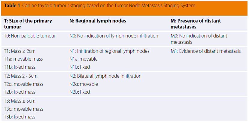
 Canine thyroid tumours: diagnosis and treatment

> Differential diagnosis

A thyroid neoplasm should be differentiated from abscess, granuloma, salivary mucocele, lymphogenic metastasis of tonsilar squamous cell carcinoma, lymphoma, carotid body tumour and sarcoma.43

> Treatment
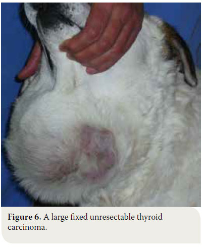
 Canine thyroid tumours: diagnosis and treatment In cases of thyroid adenoma, surgical excision of neoplastic tissue is the treatment of choice and leads to clinical cure.6,32 Regarding thyroid carcinoma, treatment options depend on factors including tumour size, the extent of surrounding tissue infiltration, the presence of metastatic lesions and the possibility of using alternative treatment modalities (radiation therapy, chemotherapy).43 In general, surgical treatment is indicated in cases of small (<7cm) and movable neoplasms or tumours with superficial infiltration of surrounding tissues, but it is contraindicated in cases of severely invasive and fixed tumours (Figure 6).6,7,10,32 In many cases, surgical exploration of the carcinoma is necessary in order to identify the extent of surrounding tissue invasion. In cases where metastatic lesions are present at the time of diagnosis, treatment is palliative and includes monotherapy or a combination of surgical excision and radiation therapy. In cases when diagnostic imaging has not demonstrated the presence of metastasis, selection of treatment is based on whether the tumour is movable or fixed. If it is movable, surgical excision by unilateral or more rarely bilateral thyroidectomy is undertaken, combined or not with radiotherapy. When the tumour is fixed, surgical treatment is contraindicated and radiotherapy and radioactive iodine administration are recommended.32

Surgical treatment

Surgical anatomy

Sound knowledge of the anatomy of the cervical region and of the thyroid gland in particular is necessary for the unimpeded and safe surgical exploration and management of thyroid tumours. The thyroid gland is formed by two elongated lobes and is located at the level of the cranial aspect of the cervical trachea. Usually the right lobe is located at the level of the first five tracheal rings, whereas the left lobe is located more caudally, extending from the third to the eighth tracheal ring. The lobes come in contact ventrally with the sternohyoid and sternocephalicus muscles, and laterally with the sternothyroid muscle. The lobes are located close to the carotid artery, the cervical sympathetic trunk and the jugular vein (Figure 7).44-45
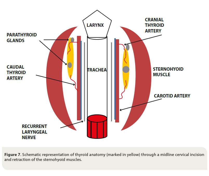
 Canine thyroid tumours: diagnosis and treatment

The parathyroid glands are closely attached to the thyroid gland and there are usually two for every thyroid lobe, one external on the cranial pole of the lobe, and one internal in the inner surface of the lobe, under the fibrous capsule or within the thyroid parenchyma. 44-45

The blood supply of the thyroid gland is provided by the cranial and caudal thyroid arteries. The cranial thyroid artery branches out from the carotid artery and the caudal thyroid artery arises from the brachiocephalic artery. Venous drainage is through the cranial thyroid vein, which drains into the jugular vein.44-45

Surgical preparation

In case of surgical treatment, the general condition of the dog must be evaluated as for every other surgical procedure through the appropriate haematology and biochemistry examinations. Moreover, due to extensive vascularisation of the thyroid and sometimes invasion of large vessels, extensive haemorrhage and sometimes severe blood loss are expected during thyroidectomy. This fact renders indispensable the hemostatic evaluation and the preparation for possible blood transfusion during or post surgery.32,43,46 Routine anaesthesia is induced considering that thyroid tumours are rarely functional.43 Furthermore, it is noted that even in cases of hyperthyroid dogs, achieving a euthyroid state prior to surgery is not necessary, in contrast to hyperthyroid cats. In any case of severe perioperative arrhythmias, however, or when severe hypertension is noted, the appropriate treatment should be instituted.31

Thyroidectomy 32,38,39,47

  1. Following induction of general anaesthesia, the dog is placed in dorsal recumbency, the front limbs are tied caudally, a folded towel is placed under the cervical region, in order to achieve mild hyperextension of the neck and a tube is placed in the oesophagus to facilitate orientation during surgery. The ventral aspect of the cervical region from caudal to the mandible to the manubrium is surgically prepared (Figure 8).
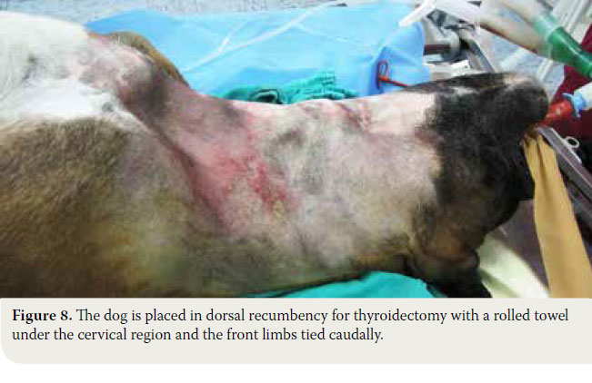
     Canine thyroid tumours: diagnosis and treatment
  2. A midline cervical incision is extended from the thyroid cartilage of the larynx to the level of the manubrium.
  3. The sphincter colli muscles are incised by scalpel to expose the paired sternohyoid muscles. Separation of the muscles is performed in the median raphe. Haemorrhage is controlled by diathermy or ligation of the small vessels that are located in the median raphe. The trachea is exposed and the thyroid glands are identified resting on the dorsal or dorsolateral aspects of the trachea near the thyroid cartilage (Figure 9).
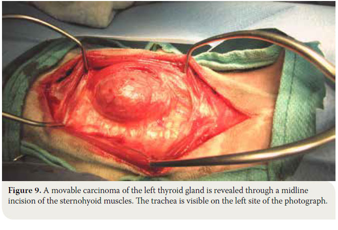
     Canine thyroid tumours: diagnosis and treatment
  4. If the thyroid gland is not visible, the pair of sternothyroid muscles is retracted from the midline, the latter being located dorsally and on the inner aspect of the sternohyoid muscles. For an unimpeded view of the surgical site retraction of the sternohyoid and sternothyroid muscles is recommended with the placement of two Gelpi retractors in the cranial and caudal end of the incision.
  5. Both thyroid lobes are carefully observed, as well as their anatomical relationship to the surrounding structures.
  6. The recurrent laryngeal nerves are exposed, which lie along the trachea at the inner surface of the gland or in the fascia dorsal to the gland. During thyroidectomy, special attention is needed so as to avoid trumatising these nerves.
  7. The blood vessels surrounding the gland are ligated. The caudal thyroid artery is ligated and once the tissues dorsal to the gland are dissected, the gland is elevated and pulled forward so that the cranial thyroid artery can be identified and ligated. Ligations are done with synthetic absorbable suture no 3/0 or with vascular clips (Figures 10, 11). In cases when extensive haemorrhage is observed due to extensive neovascularisation, temporary unilateral ligation of the carotid artery and jugular vein may be necessary, and it is well tolerated by the animal.
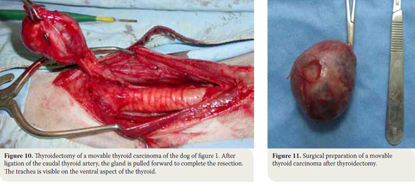
     Canine thyroid tumours: diagnosis and treatment
  8. In cases of severely invasive carcinoma infiltrating the surrounding tissues and structures, such as the corresponding jugular vein, the carotid artery, the cervical sympathetic trunk and the recurrent laryngeal nerve, all of the above are excised and removed unilaterally en bloc, with no serious problems except for the development of unilateral Horner’s syndrome. In contrast, when carcinoma seems to be infiltrating the trachea and/or oesophagus or there is extensive neovascularisation, then the tumour is considered to be unresectable.
  9. When both glands are simultaneously affected, as long as they are movable, this can be addressed with bilateral thyroidectomy with or without preservation of the parathyroid glands. In cases of total parathyroidectomy, however, it is important to manage calcium homeostasis alterations.
  10. Midline closure of the muscles is done by 3/0 absorbable suture and routine subcutaneous tissue and skin closure is performed.

Postoperative monitoring and complications

Postoperative bleeding in dogs that underwent thyroidectomy can be severe. When bleeding is minor the application of cold packs on the area and bandaging the cervical region are recommended. In cases of severe bleeding blood transfusion is necessary and/or exploration of the surgical site to control the source of the bleeding.32,43,46

During thyroidectomy - if it has not previously been identified and properly protected - the recurrent laryngeal nerve can be traumatised. During the removal of large tumours, neurapraxia may occur due retraction of the nerve; dissection of the nerve may become necessary if it has been infiltrated by the neoplasm. In such cases and as long as the damage is unilateral, there are usually no respiratory complications. Moreover, voice disorders can be noted (change in tone, dysphonia, aphonia).46 Laryngeal paralysis is usually observed after bilateral injury to the recurrent laryngeal nerves during bilateral thyroidectomy. In such cases cricoarytenoid laryngoplasty is recommended.46 In cases of vagal nerve injury unilateral Horner’s syndrome can be observed, whereas during bilateral injury and/or dissection of the vagal nerve, megaoesophagus is to be expected.32,43

Following total bilateral thyroidectomy with concurrent removal of the parathyroid glands, most of the cases develop permanent hypoparathyroidism and consequent hypocalcemia.32 If there are clinical signs of hypocalcemia (restlessness, muscle tremors, seizures) calcium gluconate 10% is intravenously administered at a slow rate. In dogs with hypothyroidism and hypocalcemia, vitamin D and calcium supplements are administered for life with the first dose of vitamin D being offered twelve hours post-operatively.48

Moreover, after bilateral thyroidectomy there is a possibility for hypothyroidism, and for that reason, thyroid hormone concentrations must be frequently measured in blood serum. In such cases hormone replacement medications are provided such as sodium L-thyroxine or levothyroxine.46 The management algorithm for thyroid tumours is summarised in Figure 12.

 Canine thyroid tumours: diagnosis and treatment

Prognosis

In cases of thyroid adenoma the prognosis is excellent, considering that total surgical extraction of the tumour results in clinical cure.

Prognosis of thyroid carcinoma is associated with the size and invasiveness of the tumour, as well as the presence of metastatic lesions, whereas no correlation has been shown between vascular density and survival time.40 In two studies of dogs with movable, noninvasive carcinomas that underwent surgical excision a median survival time of one to three years and seven months were reported respectively. 3,40 Moreover, another big study in 82 dogs with thyroid carcinoma revealed that when the twenty dogs with movable carcinoma without indications of metastasis underwent surgical excision of the tumours, median survival time was 36 months.7 In dogs with inoperable carcinoma, in which no other treatment was undertaken, a clinical study showed that out of eight dogs, five survived for about six months and two for at least one year, whereas in another study median survival time was three months.9,10 Recently in a study of fifteen dogs with movable bilateral thyroid carcinoma that underwent bilateral thyroidectomy with or without concurrent excision of the parathyroid glands it was noted that median survival time was 38.3 months; in this study eleven dogs presented with hypocalcemia during the immediate postoperative period, and at the end of the study seven received treatment for hypocalcemia, whereas in eight dogs medical treatment was administered for hypothyroidism. 49 Finally, in cases of medullary carcinoma, it is not known if prognosis is more favorable compared to thyroid adenocarcinoma. It is worthy of note that medullary carcinoma is better encapsulated and is less invasive for the surrounding tissues compared to adenocarcinoma.10

Radiation therapy

Radiation therapy can be sufficiently effective in cases of extensively invasive and inoperable tumours and it is used either as monotherapy or in combination with surgical treatment, which is undertaken later, after the tumour has regressed. There are several radiotherapy protocols for fractionated or hypofractionated radiation therapy.8,50 In a large study of 25 dogs with extensively invasive thyroid carcinoma with no metastases that underwent hypofractionated radiation therapy, survival percentages regardless of the tumour progression reached 80% one year post radiotherapy and 72% three years later. The time during which the tumours reached their smallest size varied between 8-22 months, whereas only 28% of the dogs had metastatic lesions in other sites.8 In a different smaller study performed in dogs with invasive thyroid carcinoma, which underwent fractionated radiotherapy, median survival time was two years.49 Both hypofractionated as well as fractionated protocols offer excellent results regarding survival time and the prevention of metastatic lesions. 8,50

Radioactive iodine treatment (131I)

Treatment with131I is recommended in cases of invasive carcinoma or in the presence of metastatic sites and it can be combined with surgical management or used as monotherapy. Published studies showed that it can significantly prolong survival time.9,13 In a large study, in 65 dogs with thyroid carcinoma, 43 dogs received131I (1-3 sessions of131I in a dose of 555-1850 MBq), in combination with surgery or not, whereas the rest of the dogs received no treatment. Median survival times were 34, 30, and 3 months, respectively.9

Chemotherapy

Chemotherapy is used in dogs with invasive carcinoma with or without metastasis combined with or without surgery. Its effectiveness, however, is doubted for the time being. In a study in which doxorubicin was administered as monotherapy, in 40% of dogs carcinoma had regressed to less than half its original size and median survival time reached 37 weeks.51 In another study in which cisplatin was used, 54% of dogs had regression in tumour size to less than half the original size and median survival time was 322 days. However, in most of these dogs doxorubicin had been previously administered, and this makes any estimation of the effect of cisplatin challenging. Τhe remaining 46% of the dogs had a median survival time of 98 days.14 In a recent study in 44 animals chemotherapy was instituted with several chemotherapeutic medications (carboplatin, cisplatin, doxorubicin or gemcitabine) as monotherapy or combined with surgical treatment. Median survival times of animals between these two groups did not differ significantly.52


> References

1. Brodey RS, Kelly DF. Thyroid neoplasms in the dog. A clinicopathologic study of fifty-seven cases. Cancer 1968, 22: 406-416.

2. Birchard SJ, Roesel OF. Neoplasia of the thyroid gland in the dog: a retrospective study of 16 cases. J Am Anim Hosp Assoc 1981, 17: 369-372.

3. Hararι J, Patterson JS, Rosenthal RC. Clinical and pathologic features of thyroid tumors in 26 dogs. J Am Vet Med Assoc 1986, 188: 1160-1164.

4. Wucherer KL, Wilke V. Thyroid Cancer in Dogs: An Update Based on 638 Cases (1995-2005). J Am Anim Hosp Assoc 2010, 46: 249-254.

5. Loar AS. Canine thyroid tumors. In Current Veterinary Therapy IX. Kirk RW (ed). WB Saunders: Philadelphia, 1986, pp. 1033-1039.

6. Barber LG. Thyroid tumors in dogs and cats. Vet Clin North Am Small Anim Pract 2007, 37: 755-773.

7. Klein MK, Powers BE, Withrow SJ, Curtis CR, Straw RC, Ogilvie GK, Dickinson KL, Cooper MF, Baier M. Treatment of thyroid carcinoma in dogs by surgical resection alone: 20 cases (1981-1989). J Am Vet Med Assoc 1995, 206: 1007-1009.

8. Théon AP, Marks SL, Feldman ES, Griffey S. Prognostic factors and patterns of treatment failure in dogs with unresectable differentiated thyroid carcinomas treated with megavoltage irradiation. J Am Vet Med Assoc 2000, 216: 1775-1779.

9. Worth AJ, Zuber RM, Hocking M. Radioiodide (131) I therapy for the treatment of canine thyroid carcinoma. Aust Vet J 2005, 83: 208-14.

10. Carver JR, Kapatkin A, Patnaik AK. A Comparison of medullary thyroid carcinoma and thyroid adenocarcinoma in dogs: A retrospective study of 38 cases. Vet Surg 1995, 24: 315-319.

11. Leav I, Schiller AL, Rijnberk A, Legg MA, Der Kinderen PJ. Adenomas and Carcinomas of the Canine and Feline Thyroid. Am J Pathol 1976, 83: 61-122.

12. Hayes HM, Fraumeni JF. Canine thyroid neoplasms: epidemiologic features. J Natl Cancer Inst 1975, 55: 931-934.

13. Turrel JM, McEntee MC, Bruke BP, Page RL. Sodium iodide I 131 treatment of dogs with nonresectable thyroid tumors: 39 cases (1990-2003). J Am Vet Med Assoc 2006, 229: 542-548.

14. Fineman LS, Hamilton TA, Gortari A, Bonney P. Cisplatin chemotherapy for treatment of thyroid carcinoma in dogs: 13 cases. J Am Anim Hosp Assoc 1998, 34: 109-112.

15. Benjamin SA, Saunders WJ, Angleton GM, Lee AC. Radiation carcinogenesis in dogs irradiated during prenatal and postnatal development. J Radiat Res 1991, 32: 86-103.

16. Verschueren CP, Rutteman GR, Vos JH, Van Dijk JE, De Bruin TW. Thyrotrophin receptors in normal and neoplastic (primary and metastatic) canine thyroid tissue. J Endocrinol 1992, 132: 461-468.

17. Benjamin SA, Stephens LC, Hamilton BF, Saunders WJ, Lee AC, Angleton GM, Mallinckrodt CH. Associations between lymphocytic thyroiditis, hypothyroidism, and thyroid neoplasia in beagles. Vet Pathol 1996, 33: 486-494.

18. Suarez HG, du Villard JA, Severino M, Caillou B, Schlumberger M, Tubiana M et al. Presence of mutations in all three ras genes in human thyroid tumors. Oncogene 1990, 5: 565-570.

19. Kondo T, Ezzat S, Asa SL. Pathogenetic mechanisms in thyroid follicular-cell neoplasia. Nat Rev Cancer 2006, 6: 292-306.

20. Verschueren CP. Flow-Cytometric DNA Ploidy analysis in primary and metastatic canine thyroid carcinomas. Anticancer Res 1991, 11: 1755-1761.

21. Bertazzolo W, Giudice C, Dell’Orco M, Caniatti M. Paratracheal cervical mass in a dog. Vet Clin Pathol 2003, 32: 209-212.

22. Almes KM, Heaney AM, Andrews JA. Intracardiac ectopic thyroid carcinosarcoma in a dog. Vet Pathol 2008, 45: 500-504.

23. Lantz GC, Salisbury SK. Surgical excision of ectopic thyroid carcinoma involving the base of the tongue in dogs: three cases (1980-1987). J Am Vet Med Assoc 1989, 195: 1606-1608.

24. Broome MR, Peterson ME, Walker JR. Clinical features and treatment outcomes of 41 dogs with sublingual ectopic thyroid neoplasia. J Vet Intern Med 2014, 28: 1560-1568.

25. Liptak JM, Kamstock DA, Dernell WS, Ehrhart EJ, Rizzo SA, Withrow SJ. Cranial mediastinal carcinomas in nine dogs. Vet Comp Oncol 2008, 6: 19-30.

26. Constantino-Casas F, Rodriguez-Martinez HA, Gutierrez Diaz- Ceballos ME. A case report and review: the gross, histological and immunohistochemical characteristics of a carcinoma of ectopic thyroid in a dog. Br Vet J 1996, 152: 669-672.

27. Kiupel M, Capen C, Miller M, Smedley R. Histological classification of the endocrine system of domestic animals Washington. Armed Forces Institute of Pathology, 2008, pp. 64-68.

28. Patnaik AK, Lieberman PH. Gross, histologic, cytochemical and immunocytochemical study of medullary carcinoma in sixteen dogs. Vet Pathol 1991, 28: 223-233.

29. Susaneck SJ. Thyroid tumors in the dog. Compend Cont Educ Pract Vet 1983, 5: 35-38.

30. Taeymans O, Peremans K, Saunders JH. Thyroid imaging in the dog: current status and future directions. J Vet Intern Med 2007, 21: 673-684.

31. Simpson AC, McCown JL. Systemic hypertension in a dog with a functional thyroid gland adenocarcinoma. J Am Vet Med Assoc 2009, 235:1474-1479.

32. Seguin B, Brownlee L, Walsh PJ. Endocrine system. In Veterinary Surgical Oncology. Kudnig ST, Sequin B(eds). Wiley-Blackwell: Chichester, 2012, pp. 405- 441.

33. Wisner ER, Nyland TG. Ultrasonography of the thyroid and parathyroid glands. Vet Clin North Am Small Anim Pract 1998, 28: 973-991.

34. Weber AL, Randolph G, Aksoy FG. The thyroid and parathyroid glands. CT and MR imaging and correlation with pathology and clinical findings. Radiol Clin North Am 2000, 38: 1105-1129.

35. Feldman N. Canine thyroid tumors and hyperthyroidism. In Canine and Feline Endocrinology and Reproduction. Feldman N (ed). 3rd ed. Saunders: Philadelphia, 2003, pp. 219-249.

36. Taeymans O, Penninck DG, Peters RM. Comparison between clinical, ultrasound, CT, MRI, and pathology findings in dogs presented for suspected thyroid carcinoma. Vet Radiol Ultrasound 2013, 54: 61-70.

37. Taeymans O, Schwarz T, Duchateau L, Barberet V, Gielen I, Haskins M, van Bree H, Saunders J. Computed tomographic features of the normal canine thyroid gland. Vet Radiol Ultrasound 2008, 49: 13-19.

38. Liptak JM. Canine thyroid carcinoma. Clin Tech Small Anim Pract 2007, 22: 75-78.

39. Flanders JA. Surgical therapy of the thyroid. Vet Clin North Am Small Anim Pract 1994 , 24: 607-621.

40. Kent MS, Griffey SM, Verstraete FJ, Naydan D, Madewell BR. Computerassisted image analysis of neovascularization in thyroid neoplasms from dogs. Am J Vet Res 2002, 63: 363-369.

41. Sullivan M, Cox F, Pead MJ, McNell P. Thyroid tumours in the dog. J Small Anim Pract 1987, 28: 505-512.

42. Owen LN. TNM Classification of Tumours in Domestic Animals. 1st ed. World Health Organization: Geneva, 1980, pp. 51-53.

43. Lunn KF, Page RL. Tumors of the endocrine system. In Withrow and Mac- Ewen’s Small Animal Clinical Oncology. Withrow SJ, Vail DM, Page RL (eds) 5th ed. Elsevier: St Louis, 2013, pp. 504-531.

44. Michail S. Comparative Anatomy of the Domestic Animals. 2nd edn. D Kyriakidis: Thessaloniki, 2015.

45. Hullinger RL. The endocrine system. In Miller’s Anatomy of the Dog. Evans HE (ed). 3rd ed. WB Saunders Co: Philadelphia, 1993, pp. 559-585.

46. Radlinsky MG. Thyroid surgery in dogs and cats. Vet Clin Small Anim 2007, 37: 789-798.

47. Fukui S, Endo Y, Hirayama K, Taniyama H, Kadosawa T. Identification and preservation of the parathyroid gland during total thyroidectomy in dogs with bilateral thyroid carcinma: a report of six cases. J Vet Med Sci 2015, 77: 747-751.

48. Kornegay JN. Hypocalcemia in dogs. Compend Cont Educ Pract Vet 1982, 4: 103-110.

49. Tuohy JL, Worley DR Withrow SJ. Outcome following simultaneous bilateral thyroid lobectomy for treatment of thyroid gland carcinoma in dogs: 15 cases (1994–2010). J Am Vet Med Assoc 2012, 241: 95-103.

50. Pack L, Roberts R, Dawson SD, Dookwah HD. Definitive radiation therapy for infiltrative thyroid carcinoma in dogs. Vet Radiol Ultrasound 2001, 42: 471-474.

51. Jeglum KA, Whereat A. Chemotherapy of canine thyroid carcinoma. Compend Cont Educ Pract Vet 1983, 5: 96-98.

52. Nadeau ME, Kitchell BE. Evaluation of the use of chemotherapy and other prognostic variables for surgically excised canine thyroid carcinoma with and without metastasis. Can Vet J 2011, 52: 994-998.


Exposure of a cat to human-edible mushrooms: were they toxic?


> Abstract

The owners of a 3-month-old female DSH cat witnessed her eating raw mushrooms of the species Boletus edulis, Boletus aereus and Amanita caesarea. These mushrooms are edible for humans and highly prized in various cuisines. Vomiting, hypersalivation, horizontal head oscillation and limb muscle tremor were developed within 6 hours. Two days later the cat was admitted due to depression and anorexia, while the neurologic signs had subsided. Dehydration, depression, lymphopenia, increased serum urea nitrogen concentration, proteinuria and bilirubinuria were detected. During the 5-day-hospitalisation period, treatment comprised of intravenous fluids, and per os vitamin E and hepatoprotectants (SAMe – vitamin Ε – vitamin C – silibinin complex). Due to mucohaemorrhagic diarrhoea present on the first day of hospitalisation, ampicillin and sucralfate were subsequently added. The kitten recovered completely a week later and was still healthy 8 months later. Mushrooms in general, are classified as edible or poisonous; the latter could be hepatotoxic, neurotoxic, nephrotoxic, gastroenterotoxic, muscarinic or coprinoid. This basic classification based on human experience may not apply to other species, and consequently “edible” mushroom species may be potentially toxic for animals. In addition, in many cases of mushroom ingestion in animals, the species involved remained unidentified. Thus, this case report describes presumed poisoning from three identified mushrooms, Boletus edulis, Boletus aereus and/or Amanita caesarea, which are considered edible for humans, but caused gastrointestinal, hepatic and neurologic signs in a cat. Prognosis in these cases may be favourable, if early supportive care is instituted.

> Introduction

Mushrooms are classified as edible or poisonous; the former can be edible by all species or only by humans but toxic for animals, whilst the latter can be hepatotoxic, neurotoxic, nephrotoxic, gastroenterotoxic, muscarinic or coprinoid.1-3 There is also another classification that may be useful for the clinician, based on the time of onset of clinical signs from exposure; mushrooms with a toxic latent period up to 3 hours from ingestion (self-limiting, not life-threatening), up to 6 hours from ingestion (lifethreatening) and up to 24 hours from ingestion4. The toxicity of a mushroom depends on its toxin and/or the dose consumed. Hepatotoxic mushrooms (e.g. Amanita ocreata, A. phalloides) contain mainly cyclopeptides (amatoxins) causing acute liver failure and death in humans and animals. A. phalloides (“death cap”) is considered to be the most toxic mushroom worldwide.2 Neurotoxic mushrooms contain hydrazines (e.g. Gyromitra spp.), isoxazoles (e.g. A. pantherine, A.muscaria), psilocin and psilocybin (e.g. Psilocybe spp., Panaeolus spp., Conocybe spp., Gymnopilus spp.). Nephrotoxic mushrooms (e.g. Cortinarius sp.) contain a bipyridyl toxin (orellanine), which is thought to be their main toxin. Most of the human and animal cases with Cortinarius sp. intoxication eventually develop renal failure.2 Gastroenterotoxic mushrooms (e.g. Agaricus sp., Boletus sp.) contain a variety of toxins, the majority of which have not been identified, but they mainly cause gastrointestinal (GI) signs (GI irritants). Muscarinic mushrooms (e.g. Inocybe spp., Clitocybe spp.) contain muscarine; the animals present clinical signs characterised by the acronym SLUDDE (Salivation, Lacrimation, Urination, Diarrhea, Dyspnea and Emesis).2 The coprinoid mushrooms are the coprine-containing Coprinopsis spp. Toxicosis in humans occurs with the simultaneous consumption of these mushrooms and alcohol (alcohol-induced toxicosis); thus, these poisonings occur exclusively in humans.1 Ultimately, all toxic mushrooms, regardless of their classification, may lead to multi-systemic manifestations.

This report describes a case of mushroom toxicosis in a cat where the species involved were identified as Boletus edulis, B. aereus and Amanita caesarea, which are highly regarded edible species in human cuisine. Mushroom toxicoses have not been described thoroughly in animals and especially in cats; consequently, the current case report could promote knowledge on feline mushroom intoxication, on potential feline toxic mushroom species and on mushroom toxicosis management.

Exposure of a cat to human-edible mushrooms: were they toxic?> Case report

A 3-month-old female DSH non-vaccinated cat was observed eating pieces of three different mushroom species collected by the owner for consumption (Figure 1). The mushrooms were identified as Boletus edulis, Boletus aereus, and Amanita caesarea by the owner, a chemist and experienced mushroom collector and by A. Dinopoulos DVM, PhD. A. Dinopoulos is a professor in anatomy and histology (therefore a morphologist). He has a long experience in hunting mushrooms and he is the writer of a relevant book; thus he can be considered as an expert in mushroom identification, taking into account the absence of this specialty in Greece. Within 6 hours, hypersalivation, vomiting, horizontal head oscillation and limbs muscle tremor were the initial presenting signs. On the following day, the cat developed anorexia and depression, and she did not visit her litterbox. Forty-eight hours after ingestion the cat was admitted to our university clinic due to persistent anorexia and depression. No neurologic signs were present at that time. Careful questioning of the owner to possible exposure of the kitten to other toxicants (e.g. food, plants, insecticides, pesticides, medications, detergents) did not reveal any exposure.

On physical examination, depression, dehydration and bilateral third eyelid protrusion were detected, while neurologic examination did not reveal any abnormalities concerning mental status, posture, gait, nociception, postural reactions and cranial nerve assessments.

In standard haematology and clinical chemistry testing, lymphopenia, thrombocytopenia, and elevation of serum urea nitrogen (BUN) concentration were detected (Table 1). Urinalysis revealed bilirubinuria and proteinuria. Urine specific gravity was 1.060, whilst urine sediment microscopic examination and urine protein/creatinine ratio were normal.

Serology for FeLV/FIV infection through ELISA snap test (Snap® Combo FeLV/FIV, IDEXX Laboratories Inc., Maine, USA) and an agglutination test were negative. Moreover, anal mucosal swab cytology revealed neutrophilic inflammation. Neither parasitic elements nor fungal spores were found on fecal examination (sedimentation and flotation method). Also, buffy-coat cytology was normal.

Exposure of a cat to human-edible mushrooms: were they toxic?

§Haematocrit; *White blood cells; &Platelets; ¥Albumin; $Blood urea nitrogen, ≠Creatinine;
©Glucose; €Total bilirubin; £Alkaline phosphatase; ∞Alanine transfera; **γ Glutamyl transferase;
***Urine protein/creatinine ratio; %Not done

Intravenous fluids [Half-Strength Saline (1:1 NaCl 0.9%, Dextrose 5%)], per os vitamin E (Eviol®, G.A. Pharmaceuticals Ltd., Athens, Attica, Greece) in the dose of 8mg/kg q24h and per os hepatoprotectants, a complex of S-adenosyl-methionine, vitamin Ε, vitamin C and silibinin (Samylin®, VetPlus, Lytham, UK) in the dose of 20mg/kg q24h, were administered for the dehydration as well as any potential hepatotoxicity, suspected due to billirubinuria, respectively. The cat developed mucohaemorrhagic diarrhoea on the first day of hospitalisation and intravenous ampicillin (Begalin®, Pfizer Hellas Ltd., Neo Psychiko, Attica, Greece) in the dose of 20mg/ kg q8h and per os sucralfate (Peptonorm®, Uni- Pharma S.A. Pharmaceutical Laboratories, Kifissia, Attica, Greece) in the dose of 1g/30kg q8h, were added.

The cat was discharged from the clinic five days after admission in good general condition and appetite, but still showing a mild diarrhoea. Vitamin E, hepatoprotectants, ampicillin and sucralfate were prescribed per os for 6 more days. On reexamination, 6 days after discharge, she was fully recovered. Physical, neurologic and laboratory examination (haematology, serum biochemistry, urinalysis, parasitological fecal examination) were normal and remained normal on re-examination eight months later.

> Discussion

Mushroom intoxications in animals, and especially in cats, are underreported.2 Only six feline incidents per year during a four-year period have been recorded by the American Society for the Prevention of Cruelty to Animals (ASPCA) – Animal Poison Control Center (APCC) in the USA, while canine cases in the same period number 400. Ιn the majority of feline mushroom toxicoses reported, mushrooms were characterised as of “unknown origin” and were not identified.2 In addition, the North American Mycological Association (NAMYCO) has recorded 21 feline mushroom intoxications over a forty-year period (1974 – 2016) in the USA,5-8 whilst 28 enquiries have been recorded by the Veterinary Poisons Information Service (VPIS) through an eighteenyear period (1999 – 2016) in the UK9,10 plus two short reports in the veterinary toxicology literature.1,11 Cats are potentially susceptible to toxicosis from all edible and non-edible mushrooms;2 however, there is no information to specify the toxic mushroom species for cats or their toxic doses. Indeed, there are only two detailed reports concerning three cats with mushroom intoxication, but the mushroom species in these cases were unfortunately not identified.12,13 Amanita spp. and especially A. ocreata, as well as Conocybe sp., Galerina sp. and some unknown species have been reported to cause hepatotoxicity in cats.5,8,13 Amanita pantherina and Amanita muscaria, containing ibotenic acid and muscimol, respectively, as well as Psilocybin spp. and Inocybe spp. have been recorded to be neurotoxic for cats.5,7,8,9,11 Muscimol causes intoxication in humans and cats called “pantherine-muscaria” syndrome, which is characterised by mydriasis, dryness of the mouth, ataxia, disorientation, euphoria, dizziness and tiredness occurring within 30 minutes to 2 hours after consumption, and followed by full recovery within 1 to 2 days.11 Death, however, has been reported following Amanita muscaria consumption in cats.7,9 Cortinarius orellanus may cause renal tubular epithelium damage in cats.11 Agaricus spp.1,5 and Russula spp.1 have been incriminated as gastroenterotoxic mushrooms. Russula spp. have a shellfish odor, which may make them attractive to cats.1 Moreover, consumption of Tricholomapardinum and/or Paxillusatrotomentosus 5 and Armillaria spp. (especially Armillaria gallica) have been reported to cause GI distress in animals10 possibly due to the sesquiterpene aryl ester compound of the latter. Muscarinic mushroom toxicosis was suspected in two cats with acute dyspnoea, open-mouth breathing, cyanosis and hypersalivation followed by vomiting, diarrhea, miosis, bradycardia, tachypnoea, azotaemia, and finally, full recovery.12 At last, intoxication in cats has been identified by Coprinopsis atramentaria var. crassivelata and Pluteus cinereofuscus targeting various body systems.6,8,10

Amanita caesarea has not been reported as a potentially toxic mushroom, but, given the signs caused by other Amanita species, and as was seen also in the cat of this report, it may be hypothesised that the neurologic signs could have been caused by Amanita caesarea consumption. Furthermore, amatoxins (i.e. α-amanitin), possibly contained in Amanita caesarea of our report, targets hepatocytes, crypt cells, and proximal convoluted tubules of the kidney via inhibition of protein synthesis.14 Additionally, Boletus spp. are considered to have gastroenterotoxic properties and may cause intoxication in cats. All Boletus spp., including Boletus edulis and Boletus aereus, are considered edible and highly prized by humans, however, Boletus spp. have been classified as GI irritants,2,15 because they contain substances that cause GI upset. The mechanism of action is hypothesised to be idiosyncratic or allergic.15 In addition, it is suspected that Boletus spp. contain significant amounts of muscarine.16,17 Muscarine binds to cholinergic receptors resulting in effects on smooth muscles, exocrine glands and the cardiovascular system; thus, muscarine can cause GI distress manifesting as increased gastric tract peristalsis and diarrhea.3 Consequently, gastrointestinal signs in this case may be attributed to Boletus edulis and/or Boletus aereus consumption. In the present case, the owners consumed the mushrooms overall, without presenting any clinical sign of toxicity. Rare cases of allergic reactions to B. edulis have been described in humans,18 as well as trehalose dysanexia, in which this sugar is not absorbed due to deficiency of trehalase.19 Moreover, sixteen cases of human intoxication by B. edulis have been recorded, where GI distress occurred 6-7 hours after mushroom consumption.5,7,20 Another syndrome associated with B. edulis is alcoholinduced GI distress in susceptible individuals in up to 5 hours after consumption, but dissimilar to the Coprinoid Alcohol-Induced Syndrome (also called Antabuse syndrome).21

Occasionally, spoiled mushrooms (contaminated by bacteria) produce illness3 rather than toxins present in the mushrooms. In this case, however, the mushrooms were washed and well preserved prior to intended consumption by the cat’s owners.

In general, mushrooms can cause a variety of non-specific clinical and clinicopathologic signs, which make diagnosis of a mushroom-specific toxicosis difficult. Lymphopenia, in this case, was attributed to a stress leukogram, whilst urea nitrogen elevation could have been a consequence of dehydration. Thrombocytopenia was probably the result of sampling difficulties in this kitten, an assumption supported by the evidence of platelet aggregates found on blood smear examination. Also, proteinuria could have been a false positive finding in the chromatographic dipstick since the UPC ratio was normal. Finally, although bilirubinuria has not been comprehensively studied in feline medicine, in this case it could have been the result of hepatotoxicosis or a reactive hepatic consequence of the gastrointestinal inflammation. Nevertheless liver enzyme activities were within normal limits perhaps due to their short half-life; serum half-life of alanine is aminotrasferase < 24 hours (about 3-4 hours) and of alkaline phosphatase 6h in cats.

Differential diagnosis in this case of GI distress accompanied by possible hepatic failure and neurologic signs in a young cat, include porto-systemic shunt and associated hepatic encephalopathy, bacterial gastrointestinal infection, as well as intoxication by food (including toxins such as aflatoxin, gyromitrin), plants (lily toxicosis, cocklebur, cycad palm, ricin, abrin, marijuana), pesticides (carbamates, organophosphates), microcystins in cyanobacteria, copper, zinc and acetaminophen overdose,3 and amphetamines. The cat of this study presented neurologic as well as GI and systemic signs. Of great importance was thought to be the evidence of mushrooms consumption observed by the owner, and the lack of possible exposure of the cat to other toxic substances. Furthermore, infectious diseases were ruled out grossly through laboratory investigation and the information about her lifestyle, being exclusively an indoor cat. Ultimately, response to treatment and favourable outcome excluded any congenital anomalies. Therefore, mushroom toxicosis was considered to be the most likely diagnosis. Mushroom species identification is of great importance as well and although the species involved were reliably identified, it is difficult to determine whether one or more of the species involved resulted in the presenting signs.

In mushroom poisoning, identification is undertaken by morphological (mainly sporological) and/or biochemical (toxicological) analysis. Etiologic diagnosis can be established by identification of toxins in serum or urine samples with an ELISA based method22 or highperformance liquid chromatography,23 however, these methods are not routinely performed in veterinary medicine. Amanitines are detectable in the urine of dogs for hours and in humans for up to three days after mushroom consumption; this may indicate ongoing intestinal absorption, intestinal reabsorption or reduced renal amanitin elimination due to toxic kidney damage.24 In plasma, the half-life of amanitines is 25-50 minutes, while it cannot be detected 24 hours after exposure.14 Consequently, any vomitus should be examined for the presence of mushrooms,2 while mushroom samples should be placed in paper (not plastic bags) or ideally wrapped in wax paper.1 Muscarine can be detected in urine and GI content, but analysis is not routinely offered by veterinary diagnostic laboratories. However, a positive response to a therapeutic test with atropine is of great diagnostic importance.3 In this case, an etiologic diagnosis based on the detection of mushroom toxins by specific techniques could not be established due to the delayed admission of the kitten to our clinic and the lack of a mushroomspecific toxicological laboratory in Greece. Also, due to the delayed presentation, sporological examination based on microscopic examination of the clinical material was not performed in our case.

For the majority of mushroom toxins, there are no antidotes with the exception of muscarine, for which atropine reverses the cholinergic effects. Treatment is supportive with management of hypovolemic shock, dehydration, hepatotoxicity, neurotoxicity or other clinical signs. In general, activated charcoal may not increase decontamination due to the rapid onset of clinical signs.3 Silibinin was administered in our case because of suspected hepatotoxicity; it is the main component of silymarin, extracted from the common milk of thistle, Silybum marianum, and it reduces the uptake of amanitines into hepatocytes. A complex of silibinin with phosphatidyl choline (lecithin), known as silipide, has been suggested for amatoxin poisoning. It has four to ten times better oral bioavailability than pure silibinin, but has not been tested in clinical cases in animals.25

Prognosis depends on a variety of factors such as the age of the patient, the quantity of mushroom ingested, the mushroom species and time of treatment initiation, as well as the specific measures undertaken.3 In this report, despite the young age of this cat, which was a poor prognostic factor, the cat survived and responded favourably to the supportive treatment, perhaps reflecting the small amount of mushroom consumed and the appropriate management protocol instituted. It is important to note that gastroenterotoxic mushroom intoxication is rarely fatal.3

In conclusion, this is the first report of ingestion of the edible mushroom species Boletus edulis, Boletus aereus and Amanita caesarea causing toxicosis in cats. Until more information is available, any mushroom species should be considered potentially toxic for cats. The need for a global or national mushroom toxicosis case registry database for humans and animals should be emphasised. Owners should be aware of the fact that not all human edible foods are safe to be consumed by their pets. Thus, care should be taken in food preparation, in order to prevent accidental ingestion by pets.

> Acknowledgements

Authors would like to thank professor Athanasios Dinopoulos, DVM, PhD, Laboratory of Anatomy, Histology & Embryology, School of Veterinary Medicine, Faculty of Health Sciences, Aristotle University of Thessaloniki, for his contribution to mushroom identification.


> Referenses

1. Spoerke D. Mushrooms. In: Small Animal Toxicology. Peterson ME, Talcott PA (eds). 2nd edn. WB Saunders Co: Philadelphia, 2006, pp. 860-884.

2. Puschner B, Wegenast C. Mushroom Poisoning Cases in Dogs and Cats: Diagnosis and Treatment of Hepatotoxic, Neurotoxic, Gastroenterotoxic, Nephrotoxic and Muscarinic Mushrooms. Vet Clin North Am Small Anim Pract 2012, 42: 375-387.

3. Puschner B. Mushrooms. In: Small Animal Toxicology. Peterson ME, Talcott PA (eds). 3rd edn. Elsevier Saunders: Missouri, 2013, pp. 659-676.

4. Brownie C. Poisonous mushrooms. 2006, http://www. merckvetmanual.com/toxicology/poisonous-mushrooms/overview-ofpoisonous- mushrooms, (accessed 14 January 2017).

5. Beug M, Shaw M, Cochran K. Thirty-plus years of mushroom poisonings: summary of the approximately 2,000 reports in the NAMA Case Registry. McIlvainea 2006, 16(2): 47-68.

6. Beug M. 2008 NAMA Toxicology Committee report: North American Mushroom Poisonings. McIlvainea 2009, 18: 45-54.

7. Beug M. 2013 NAMA Toxicology Committee report: North American Mushroom Poisonings. McIlvainea 2014, 24: 1-13.

8. Beug M.2014 NAMA Toxicology Committee report: North American Mushroom Poisonings. McIlvainea 2015, 25: 1-16.

9. Veterinary Poisons Information Service (VPIS). VPIS Annual Report 2014. 2014, https://vpisglobal.com/our-research/, (accessed 20 January 2017).

10. Bates N, Edwards N, Dentiger B, Ainsworth A. Fungal ingestion in companion animals. Vet Rec 2014, 175: 179-180.

11. Ridgway R. Mushroom (Amanita pantherina) poisoning. J Am Vet Med Assoc 1978, 172: 681-682.

12. Herreria-Bustillo VJ, Saiz-Alvarez R, Jasani S. Suspected muscarinic mushroom intoxication in a cat. J Feline Med Surg 2012, 15(2): 160-162.

13. Tokarz D, Poppenga R, Kaae J, Filigenzi M, Lowenstine LJ, Pesavento P. Amanitin Toxicosis in Two Cats with Acute Hepatic and Renal Failure. Vet Path 2012, 49(6): 1032-1035.

14. Palm C, Kanakubo K. Blood purification for intoxications and drug overdose. In: Small Animal Critical Care Medicine. Silverstein D, Hopper K (eds). 2nd edn. Elsevier Saunders: Missouri, 2005, pp. 390-394.

15. Cope RB. Toxicology Brief: Mushroom poisoning in dogs. Vet Med 2007, Feb: 95-100.

16. Turner N, Szczawinski A. Common poisonous plants and mushrooms of North America. 1st edn. Timber Press: Portland (OR), 1991.

17. Benjamin DR. Mushrooms: poisons and panaceas. 1stedn. WH Freeman & Co: New York, 1995.

18. Torricelli R, Johansson S, Wuthrich B. Ingestive and inhalative allergy to the mushroom Boletus edulis. Allergy 1997, 52: 747-751.

19. Roncarolo D, Minale P, Mistrello G, et al. Food allergy to Boletus edulis. J Allergy Clin Immunol 1998, 101: 850-851.

20. Beug M. Mushroom Poisoning in North America: Summary of Voluntary Reporting and News Articles for 2015 and 2016. McIlvainea 2017, 26.

21. Armes, IA. Mushroom poisoning syndromes. 2017, www.namyco. org/mushroom_poisoning_syndromes.php, (accessed 24 January 2017).

22. Butera R, Locatelli C, Coccini T, Manzo L. Diagnostic accuracy of urinary amanitin in suspected mushroom poisoning: a pilot study. J Toxicol Clin Toxicol 2004, 42 (6): 901-912.

23. Jehl F, Gallion C, Birckel A, Jaeger A, Flesch F, Minck R. Determination of α-amanitin and β-amanitin in human biological fluids by highperformance liquid chromatography. Analyt Biochem 1985, 149 (1): 35-42.

24. Faulstich H, Talas A, Wellhoner H. Toxicokinetic of labeled amatoxins in the dog. Arch Toxicol 1985, 56: 190-194.

25. Beug M. Amatoxin Mushroom Poisoning in North America 2015- 2016. McIlvainea 2017, 26.


Quick Links

Social Media

Contact us

Hellenic Journal of
Companion Animal Medicine

Apollo Tower
64 Louizis Riankour Street,
115 23 Athens
Tel.: +30 210 7759727
Fax.: +30 210 7753460
iatrikizs@hcavs.gr

Sponsor

 
diagnovet