Scientific Journal

Scientific Journal of the Hellenic Companion Animal Veterinary Society (HCAVS)


Hellenic Journal of Companion Animal Medicine - Volume 7 - Issue 1 - 2018

Table of Contents

  • Bullet71 1

    Editorial

  • Bullet71 2

    Protein-losing enteropathy in the dog: a report of two clinical cases

  • Bullet71 3

    Study of the bacterial population of the duodenum and presence of bacteria in the bile of cats with chronic inflammatory bowel disease, cholangitis, pancreatitis, triaditis and small intestinal lymphoma, in comparison to healthy cats

  • Bullet71 4

    Diagnostic dilemma: neurological or orthopaedic case?

  • Bullet71 5

    Canine anal furunculosis: is there a place for surgery?

  • Bullet71 8

    Instructions for authors


Editorial

Military Veterinary Medicine in the 21st Century

Hellenic Journal of Companion Animal Medicine - Volume 7 - Issue 1 - 2018Military Veterinary Medicine is inextricably linked with the history of the modern veterinary science. In 1774, just 13 years after the establishment of the First School of Veterinary Medicine in Lyon, the first veterinarians were appointed in the French Army. In Greece, the first veterinarian, the Bavarian Georg Horsch, emerged in 1833 and within the next years (1844), the first Greek Veterinary Officers became widely known, such as Emmanouel Pyllas and Nikolaos Kordikas, with the latter writing in 1853 the first Greek book under the title “Equidae Treatment”. In 1875, Georgios Pilavios was employed by the Greek Army. He is considered as the founder of the Military Veterinary Service.

The main task of military veterinarians was, initially, to provide veterinary services to equidae (the so-called “Equidae Medicine”), while it was just in the early 20st century when the importance of food control was made clear. Since then, the food and water hygiene, the macroscopic, as well as their check in the veterinary laboratories of the Hellenic Army, have been an integral part of the veterinary support provided by the Armed Forces, both inside and outside the borders of the country, for example, in peacekeeping missions. The veterinarians played a decisive role in defending the Public Health through the design and the control of food management and safety systems application in the military units. As far as places of particular health interest is concerned, such as military hospitals and recruits classification centers, these systems are ISO certified.

The year 1989 was a milestone in the Military Veterinary, as the Greek Air Force begins to use military dogs in order to reinforce the defence and guarding of its Units. Today, 29 years later, besides the defence and guarding, these dogs are also used in explosives and drugs detection tasks as rescuers, and as therapy dogs as well. In 1999 the USA started to use specifically trained hawks for the impact reduction of birds on aircrafts. Their healthcare and treatment, the controlled reproduction, as well as the supervision of training and wellbeing processes, were the duties of the military veterinarians.

The modern geopolitical environment that has been created during the 21st century requires the close cooperation between the military veterinarians and the broader public and private sector. Thus, the Armed Forces veterinarians participated in the investigation of the possible environmental impact of the use of depleted uranium in the war in Bosnia, the support of victims of earthquakes, the organisation of the Olympic Games, as well as the food and water control in the hotspots for refugees and immigrants during the recent refugee crisis. Another significant factor has been their contribution to the registration of livestock loss in the disastrous fires in 2008, to dealing with the ruminants’ spongiform encephalopathy, the foot- and –mouth disease, the avian influenza, rabies and, recently, the epizootic smallpox disease of small ruminants in Lesvos. Within the framework of the cooperation with the law enforcement bodies, the military veterinarians provide medical assistant to the dogs of the Hellenic Police, the Fire Service, the Harbour Police Corps and the Ministry of Finance as well.

The subjects of the veterinary support provided by the Armed Forces in the 21st century are more and more complex than ever before. The veterinary officers show particular commitment in carrying out their daily task, not only by just keeping pace with developments, but also by participating actively in them and, often, by leading them!

We are present in all the difficult situations…

Study of the bacterial population of the duodenum and presence of bacteria in the bile of cats with chronic inflammatory bowel disease, cholangitis, pancreatitis, triaditis and small intestinal lymphoma, in comparison to healthy cats


> Abstract

The etiopathogenetic relationship between the intestinal flora and the presence of bacteria in bile in feline gastrointestinal disorders has not been studied previously. The aim of the study was the bacteriological analysis of duodenal juice and bile in cats with chronic inflammatory bowel disease (IBD), cholangitis, pancreatitis, and their combinations (triaditis), as well as in cats with intestinal lymphoma. In this prospective study 49 sick cats were included, 45 (25 symptomatic, 20 asymptomatic) with histopathological evidence of IBD, and/or cholangitis, and/or pancreatitis and four with intestinal lymphoma, as well as eight healthy cats. Samples of duodenal juice and bile were collected during exploratory laparotomy and cultured under aerobic, anaerobic and microaerobic conditions in order to isolate, enumerate and identify bacteria following standard microbiological guidelines. Comparisons of the bacterial populations of the duodenum among the groups of cats of the study regarding the growth of aerobic (P=0,831), anaerobic (P=0,406) and the total population of bacteria (P=0,752) did not outline any statistically significant differences. A statistically significant difference was noted in cats with triaditis regarding the growth of anaerobic Clostridium spp. (P=0,055). Τhe bile samples of the normal and most (48/49, 98%) of the sick cats were bacteriologically negative. However, growth of a strain of Enterobacter cloacae was noted in a bile sample of a cat with IBD and pancreatitis. Inflammatory disorders of the small intestine, the liver, and the pancreas are not related to bacterial growth in the bile. In order to confirm the possibility of triaditis being correlated with an overgrowth of anaerobic intestinal, such as Clostridium spp., further research using sensitive molecular diagnostics will be necessary.

> Introduction

Canine and feline intestinal flora is composed of several hundreds to thousands of species of aerobic, microaerophilic and obligate anaerobic bacteria, the composition of which is specific and characteristic to each animal with differences even between individuals of the same species. However, its basic composition has not been fully revealed. 1 According to studies based on bacterial culturing,2,3,4 the main species of bacteria prevalent in the feline small intestine are Escherichia coli and strains of the genera Bacteroides, Lactobacillus, Streptococcus, Enterococcus, Staphylococcus and Clostridium, in various percentages depending on the segment of the gastrointestinal tract and its distance from the large intestine. In the stomach bacterial populations ranging from 101 to 106 cfu/g have been reported, whereas in the duodenum and the jejunum bacterial populations from 105 up to 109 cfu/ml have been noted in some cats. The number and range of bacterial strains increases in the ileum (107 cfu/ ml) and even more in the colon (>109 cfu/ml).2,14 Αerobes are detected in higher number in the cranial segments of the intestinal tract, whereas anaerobes predominate in the colon. In cats, however, the number of anaerobic bacteria colonising the small intestine appears to be higher than in dogs.2,4,5

In recent years it has been proven that, similar to humans, in dogs and cats, alterations in the composition of intestinal flora are implicated in chronic enteropathies.6-13 Higher than normal colonisation of enteric bacteria in the proximal segment of the small intestine characterise the bacterial overgrowth syndrome, which is involved in the development of chronic gastrointestinal signs. The standard diagnostic procedure includes culturing intestinal juice collected from the duodenal lumen under aerobic and anaerobic conditions.14,15 Bacterial overgrowth in the cat is defined as an increase in the bacterial population of the proximal small intestine higher than 1,1x10⁹ cfu/ml of intestinal content.4,14,15 In healthy cats the total bacterial population in the proximal small intestine shows high variation and it may usually surpass the numbers initially set as bacterial overgrowth. Moreover, apart from alterations in the number of bacteria, changes in the bacterial species comprising the intestinal flora are also of great significance. This disorder is described by the term “intestinal dysbiosis”.5,15

Alterations in the composition of intestinal flora in the proximal small intestine, and the presence of bacteria in bile and their relationship with the etiopathogesis of inflammatory disorders of the feline gastrointestinal tract still remain unclear. The aim of the present study was to reveal the duodenal bacterial composition and the presence of bacteria in the bile of cats with IBD, cholangitis, pancreatitis, or combinations of the aforementioned, including the clinical syndrome of triaditis (IBD, cholangitis and pancreatitis), or with intestinal lymphoma, and to compare all of the above with normal cats. The present study exists in continuation of a previous research project about the clinical, laboratory and histopathologic presentation of the feline triaditis complex,16 in order to investigate the etiopathogenesis of inflammatory bowel disorders.

> Materials and methods

- Study design

This prospective study involved domestic cats, which were examined in the Department of Internal Medicine of the Companion Animal Clinic, School of Veterinary Medicine, A.U.Th. (February 2008 - February 2011). Τhe study protocol was approved by the Department of the School of Veterinary Medicine (Clinical research ethical approval: Special General Assembly of the Department of Veterinary Medicine no. 430/20-11-2007) and by the appropriate National Department (Approval of clinical research: Veterinary Administration Office of Thessaloniki, protocol no. 13/3657/29.03.2010). No procedure was undertaken in the cats of the study without signed owner consent. For study purposes, two categories of cats were examined: symptomatic cats, presented to the Companion Animal Clinic with chronic clinical signs, which could be attributed to inflammatory bowel disease, including triaditis or intestinal lymphoma (in particular they had persistent or recurrent one or some combination of the following clinical signs: depression, increased or decreased appetite, vomiting, fecal consistency abnormalities, jaundice, weight loss), as well as asymptomatic cats presented for ovariohysterectomy. At least two weeks prior to diagnostics, all of the clinically healthy cats were admitted to a separate area of the hospitalisation ward of the Department of Internal Medicine of the Companion Animal Clinic in order to adapt to their surroundings, be fed exclusively with commercial high quality dry food (base components: 33.8-34.2% protein, 21.9-22.3% fat, 36.9-38.1% carbohydrates, 1.1-1.3%, fibre and 0.80-0.88% calcium in dry matter) (Feline Adult Optimal Care™ Chicken-Dry, Science Plan™, Hill’s™) to be monitored and diagnostic tests to be performed.

The selection of cats for the study was made based on the following inclusion criteria: (1) age over one year of both genders and of various breeds, (2) diet with commercial cat food (dry and/or canned) for at least eight weeks prior to the initial physical examination, (3) written owner consent for the exploratory laparotomy, biopsy sampling and collection of biological materials, (4) histopathological evidence of inflammation (enteritis, cholangitis, pancreatitis, or a combination of the above) or intestinal lymphoma with or without compatible clinical signs at the time of physical examination (study group) or normal clinical and histopathological findings (control group).

Exclusion criteria were as follows: (1) presence of clinical or laboratory findings of other pathological conditions, which could affect the liver, the pancreas, or the small intestine, (2) presence of histopathological findings in the liver, the pancreas and the small intestine other than those investigated for the purposes of the study, (3) positive results in fecal parasitological analysis, (4) positive results in serological detection of antibodies against feline immunodeficiency virus (FIV), antigen of feline leukemia virus (FeLV) and antibody against feline coronavirus (FCoV) and feline infectious peritonitis (FIP), (5) abnormal results of total or free thyroxin concentrations in blood serum (T4, Free T4), (6) administration of drugs such as antimicrobials, anti-inflammatories, or immunosuppressants, in the last two weeks prior to admission.

Symptomatic cats: During the study, 302 cats with clinical signs were evaluated, 82 of which fulfilled the inclusion criteria. Based on owner consent for biopsy sampling, 39 cats were fully investigated, 25 of which were included in the study based on the predetermined inclusion criteria.

Αsymptomatic cats: During the same time period, 39 cats without clinical signs were fully investigated, following the same diagnostic protocol with symptomatic cats. Following histopathology results, eleven cats were excluded through the predetermined criteria, eight were found to be normal, whereas in twenty cats histopathological evidence of inflammation was uncovered in the organs under investigation.

Following histopathological results all cats with inflammatory lesions in the intestine, the liver, and the pancreas regardless of the presence of clinical signs at the time of sampling, as well as cats with small intestinal lymphoma, were included in the study group of cats with abnormal findings intended for diagnostic investigation. Asymptomatic cats, with normal histopathological findings in the liver, pancreas, and the intestine constituted the control group.

Thus, in our study, 57 cats were included in total: 49 cats with abnormal findings (45 cats with histopathologicaly evidence of various combinations of IBD, cholangitis, pancreatitis, 4 with intestinal lymphoma) and 8 cats as normal controls.

- Groups of cats

Based on histopathological results, the cats of the study were classified into eight groups, which are presented in Table 1. In one cat with cholangitis, one cat with IBD and cholangitis, two cats with simultaneous IBD, cholangitis and pancreatitis, two cats with IBD and pancreatitis, three cats with IBD and a cat with intestinal lymphoma,duodenal juice was not sampled for analysis.
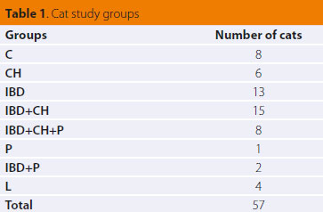
Study of the bacterial population of the duodenum and presence of bacteria in the bile of cats with chronic inflammatory bowel disease C: controls
CH: cats with histopathological evidence of cholangitis
IBD: cats with histopathological evidence of chronic inflammatory bowel disease
IBD+CH: cats with histopathological evidence of chronic inflammatory bowel disease and cholangitis
IBD+CH+P: cats with histopathological evidence of chronic inflammatory bowel disease, cholangitis and pancreatitis
P: cats with histopathological evidence of pancreatitis
IBD+P: cats with histopathological evidence of chronic inflammatory bowel disease and pancreatitis
L: cats with histopathological evidence of small intestinal lymphoma


- Histopathological characteristics of study groups

IBD: In total, thirty eight cats had histopathological evidence of the lymphocytic/plasmacytic type of IBD. In all of the latter there was infiltration of lymphocytes, plasmacytes and macrophages in the intestinal mucosa, whereas neutrophils were observed in variable numbers and more rarely, occasional eosinophils. The infiltrations, combined with architectural lesions in the intestinal epithelium, extended in all entire parts (duodenum, jejunum, ileum) with a variable degree of severity.

Cholangitis: Twenty nine cats had histopathological evidence of cholangitis. The of majority lesions was defined by infiltration of portal areas consisting primarily of lymphocytes and to a lesser extent by plasmacells, fibrosis, and hyperplasia of the bile ducts (lymphocytic type of cholangitis). In a small number of cats (5/29), in addition to mononuclear cells neutrophils were present (chronic neutrophilic cholangitis).

Pancreatitis: Eleven cats showed lesions of chronic pancreatitis, characterised by mononuclear cell infiltration and fibrosis. In five of them, there was also a considerable number of neutrophils (three cases were classified as chronic-active pancreatitis and the other two, where necrotic lesions were present, as acute necrotic pancreatitis in combination with chronic pancreatitis).

Lymphoma: Four cats had histopathological findings compatible with intestinal lymphoma. The latter constituted of diffuse aggregations of a uniform population of lymphocytes in the connective tissue and submucosal layer with multifocal infiltrations of the muscularis, whereas in some cases they were also present in the innermost mucosa. Very small numbers of the other types of inflammatory cells were observed, whereas micro-erosions and erosions of the epithelial surface were noted. In one cat an increased number of neutrophils was noted in the connective tissue layer.

- Physical and Diagnostic examinations

In all 57 cats included in the study the history was initially obtained and a general physical examination was performed. Diagnostic procedures ((<3 days prior to exploratory laparotomy) for the 57 cats included fecal parasitological examination and the detection of Giardia spp. antigen, standard urinalysis, complete blood count, biochemistry in blood serum including: albumin (ALB), blood urea nitrogen (BUN), creatinine (CREA), alkaline phosphatase (ALP), alanine aminotransferase activity (ALT), g-glutamine transferase activity (γGT), aspartate aminotransferase activity (AST), total bilirubin (TBIL), lipase activity, ionised calcium (Ca), phosphorus (P), potassium (Κ), sodium (Na), blood coagulation profile including prothrombin time (PT) and partial thromboplastin time (PTT) (52/57), serum total thyroxin (Τ4) and free thyroxin (Free T4), serum feline immunoreactivity of pancreatic lipase (fPLI measured by Spec fPL®)17 (56/57) trypsin like immunoreactity (fTLI) and testing for viral origin disorders including FIV, FeLV, and coronavirus for FIP. Diagnostic imaging included thoracic and abdominal radiographs, (49/57) and abdominal ultrasonography (56/57). Full thickness biopsy samples were collected for histopathological diagnosis (at least five from each cat: one from the liver, the pancreas, the duodenum, the jejunum, and the ileum) via exploratory laparotomy and were evaluated in a blinded fashion by a specialised veterinary pathologist (P. T.), based on internationally acceptable histopathological criteria.18-20

- Intestinal content and bile sampling

Prior to intestinal biopsy, a sample of duodenal juice was obtained from the duodenum. To this purpose, the duodenum was located and by “massaging” its full length any contents present were collected in the middle segment. Duodenal juice was removed by suction through plastic intravenous catheter (Abbocath-T I.V. Catheter 20 G x 1,25”, VenisystemsTM, Abbott, Ireland) attached on a 20 ml syringe.15 Τhe sample of duodenal juice was immediately placed in a sterile, glass vacuum blood collection vial without anticoagulant.

During exploratory laparotomy, 1 ml of bile was obtained from the gall bladder, by use of sterile 1 ml syringe and a 25 G needle. In cases when bile could not be aspirated (e.g. increased viscosity), a wider bore needle was used (23-21 G). The samples were immediately transfused to a sterile vacuum glass vial for blood collection, without anticoagulant (Venoject®, Terumo Europe N.V., Leuven, Belgium).

Vials containing bile and intestinal content were immediately placed in a transportation refrigerator (temperature levels of 4-6 °C) and within one hour from sampling they were transported to the laboratory of microbiology, where they were inoculated in the appropriate growth mediums under specific aerobic, microaerophilic and anaerobic conditions, aiming in growth of any bacteria in the samples.

- Culturing of duodenal juice and bile

Isolation and enumeration of bacteria

For the isolation and enumeration, as well as the identification of bacteria standard microbiological guidelines were employed.21 For the isolation of aerobic and facultative anaerobic bacteria, such as Enterobacteriaceae spp., Lactobacillus spp., Staphylococcus spp., Streptococcus spp., Enterococcus spp. and Pseudomonas spp., the mediums Blood Agar, MacConkey Agar, Rogosa Agar and Bile Esculin Agar were used. For the isolation of anaerobic bacteria, such as Clostridium spp., Bacteroides spp., Peptostreptococcus spp. and Eubacterium spp., the following mediums were used: Anaerobic Agar acc. to Brewer and TSC-Agar (Tryptose Sulfite Cycloserine Agar, Perfringens Agar). For the isolation of microaerophilic bacterial strains, such as Campylobacter spp., the special medium Campylobacter Selective Agar was used.

In order to calculate the bacterial population of samples, the technique of serial dilutions (10-1 up to 10-6) and inoculation of every dilution in agar plates with the spread plate method was used. From each dilution usually two agar plates were inoculated for every growth medium. Agar plates were incubated in 37°C in aerobic, anaerobic and microaerophilic conditions, depending on the inoculated substrate. For incubation in anaerobic conditions specialised containers were used (GasPakTM EZ Anaerobe Pouch System, Anaerobe Gas Generating pouch system with indicator, Becton Dickinson, NJ, USA), as well as for incubation of microaerophilic bacteria (GasPakTM Campy Pouch System, BBLTM Microaerophilic Campy pouch system, Becton Dickinson, NJ, USA). Τhe agar plates were daily monitored for the presence of bacterial growth up to 48 hours for aerobic cultures and up to six days for anaerobic and microaerophilic cultures.

In order to calculate the population of bacteria, the colonies that were observed in aerobic, anaerobic and microaerophilic conditions were counted, including the agar plates containing 30-300 colonies. The total of visible colonies from both agar plates that had been inoculated from each dilution and the median of both agar plates were calculated. Finally, the number of bacteria capable of forming colonies was expressed per 1 ml of initial sample (colony forming units, cfu/ml).

Bacterial identification

For the identification of bacterial strains the following were evaluated: (1) growth in special media for isolation and identification of bacteria [(i) Bile Esculin agar (BBLTM Bile Esculin Agar, Becton Dickinson, Maryland, USA): for isolation of Enterococcus spp. and differentiation from Streptococcus spp. (ii) Campylobacter Selective agar: for isolation of Campylobacter spp. (iii) TSC agar: for isolation of Clostridium spp. (iv) Rogosa agar: for isolation of Lactobacilli (v) Anaerobic agar acc. to Brewer: for isolation of Clostridium spp. and other anaerobic or microaerophilic bacteria.] (2) the morphological characteristics of bacteria post staining (Gram stain). (3) the initial biochemical characteristics: catalase testing, oxidase testing, positive or negative growth in MacConkey agar, indole testing, nitric salt induction, production of lecithinase, production of lipase. (4) sensitivity or not to the antimicrobial substance vancomycin, as an additional test beyond Gram staining, for the differentiation between Gram positive and Gram negative bacteria. (5) For specific identification of Enterobacteriaceae spp. the API 20 E system was used (API® bio- Mérieux Inc., Durham NC, USA).

> Statistical analysis

The cats of the study were classified into groups using histopathological diagnosis as a criterion. Data processesing and comparisons were made among study groups. Cats with pancreatitis alone, with IBD lesions in combination with pancreatitis and cats with intestinal lymphoma were excluded from statistical analysis, due to small sample size. For the synoptic presentation of statistical results absolute and relative frequencies (percentages %), measures of central tendency (mean, median) and measures of spread-dispersion [(minimum (min)-maximum (max) values and standard deviation] were calculated. For comparisons of means and medians the Kruskal-Wallis and Mann-Whitney tests were employed. For comparisons of proportions (percentages %) z-test was used with Bonferroni correction to the significance level. In all statistical analyses the observed significance level (P-value) was estimated, as appropriate, either with the Exact Method, or a Monte-Carlo simulation based on 10.000 resampling cycles. 22 Τhe level of statistical significance was set at α=0,05 (P≤0,05). Statistical analyses were performed by the IBM SPSS v. 20.0 statistical package (USA, Chicago: Illinois) with the Exact Tests subsystem installed. (Statistical pack IBM SPSS v.20.0)

> Results

Duodenal bacterial population

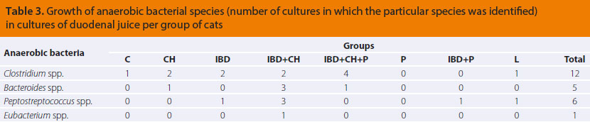
Aerobic and anaerobic bacterial species isolated during culture of duodenal juice per group of cats, are described in Tables 2 and 3. No growth of microaerophilic bacteria of the genus Campylobacter spp. was observed in any of the duodenal juice cultures from the cats in this study.

v7i1 bacterial population img2 en

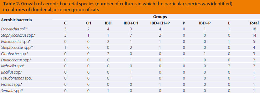
* facultative anaerobes
C: controls (n=8)
CH: cats with histopathological evidence of cholangitis (n=5)
IBD: cats with histopathological evidence of chronic inflammatory bowel disease (n=10)
IBD+CH: cats with histopathological evidence of chronic inflammatory bowel disease and cholangitis (n=14)
IBD+CH+P: cats with histopathological evidence of chronic inflammatory bowel disease, cholangitis and pancreatitis (n=6)
P: cats with histopathological evidence of pancreatitis (n=1)
IBD+P: cats with histopathological evidence of chronic inflammatory bowel disease and pancreatitis (n=1)
L: cats with histopathological evidence of small intestinal lymphoma (n=3)

v7i1 bacterial population img3 en

C: controls (n=8)
CH: cats with histopathological evidence of cholangitis (n=5)
IBD: cats with histopathological evidence of chronic inflammatory bowel disease (n=10)
IBD+CH: cats with histopathological evidence of chronic inflammatory bowel disease and cholangitis (n=14)
IBD+CH+P: cats with histopathological evidence of chronic inflammatory bowel disease, cholangitis and pancreatitis (n=6)
P: cats with histopathological evidence of pancreatitis (n=1)
IBD+P: cats with histopathological evidence of chronic inflammatory bowel disease and pancreatitis (n=1)
L: cats with histopathological evidence of small intestinal lymphoma (n=3)

The numerical estimation of the total of aerobes, anaerobes as well as the entire bacterial population from cultures of duodenal juice of cats in this study are reported per group in Table 4.

v7i1 bacterial population img4 en 
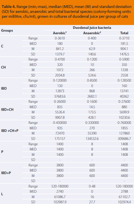
1aerobic and facultative anaerobic bacteria
2strictly anaerobic bacteria
C: controls (n=8)
CH: cats with histopathological evidence of cholangitis (n=5)
IBD: cats with histopathological evidence of chronic inflammatory bowel disease (n=10)
IBD+CH: cats with histopathological evidence of chronic inflammatory bowel disease and cholangitis (n=14)
IBD+CH+P: cats with histopathological evidence of chronic inflammatory bowel disease, cholangitis and pancreatitis (n=6)
P: cats with histopathological evidence of pancreatitis (n=1)
IBD+P: cats with histopathological evidence of chronic inflammatory bowel disease and pancreatitis (n=1)
L: cats with histopathological evidence of small intestinal lymphoma (n=3)


Comparison of the bacterial population of the duodenum between the feline study groups regarding the growth of aerobics (P=0,831), anaerobics (P=0,406) and the total bacterial population (P=0,752) did not reveal statistically significant differences.

The numerical estimation of the most common aerobic and anaerobic bacterial growth in cultures of duodenal juice of cats in this study is presented per group in Table 5.
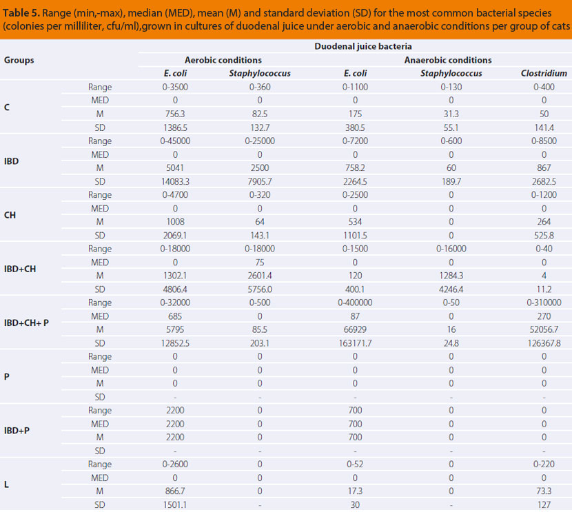
v7i1 bacterial population img5 en

C: controls (n=8)
CH: cats with histopathological evidence of cholangitis (n=5)
IBD: cats with histopathological evidence of chronic inflammatory bowel disease (n=10)
IBD+CH: cats with histopathological evidence of chronic inflammatory bowel disease and cholangitis (n=14)
IBD+CH+P: cats with histopathological evidence of chronic inflammatory bowel disease, cholangitis and pancreatitis (n=6)
P: cats with histopathological evidence of pancreatitis (n=1)
IBD+P: cats with histopathological evidence of chronic inflammatory bowel disease and pancreatitis (n=1)
L: cats with histopathological evidence of small intestinal lymphoma (n=3)

Comparisons of the duodenal bacterial population of the cat study groups, regarding the growth of Εscherichia coli, which was evaluated in aerobic (P=0,317) as well as anaerobic conditions (P=0,313), and Staphylococcus spp., in both aerobic (P=0,332) and anaerobic conditions (P=0,279), did not show any statistically significant differences among groups. Statistically significant differences were revealed during comparison of results among groups in regard to growth of anaerobic Clostridium spp., as they are presented in Table 6.

v7i1 bacterial population img6 en
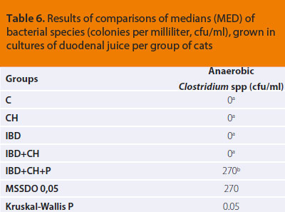
ΜSSDO: Minimum Statistically Significant Difference Observed at a significance level of P=0,05
a, b: In the same column of the table medians followed by common letter (superscript) do not differ significantly according to the results of a series of Mann-Whitney tests. A statistically significant difference exists between medians with different superscript letter.
C: controls (n=8)
CH: cats with histopathological evidence of cholangitis (n=5)
IBD: cats with histopathological evidence of chronic inflammatory bowel disease (n=10)
IBD+CH: cats with histopathological evidence of chronic inflammatory bowel disease and cholangitis (n=14)
IBD+CH+P: cats with histopathological evidence of chronic inflammatory bowel disease, cholangitis and pancreatitis (n=6)

Presence of bacteria in bile

Bile cultures of the control group were negative for bacterial growth. From the cat groups with abnormal findings, only one bile culture was positive from the IBD+P group, in which Enterobacter cloacae was isolated (Table 7).
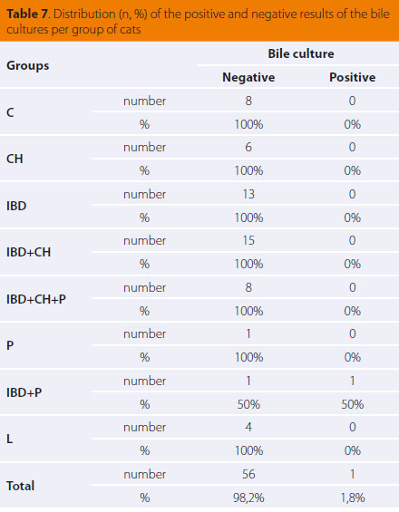
v7i1 bacterial population img7 en

C: controls (n=8)
CH: cats with histopathological evidence of cholangitis (n=6)
IBD: cats with histopathological evidence of chronic inflammatory bowel disease (n=13)
IBD+CH: cats with histopathological evidence of chronic inflammatory bowel disease and cholangitis (n=15)
IBD+CH+P: cats with histopathological evidence of chronic inflammatory bowel disease, cholangitis and pancreatitis (n=8)
P: cats with histopathological evidence of pancreatitis (n=1)
IBD+P: cats with histopathological evidence of chronic inflammatory bowel disease and pancreatitis (n=2)
L: cats with histopathological evidence of small intestinal lymphoma (n=4)

> Discussion

There was wide variability in the duodenal bacterial populations among the cat groups. However, comparisons did not reveal statistically significant differences in the total bacterial populations, as well as in the subgroups of aerobic and anaerobic duodenal bacteria between controls (C group) and cats with abnormal findings of all groups (IBD, Ch, IBD+Ch, IBD+Ch+P, Tables 4, 5 & 6). By reviewing our findings and in comparison to the referred as the feline normal intestinal flora, intestinal bacterial overgrowth was not substantiated in any of the cats in our study. In the control group the small intestinal bacterial population ranged from 0 to 3,7x10³ cfu/ ml (mean 9x10²). Among the groups of sick cats, the most numerous bacterial populations were noted in the triaditis group (IBD+Ch+P, Table 4), ranging from 0 to 7,6x10⁵ cfu/ml (mean 1,2x10⁵).

Both the mean and maximum values of bacteria observed in all study groups were found to be within the previously published reference range for the normal feline bacterial flora (105-108 cfu/ ml).2,14,15 However, in our study a predominance of anaerobic species of the genus Clostridium was observed in cats of the triaditis group (IBD+Ch+P, Table 5) compared to the rest of the study groups. Clostridium spp. (division Firmicutes, family Clostridiaceae, including at least 70 different species) constitute most of the cecal flora, however, they can be detected in other intestinal segments performing different functions.5 They are therefore part of the normal feline small intestinal flora despite their anaerobic nature.2,15

The etiopathogenetic connection between abnormal variations in the intestinal flora and induction of inflammation has not yet been clarified.1 An increase in several bacterial strains of Proteobacteria, such as Escherichia coli, and a reduction the Firmicutes and especially of the diversity of certain Clostridium spp. have both been reported as common disorders.1,10-13 In dogs with IBD a reduction in the diversity of small intestinal bacterial flora has been observed.23 Research in cats with IBD has proven the existence of «intestinal dysbiosis» in sick cats, with the Enterobacteriaceae spp., Clostridium spp., Bacteroides spp. and Streptococcus spp. corresponding to 91% of bacteria attached to the intestinal mucosa and Εscherichia coli comprising 30% of the Enterobacteriaceae spp..8 A different study indicated that strains from the genus Desulfovibrio predominated in the intestinal flora of cats with IBD, whereas strains of the Bifidobacterium and Bacteroides genera predominated in the bacterial populations of healthy cats.7 In our study, at first, it seemed like a paradox that even though cats of the triaditis group (IBD+CH+P, Table 5) had increased populations of Clostridium spp. in the duodenum, similar increase was not observed in the rest of the groups. Τhis fact underlines the complicated nature of the etiopathogenetic connection between the three pathological conditions, as well as “chronologically placing” their coexistence from an evolutionary perspective in more advanced stages compared to their combinations in pairs. In the future, sensitive molecular methods could give answers, regarding the increased populations of Clostridium spp. evidenced in our study, diversity and their contribution to the pathogenesis of triaditis.

The microbiological analysis of bile, concerning the diagnosis of cholangitis, usually includes culture in aerobic and anaerobic conditions as well as antibiotic sensitivity testing in order to indicate the proper therapeutic regimen.24-27 Culturing bile is preferred to culturing liver biopsy samples or gall bladder wall samples, because of improved rates of microorganism detection.28 Despite the ruling hypothesis that bile in healthy cats is microbiologically sterile,25,29 some researchers claim that bacterial translocation from duodenum to bile can occur in healthy as well.28 In our study, however, no bacterial growth was noted in bile cultures from healthy controls.

Regarding the types of feline cholangitis, in the acute neutrophilic cholangitis, isolation of mostly Enterobacteriaceae spp. originating from the duodenal flora in the bile is a common occurrence and confirms the diagnosis.24,25,28,30-35 Bacterial translocation from the intestinal tract to the gall bladder can occur either through reflux of bile from the duodenum, or through the hematogenous or lymphic routes.25 It is maintained that inflammatory bowel disease and pancreatitis can predispose to cholestasis, resulting in reflux of pancreatic secretions and/or bacteria towards the liver.32,36 In chronic neutrophilic cholangitis, bile culture is negative in most cases. It is theorised that this occurs due to i) either the bacteriostatic properties of bile, ii) or because the initial bacterial invasion was restricted by the immune system, iii) or due to previous use of antimicrobials, and iv) in cases when bacteria are not the immediate cause of the inflammatory disorder.24,37-39 However, even in chronic cholangitis of non-bacterial origin, chronic infiltration of bile ducts by inflammatory cells results in a risk for secondary hepatic infection by Enterobacteriaceae spp., such as Εscherichia coli.26

The lymphocytic type of cholangitis appears to originate from an immune-mediated aetiopathogenetic mechanism.40-42 However, there is also a theory that this particular type of cholangitis represents the chronic stage of acute neutrophilic cholangitis or an ascending (originating from the duodenum) bacterial infection.31,37,38 Τhere is only a small amount of data on which the hypothesis of a primary bacterial infection can be based.42 Two studies have been published concerning a small group of cats with cholangitis/cholangiohepatitis in which bacterial DNA of the Helicobacter genus has been detected, although the pathophysiological significance of this finding has yet to be clarified.43,44 It is worthy of note that until the present day, there is no evidence to support the involvement of Helicobacter spp. to IBD and pancreatitis in cats.8,45 Furthermore, in an experimental study, moderate inflammation in zone 1 of the feline liver was caused after infection with Bartonella spp.46 Even though there is considerable evidence of an immune-mediated mechanism causing cholangitis, the actual etiopathogenesis of the disorder remains a mystery.42

Τhe results of our study do not support the hypothesis of a primary microbial infection, considering that all the bile samples from cats with cholangitis were found to be bacteriologically sterile. From cats lacking histopathological evidence of cholangitis, only a single bile sample was found positive with growth of the bacterium Εnterobacter cloacae. This was a cat with pancreatitis and IBD (Table 7, IBD+P group) together with cholestasis, without histopathological evidence of cholangitis or feline hepatic lipidosis. As previously mentioned, in this particular case cholestasis, as a result of obstruction in bile flow due to pancreatitis, became a risk factor for the translocation of Εnterobacter cloacae, which is part of the normal small intestinal flora, toward the gall bladder. Unfortunately, culture of duodenal content was not performed on this cat; therefore its intestinal flora is unknown. Such a hypothesis, however, cannot explain the inflammation in the bile duct system observed in 29 cats with histopathological evidence of cholangitis in our study, in which bile cultures were negative. The possibility of bacterial translocation toward the liver and consequently the pancreas via the common bile and pancreatic duct, does not exclude the theory of an immune response to such bacterial invasion.

To summarise, the present study indicated that inflammatory disorders of the gastrointestinal tract relating to triaditis as well as intestinal lymphoma do not seem to be pathogenetically related to the bacterial flora of the duodenum or any presence of bacteria in bile. The intestine and the liver play a particularly significant role in immunity. This complicated system of the intestinal flora may affect several functions as well as the global health and every disruption in its interactions with the local intestinal immune mechanisms could lead to gastrointestinal disease.1,6-10 In conclusion, an immune-mediated mechanism could be involved in the development of triaditis in cats.42,47 Modern molecular methods of analysis are expected to give more answers in the investigation of any correlations between intestinal flora and its variability with histopathological lesions of inflammation in the intestine, liver and pancreas.

> Acknlowledgements

The authors would like to acknowledge the contribution of Dr. G. Menexes, Assistant Professor of Biometrics and Agricultural Experimentation, Faculty of Agriculture, Forestry and Natural Environment, School of Agriculture, Α.U.Th. for performing the statistical analysis of the results of this study. They would also like to thank the cat owners, veterinarians, students and the staff of the Companion Animal Clinic that became a part of this research project.

The first author (F.F.) was supported by the State Scholarships Foundation (code no 5321) in order to realise part of the present study.

> References

1. Suchodolski JS. Companion Animals Symposium: Microbes and gastrointestinal health of dogs and cats. J Anim Sci 2011, 89: 1520-1530.

2. Johnston K, Lamport A, Batt RM. An Unexpected Bacterial- Flora in the Proximal Small-Intestine of normal cats. Vet Rec 1993, 132: 362-363.

3. Sparkes AH, Papasouliotis K, Sunvold G, Werrett G, Clarke C, Jones M, Gruffydd-Jones TJ, Reinhart G. Bacterial flora in the duodenum of healthy cats, and effect of dietary supplementation with fructo-oligosaccharides. Am J Vet Res 1998, 59: 431-435.

4. Johnston KL, Swift NC, Forster-van Hijfte M, Rutgers HC, Lamport A, Ballevre O, Batt. RM. Comparisοn of the bacterial flora of the duodenum in healthy cats and cats with signs of gastrointestinal tract disease. J Am Vet Med Assoc 2001, 218: 48–51.

5. Suchodolski JS. Gastrointestinal Microbiota. In: Canine and Feline Gastroenterology. Washabau RJ and Day MJ (eds). Elsevier Saunders: Missouri, 2013, pp. 32-41.

6. German AJ, Day MJ, Ruaux CG, Steiner JM, Williams DA, Hall EJ. Comparison of direct and indirect tests for small intestinal bacterial overgrowth and antibiotic-responsive diarrhea in dogs. J Vet Intern Med 2003, 17: 33-43.

7. Inness VL, McCartney AL, Khoo C, Gross KL, Gibson GR. Molecular characterisation of the gut microflora of healthy and inflammatory bowel disease cats using fluorescence in situ hybridisation with special reference to Desulfovibrio spp. J Anim Physiol Anim Nutr (Berl) 2007, 91: 48-53.

8. Janeczko S, Atwater D, Bogel E, Greiter-Wilke A, Gerold A, Baumgart M, Bender H, McDonough PL, McDonough SP, Goldstein RE, Simpson KW. The relationship of mucosal bacteria to duodenal histopathology, cytokine mRNA, and clinical disease activity in cats with inflammatory bowel disease. Vet Microbiol 2008, 128: 178-193.

9. Xenoulis PG, Palculict B, Allenspach K, Steiner JM, Van House AM, Suchodolski JS. Molecular-phylogenetic characterization of microbial communities imbalances in the small intestine of dogs with inflammatory bowel disease. FEMS Microbiol Ecol 2008, 66: 579-589.

10. Packey CD, Sartor RB. Commensal bacteria, traditional and opportunistic pathogens, dysbiosis and bacterial killing in inflammatory bowel diseases. Curr Opin Infect Dis 2009, 22: 292-301.

11. Craven M, Dogan B, Schukken A, Volkman M, Chandler A, McDonough PL, Simpson KW. Antimicrobial resistance impacts clinical outcome of granulomatous colitis in boxer dogs. J Vet Intern Med 2010, 24: 819-824.

12. Suchodolski, JS, Xenoulis PG, Paddock CG, Steiner JM, Jergens AE. Molecular analysis of the bacterial microbiota in duodenal biopsies from dogs with idiopathic inflammatory bowel disease. Vet Microbiol 2010, 142: 394-400.

13. Suchodolski JS, Dowd SE, Wilke V, Steiner JM, Jergens AE. 16S rRNA Gene Pyrosequencing Reveals Bacterial Dysbiosis in the Duodenum of Dogs with Idiopathic Inflammatory Bowel Disease. Plos One 2012, 7:e39333. doi: 10.1371/journal.pone.0039333.

14. Johnston KL. Small intestinal bacterial overgrowth. Vet Clin North Am Small Anim Pract 1999, 29: 523-550.

15. Johnston KL, Lamport A, Ballevre O, Batt RM. A comparison of endoscopic and surgical collection procedures for the analysis of the bacterial flora in duodenal fluid from cats. Vet J 1999, 157: 85-89.

16. Fragkou FC, Adamama-Moraitou KK, Poutahidis T, Prassinos NN, Kritsepi-Konstantinou M, Xenoulis PG, Steiner JM, Lidbury JA, Suchodolski JS, Rallis TS. Prevalence and Clinicopathological Features of Triaditis in a Prospective Case Series of Symptomatic and Asymptomatic Cats. J Vet Intern Med 2016, 30: 1031-1045.

17. Xenoulis PG, Steiner JM. Canine and feline pancreatic lipase immunoreactivity. Vet Clin Pathol 2012, 41: 312–324.

18. Van den Ingh TS, Cullen JM, Twedt DC, Van Winkle T, Desmet VJ, Rothuizen J. Morphological classification of biliary disorders of the canine and feline liver. In: WSAVA Standards for Clinical and Histological Diagnosis of Canine and Feline Liver Diseases. Rothuizen J, Bunch SE, Charles JE, Cullen JM, Desmet VJ, Szatmari V, Twedt DC, Van den Ingh TS, Van Winkle T, Washabau RJ (eds). Elsevier: Philadelphia, 2006, pp. 68-71.

19. De Cock HEV, Forman MA, Farver TB, Marks SL. Prevalence and histopathologic characteristics of pancreatitis in cats. Vet Pathol 2007, 44: 39-49.

20. Washabau RJ, Day MJ, Willard MD, Hall EJ, Jergens AE, Mansell J, Minami T, Bilzer TW. Endoscopic, biopsy, and histopathologic guidelines for the evaluation of gastrointestinal inflammation in companion animals. J Vet Intern Med 2010, 24: 10-26.

21. Quinn PT, Carter ME, Markey BQ, Carter GR. Clinical Veterinary Microbiology. Mosby, 1999.

22. Mehta CR, Patel NR. Exact logistic regression: theory and examples. Stat Med 1995, 14: 2143-2160.

23. Xenoulis PG, Palculict B, Allenspach K, Steiner JM, Van House AM, Suchodolski JS. Molecular-phylogenetic characterization of microbial communities imbalances in the small intestine of dogs with inflammatory bowel disease. FEMS Microbiology Ecology 2008, 66: 579-589.

24. Hirsch VM, Doige CE. Suppurative cholangitis in cats. J Am Vet Med Assoc 1983, 182: 1223-1226.

25. Brain PH, Barrs VR, Martin P, Baral R, White JD, Beatty JA. Feline cholecystitis and acute neutrophilic cholangitis: clinical findings, bacterial isolates and response to treatment in six cases. J Feline Med Surg 2006, 8: 91.

26. Rothuizen J. Liver-Diseases of the biliary system in cats. In: Small Animal Gastroenterology. Steiner JM (ed). Schlütersche: Hannover, 2008, pp. 275-278.

27. Harvey AM, Gruffydd-Jones TJ. Feline Inflammatory Liver Disease In: Textbook of Veterinary Internal Medicine: diseases of the dog and cat. Ettinger SJ and Feldman EC (eds). 7th edn. Saunders, Elsevier: St Louis, 2010, pp. 1643-1709.

28. Wagner KA, Hartmann FA, Trepanier LA. Bacterial culture results from liver, gallbladder or bile in 248 dogs and cats evaluated for hepatobiliary disease: 1998-2003. J Vet Intern Med 2007, 21: 417-424.

29. Savary-Bataille KCM, Bunch SE, Spaulding KA, Jackson MW, Law JM, Stebbins ME. Percutaneous ultrasound guided cholecystocentesis in healthy cats. J Vet Intern Med 2003, 17: 298.

30. Kaufman AC. Infectious causes of feline hepatobiliary disease. Vet Med 1994, 89: 869-873.

31. Day DG. Feline cholangiohepatitis complex. Vet Clin North Am Small Anim Pract 1995, 25: 375-385.

32. Center SA. Diseases of the gall bladder and biliary tree. In: Small Animal Gastroenterology. Guilford WG, Center SA, Strombeck DR, Williams DA, MeyerDJ (eds). 3rd edn. Saunders: Philadelphia, 1996, pp. 860-888.

33. Center SA. Hepatobiliary infections. In: Infectious Diseases of the Dog and Cat. Greene CE (ed). 2nd edn. WB Saunders: Philadelphia, 1998, pp. 615-625.

34. Lapointe JM, Higgins R, Barrette N, Milette S. Enterococcus hirae enteropathy with ascending cholangitis and pancreatitis in a kitten. Vet Pathol 2000, 37: 282-284.

35. Mayhew PD, Holt DE, McLear RC, Washabau RJ. Pathogenesis and outcome of extrahepatic biliary obstruction in cats. J Small Anim Pract 2002, 43: 247-253.

36. Weiss DJ, Armstrong PJ, Gagne J. Inflammatory liver disease. Semin Vet Med Surg (Small Anim) 1997, 12: 22-27.

37. Zawie DA, Garvey MS. Feline hepatic disease. Vet Clin North Am Small Anim Pract 1984, 14: 1201.

38. enter SA, Rowland PH. The cholangitis/cholangiohepatitis complex in the cat. In: Congress Proceedings of ACVIM Forum. San Francisco, 1994, pp. 766-771.

39. Rallis TS. Liver diseases. In: Canine and Feline Gastroenterology. Rallis TS (ed). 2nd edn. University Studio Press: Thessaloniki, 2006, pp. 227-310.

40. Prasse KW, Mahaffey EA, De Novo R, Cornelius L. Chronic lymphocytic cholangitis in three cats. Vet Pathol 1982, 19: 99-108.

41. Day MJ. Immunohistochemical characterization of the lesions of feline progressive lymphocytic cholangitis/cholangiohepatitis. J Comp Pathol 1998, 119: 135-147.

42. Warren Α, Center S, McDonough S, Chiotti R, Goldstein R, Meseck E, Jacobsen M, Rowland P, Simpson K. Histopathologic features, Immunophenotyping, Clonality, and Eubacterial Fluorescence In Situ Hybridization in Cats with Lymphocytic Cholangitis/ Cholangiohepatitis. Vet Pathol 2011, 48: 627-641.

43. Boomkens SY, Kusters JG, Hoffmann G, Pot RG, Spee B, Penning LC, Egberiink HF, van den Ingh TS, Rothuizen J. Detection of Helicobacter pylori in bile of cats. FEMS Immunol Med Microbiol 2004, 42: 307-311.

44. Greiter-Wilke A, Scanziani E, Soldati S, McDonough SP, Mc- Donough PL, Center SA, Rishniw M, Simpson KW. Association of Helicobacter with cholangiohepatitis in cats. J Vet Intern Med 2006, 20: 822-827.

45. Simpson KW. Is there a direct link between IBD, Cholangitis, and Pancreatitis in cats? (abstract). In: Congress Proceedings of ECVIM-CA. Maastricht, The Netherlands, 2012, pp. 169-171.

46. Kordick DL, Brown TT, Shin K, Breitschwerdt EB. Clinical and pathologic evaluation of chronic Bartonella henselae or Bartonella clarridgeiae infection in cats. J Clin Microbiol 1999, 37: 1536-1547.

47. Simpson KW. Pancreatitis and triaditis in cats: causes and treatment. J Small Anim Pract 2015, 56: 40-49.


Canine anal furunculosis: is there a place for surgery?


> Abstract

Anal furunculosis is a chronic progressive canine inflammatory disorder affecting the anus and the perianal region, resulting in ulcers and blind fistula in the skin and subcutaneous tissues of the perianal region. German shepherds are predisposed to this condition. The origins of the disorder are unknown but it can be immune- mediated. Treatment can be medical or surgical, but complete cure is uncommon. Medical treatment with immunomodulatory drugs, mostly cyclosporine or cyclosporine and ketoconazole usually provides satisfactory results. Surgical treatment is employed in cases with no response to medical management, due to increased cost or prolonged duration of the latter and includes complete anoplasty and simultaneous anal sacculectomy.

Canine anal furunculosis

Canine anal furunculosis

Canine anal furunculosis> Introduction

Anal furunculosis (AF) or perianal fistulae is a chronic progressive canine inflammatory disorder of dogs affecting the anal orifice and the perianal region. It is characterised by the presence of ulcerative lesions and blind fistulae in the skin and subcutaneous tissues of the perianal and perineal regions.1 The sinus tracts can be single or multiple and they can extend 3600 circumferentially in the perianal area (Figures 1,2,3,4).2 AF affects dogs with a median age of six years and of both genders. German shepherds are predisposed to the disorder even though AF occurs in other middle or large sized breeds as well as mixed breed dogs (Table 1).1,3-5


> Pathogenesis

The precise origin of the disorder is not fully known. Recent studies implicated an inflammatory response with presence of T-lymphocytes, other inflammatory cells, cytokines, enzymes, and other intermediaries of the inflammatory cascade and histological similarities between AF and Crohn’s disease in people, resulting in the theory of a possible immune-mediated origin.6-11 This theory is supported by an impressive response following administration of immune-mediated drugs.2 German shepherd breed predisposition to AF is probably of genetic origin, contributing to the pathogenesis of AF.12

Originally, AF manifests as a subtle inflammatory response without the presence of ulcers. At this time, ulcerative lesions and sinus tracts are formed, which are lined by squamous epithelium and they are infiltrated by lymphocytes, plasmacytes, macrophages, neutrophils and eosinophils. The evolution of the inflammatory response is characterised by the presence of Τ-lymphocytes with extensive formation of granulomatous and fibrous tissue (fibrosis). The normal anatomy of the area is disrupted with the formation of fistulae originating from the anal glands to the anocutaneous junction, as well as lesions in the anal sacs (Figure 5). The contribution of the anal sacs to the inflammatory response is secondary resulting in abscessation and rupture. The formation of fibrous tissue in the area of the outer anal sphincter in dogs with extensive and severe lesions can result in stricture. 3,13

Canine anal furunculosis

Canine anal furunculosis>Clinical signs

Clinical signs in dogs with AF vary and are usually connected with pain in the perineal area and disorders of defecation (Table 2).1,14-16 Several dogs already have severe lesions at the time of diagnosis.14

> Diagnosis

Reaching a diagnosis of AF is mostly based on the history and physical examination findings. It is necessary to perform a physical examination, both with the dog in an alert state and under general anaesthesia. In the alert state, other than external examination of the anus and the perianal area, the external anal spincter tone is also tested. Most of the times, however, physical examination in the alert state is difficult or even impossible due to severe pain elicited both by palpation of the area as well as from handling and elevating the tail. Prior to administrating general anaesthesia, it is necessary to obtain a complete blood count, serum biochemistry analysis and urinalysis, as part of the preanaesthetic evaluation, as well as to exclude other conditions with similar clinical signs. Then the perianal region is visually observed to assess whether the diseased areas may in part (0ο- 270ο) or entirely (360ο) affect the region. Rectal digital palpation is necessary in order to locate and evaluate the formation of fibrous tissue and the degree of rectal fibrosis. Stricture formation may lead to constipation and obstipation (Figure 6). Palpation of the anal sacs is also performed by digital rectal palpation and patency of their excretory ducts is determined; any sinus tracts connecting the perianal region with the anal glands are detected and the contribution of the anal glands to the inflammatory process is assessed (Figure 7). The depth of the sinus tracts is determined by using a probe. Colonoscopy and biopsy should be performed in some dogs with clinical signs of colitis, as a strong correlation has been found between chronic inflammatory disease of the large intestine and AF.17-20

Canine anal furunculosis

> Differential diagnosis

AF should be differentiated from abscessation and rupture of the anal glands, anal gland adenocarcinoma, adenoma and adenocarcinoma of the perianal glands, squamous cell carcinoma of the perianal region, chemical burns, trauma and subcutaneous mycosis (Figure 8).2 In such cases and when in doubt, cytological and histopathological examinations are recommended.

> Treatment

Treatment for AF can be medical or surgical, but it is uncommon to lead to complete and permanent cure. Surgical management of AF has been the treatment of choice for several years. A multitude of surgical techniques have been used in the past, such as surgical excision of fistulae, surgical debridement combined with chemical cauterisation, cryosurgery, cauterisation by electrodiathermy, complete tail amputation (given that a wide tail was implicated as a predisposing factor for AF) and excision by ND:YAG laser. The high percentage of relapsing and complications, including faecal incontinence, surgical wound dehiscence and anal stricture, have led to abandonment of surgical treatment as a single management strategy for AF. Moreover, many dogs required several surgical procedures in order to correct any complications.1,4,14,15,21-26

> Medical management

Nowadays medical management with immunomodulatory drugs is considered to be the treatment of choice due to highly satisfactory results.2 Medical treatment aims in suppressing the pain and lesions of the anal orifice and perianal area both in the short and long-term, as relapses of the disease after treatment is withdrawn are not uncommon. Medical management includes the acute phase, the maintenance phase, a specialised clinical diet and local perineal hygiene-antimicrobial treatment. For the acute and maintenance phase cyclosporine has been used as monotherapy or in combination with ketoconazole, glucocorticoids, azathioprine and local application of tacrolimus (Table 3).2,27-39 Τhis medication can lead to remission and, in some cases, elimination of lesions and clinical cure.27-37

Canine anal furunculosis

Glucocorticoids mostly suppress cellular immunity, are inexpensive but their administration usually results in adverse effects (polyuria, polydipsia, and polyphagia). Initially they had been used combined with a specialised diet in the management of AF. In 27 German shepherds prednisone was administered (2 mg/kg/SID [/24 hours] PO [orally]) for 2 weeks, then 1mg/kg for 4 weeks and then was reduced to at a dose of 1mg/kg/48h for 8-16 weeks. Clinical cure was achieved in 33.3% of dogs, improvement in 33.3% and the rest did not respond to medical treatment.27

Azathioprine has been used successfully in the management of AF. Due to the long period of time that is necessary for this drug to be fully effective, it is recommended to combine it with glucocorticoids, at least during the acute phase.38 In a prospective study with 14 dogs with AF a combination of azathioprine and prednisone was administered. Permanent cure was achieved in 57% of cases, partial improvement in 7% and no response in 36% of dogs.40

Cyclosporine is a powerful immunomodulatory agent, which suppresses the production of inflammatory cytokines, which are related to the activation of T-lymphocytes. In particular, cyclosporine suppresses mostly the production of interleukin-2, which is necessary for the differentiation and proliferation of T-lymphocytes.39 The administration of cyclosporine is considered the most effective treatment of AF with a success rate of 50-85%.29,30,33,35,36

The mean duration of treatment until the eradication of lesions, according to one study, was 8.8 weeks.35 However, discontinuation of treatment may result in relapse usually necessitating continuation of life-long treatment at the smallest dose possible. 28 Cyclosporine was initially administered at a dose regimen of 4-8 mg/kg SID PO for 2-4 months, until the eradication of lesions, and then the dose was gradually tapered by 25% every 4-8 weeks or alternatively the initial challenge dose was administered every other day after remission of clinical signs (Figures 9, 10).2,35,38 Cyclosporine treatment is costly and can be associated with adverse effects including vomiting, diarrhoea, anorexia, lethargy, aggression, hypertrichosis or trichorrhoea.29,33,35,36

Canine anal furunculosis

Τhe high cost of cyclosporine directed many researchers to investigate alternate methods of treatment. The combination of cyclosporine with ketoconazole reduces the cost of treatment with no change in effectiveness, compared to monotherapy with cyclosporine.41 Ketoconazole, an antifungal agent, affects the metabolism of cyclosporine by inhibiting the effect of cytochrome P450 3A oxidase, resulting in the increase of cyclosporine blood serum levels.41-43 Cyclosporine was administered at a dose regimen ranging from 0.5 mg/kg BID (/12 hours) up to 5 mg/kg PO SID and ketoconazole in a dose of 5-10 mg/kg PO SID.2,38,41-43 According to a study in 19 dogs with AF, the combination of cyclosporine and ketokonazole was successful in eradicating lesions when used for a duration of 3-10 weeks in 100% of dogs, whereas relapse was observed in 37% of dogs after a period of 1-6 months after the initial treatment.41

Tacrolimus is a topically applied macrolide with a similar immunomodulatory effect to that of cyclosporine, which has been used successfully in the management of AF.2,31,37 In a study of 10 dogs with AF, the topical application of tacrolimus SID or BID for 16 weeks resulted in remission of lesions in 90% of dogs, among which 50% were completely cured.31 In a different recent study of a total duration of two years, in 19 dogs with AF, 0.1% tacrolimus ointment was simultaneously administered with prednisone (2 mg/kg SID over two weeks, 1mg/kg SID for four weeks and 1mg/kg/48h for ten weeks) in combination with “hypoallergenic” diet. Moreover, metronidazole was administered PO at a dose of 10mg/kg BID for two weeks. After the completion of 16 weeks, 79% of dogs were cured and in 21% significant improvement was observed. During the next two years, the maintenance regimen was adhered to with tacrolimus and prednisone, and 86.6% of dogs apparently remained in remission.37 Tacrolimus is recommended mostly for long-term maintenance, following initial remission of clinical signs with administration of cyclosporine, when applicable, every 24-72 hours aiming at the prevention of relapses.2,38 Τhis treatment is considered to be costly.2,37,38

> Clinical Diet

The combination of AF and colitis prompted several authors to recommend a specialised “hypoallergenic” diet with hydrolysed protein or protein to which the dog has never been exposed previously. Thirty three dogs with AF were fed a diet based on fish and potatoes, for 1-180 days prior to surgery (en bloc resection of lesions and anoplasty) and one year later 87.9% of dogs experienced a complete eradication of lesions, whereas only 20.7% manifested any clinical signs.16 A specialised “hypoallergenic” diet is therefore recommended during the maintenance phase, especially in cases of lesion recurrence.2,38

> Local hygiene and systemic antimicrobial treatment

Local cleansing of the perineal region and hygiene measures such as clipping of the hair coat and lavage with antiseptic solutions along with systemic and topical administration of antibiotics, following culture and sensitivity testing, could aid in reducing the local bacterial flora and managing the secondary bacterial infections that are always present.2,38

> Preoperative immunomodulatory treatment

Preoperative immunomodulatory treatment results in remission of lesions allowing for a more conservative approach during surgical resection and reduction of complications to a minimum.30 Various treatment regimens have been employed, such as the administration of azathioprine (50 mg PO) and metronidazole (400 mg PO) for a duration of 4-6 weeks preoperatively and two weeks postoperatively. A significant clinical improvement was noted during the first two weeks, but after the advent of 4-6 weeks there was minimal further improvement. Dogs that received the above regimen did not relapse in the following 7-10 months.19 The adverse effects of azathioprine include gastrointestinal disorders, bone marrow suppression, hepatotoxicity, and pancreatitis whereas metronidazole side effects include anorexia, central nervous system toxicity and hepatotoxicity.2 In a study of 25 dogs with AF, cyclosporine was administered (2.5-5 mg/kg BID PO) as monotherapy, cyclosporine (1-1.5 mg/kg BID PO) in combination with ketoconazole (12.5 mg/kg SID PO), or azathioprine (1-2 mg/kg SID PO) in combination with prednisolone (1 mg/kg BID PO for 2 weeks and then reduced to 0.5 mg/kg BID PO) for less than 12 weeks. Surgical management included resection of all fistulae, cryptectomy of the transitional zone of the rectum up to the anocutaneous line and bilateral anal sacculectomy. None of the dogs relapsed after a follow-up time of 9 months after surgery.30

> Surgical management

Indications of surgical management include cases nonresponsive to medical treatment, cases with AF affecting the anal glands, and cases in which the cost of medical treatment is significant and its duration is prolonged. Surgical resection of all of the affected tissues is a prerequisite for successful results. 1,5,16 Good knowledge of anal canal anatomy is necessary in order to prevent severe complications. Surgical resection includes the removal of all perianal fistulae and their sinus tracts, as well as bilateral anal sacculectomy due to anal sac implication in the pathogenesis of the disorder.5,16 Currently, in cases of extensive lesions in the perianal region, complete 3600 anoplasty is recommended with simultaneous bilateral sacculectomy.5,16 Following surgical resection of the skin around the anus, all of the lesions are removed including the subcutaneous tissue, the muscles and fascia as well as the anal orifice. If necessary, partial or complete resection of the external anal sphincter is performed. Dead space is eliminated and the subcutaneous tissue is approximated to the serosa and muscular layer of the rectum by 3/0 polydioxanone simple interrupted sutures. Finally, the mucosal layer and submucosa of the rectum are sutured to the skin by 3/0 nylon sutures (Figures 11,12, 13, 14, 15).5,16 In cases when surgical wound closure is impossible, due to increased tension, healing by secondary intention is preferred. In a study of 51 dogs with AF which underwent 3600 anoplasty combined with bilateral anal sacculectomy, after a median follow-up of 18 months, 2% of cases relapsed, 13% manifested anal stenosis and 4% had fecal incontinence.5


Canine anal furunculosis

Canine anal furunculosis

Canine anal furunculosis

Treatment of AF is summarised in Figure 1.

Canine anal furunculosis

> References

1. Harvey CE. Perianal fistula in the dog. Vet Rec 1972, 91: 25-33

2. Patterson AP, Campbell KL. Managing anal furunculosis in dogs. Compend Contin Educ Pract Vet 2005, 27: 339-355

3. Day MJ, Weaver BMQ. Pathology of surgically resected tissue from 305 cases of anal furunculosis in the dog. J Small Anim Pract 1992, 33: 583-589

4. Vasseur PB. Results of surgical excision of perianal fistulas in dogs. J Am Vet Med Assoc 1984, 185: 60-62

5. Milner HR. The role of surgery in the management of canine anal furunculosis. A review of the literature and a retrospective evaluation of treatment by surgical resection in 51 dogs. New Zealand Vet J 2006, 54: 1-9

6. House A, Gregory SP, Catchpole B. Expression of cytokine mRNA in canine anal furunculosis lesions. Vet Rec 2003, 153: 354-358

7. House AK, Catchpole B, Gregory SP. Matrix metalloproteinase mRNA expression in canine anal furunculosis lesions. Vet Immunol Immunopathol 2007, 115: 68-75

8. House AK, Gregory SP, Catchpole B. Pattern- recognition receptor mRNA expression and function in canine monocyte/macrophages and relevance to canine anal furunculosis. Vet Immunol Immunopathol 2008, 124: 230-240

9. Tivers MS, Catchpole B, Gregory SP, House AK. Interleukin-2 and interferon-gamma mRNA expression in canine anal furunculosis lesions and the effect of ciclosporin therapy. Vet Immunol Immunopathol 2008, 125: 31-36

10. 1House AK, Binns MM, Gregory SP, Catchpole B. Analysis of NOD1, NOD2, TLR1, TLR2, TLR4, TLR5, TLR6 and TLR9 in anal furunculosis of german shepherd dogs. Tissue Antigens 2008, 73: 250-254

11. Barnes A, O’Neil T, Kennedy LJ, Short AD, Catchpole B, House A, Binns M, Fretwell N, Day MJ, Ollier WER. Association of canine anal furunculosis with TNFA is secondary to linkage disequilibrium with DLA-DRB1. Tissue Antigens 2008, 73: 218-224

12. Kennedy LJ, O’Neil T, House A. Barnes A, Kyöstilä K, Innes J, Fretwell N, Day MJ, Catchpole B, Lohi H, Ollier WE. Risk of anal furunculosis in German shepherd dog is associated with the major histocompatibility complex. Tissue Antigens 2008, 71: 51-56

13. Killingsworth CR, Walshaw R, Dunstan RW, Rosser EJ. Bacterial population and histologic changes in dogs with perianal fistula. Am J Vet Res 1988, 49:1736-1741

14. Robins GM, Lane JG. The management of anal furunculosis. J Small Anim Pract 1973, 14: 333-342

15. Houlton JEF. Anal furunculosis: a review of seventy cases. J Small Anim Pract 1980, 21: 575-584

16. Lombardi RL, Marino DJ. Long- Term Evaluation of canine perianal fistula disease treated with exclusive fish and potato diet and surgical excision. J Am Anim Hosp Assoc 2008, 44: 302-307

17. Jamieson PM, Simpson JW, Kirby BM, Else RW. Association between anal furunculosis and colitis in the dog: preliminary observations. J Small Anim Pract 2002, 43:109-114.

18. Massey J, Short AD, Catchpole B, House A, Day MJ, Lohi H, Olier WE, Kennedy LJ. Genetics of canine anal furunculosis in the German shepherd dog. Immunogenetics 2014, 66: 311-324.

19. Tisdall PLC, Hunt GB, Beck JA, Malik R. Management of perianal fistulae in five dogs using azathioprine and metronidazole prior to surgery. Aust Vet J 1999, 77: 374-378

20. Lombardi RL, Marino DJ. Long- term evaluation of canine perianal fistula disease treated with exclusive fish and potato diet and surgical excision. J Am Anim Hosp Assoc 2008, 44: 302-307

21. Lane JG, Burch DGS. The cryosurgical treatment of canine anal furunculosis. J Small Anim Pract 1975; 16: 387-392

22. Houlton JEF. Canine anal furunculosis: a modified approach. J Small Anim Pract 1980, 21: 585-593

23. Elkins AD, Horbson HP. Management of perianal fistulae a retrospective study of 23 cases. Vet Surg 1982, 11: 110-114

24. Goring RL, Bright RM, Stancil ML. Perianal fistulas in the dog. Retrospective evaluation of surgical treatment by deroofing and fulguration. Vet Surg 1986, 15: 392-398

25. Van Ee RT, Palmitieri A, Tail amputation for treatment of perianal fistulas in dogs. J Am Anim Hosp Assoc 1987; 23: 95-100

26. Ellison GW, Bellah JR, Stubbs WP, Gilder JV. Treatment of perianal fistulas with ND: YAG laser- results in twenty cases. Vet Surg 1995, 24: 140-147

27. Harkin KR, Walshaw R, Mullaney TP. Association of perianal fistula and colitis in the German shepherd dog: response to high-dose prednisone and dietary therapy. J Am Anim Hosp Assoc 1996, 32: 515-520.

28. Mathews KA, Ayres SA, Tano C, Riley SM, Sukhiani HR, Adams C. Cyclosporin treatment of perianal fistulas in dogs. Can Vet J 1997, 38: 39-41

29. Mathews KA, Sukhiani HR. Randomized controlled trial of cyclosporine for treatment of perianal fistulas in dogs. J Am Vet Med Assoc 1997, 211: 1249-1253

30. Klein A, Deneuche A, Fayolle P, Hidalgo A, Scotti S, Zylberstain L, Desbois C, Tessier D, Moissonnier P, Viateau V. Preoperative immunosuppressive therapy and surgery as a treatment for anal furunculosis. Vet Surg 2006, 35: 759-768

31. Misseghers BS, Binnington AG, Mathews KA. Clinical observations of the treatment of canine perianal fistulas with topical tacrolimus in 10 dogs. Can Vet J 2000, 41: 623-627

32. Griffiths LG, Sullivan M, Borland WW. Cyclosporine as the sole treatment for anal furunculosis: preliminary results. J Small Anim Pract 1999, 40: 569-572

33. Doust R, Griffiths LG, Sullivan M. Evaluation of once daily treatment with cyclosporine for anal furunculosis in dogs. Vet Rec 2003, 152: 225-229

34. O’Neill T, Edwards GA, Holloway S. Efficacy of combined cyclosporine A and ketoconazole treatment of anal furunculosis. J Small Anim Pract 2004, 45: 238-243

35. Hardie RJ, Gregory SP, Tomlin J, Sturgeon C, Lipsomb V, Ladlow J. Cyclosporine treatment of anal furunculosis in 26 dogs. J Small Anim Pract 2005, 46: 3-9

36. House AK, Guitian J, Gregory SP, Hardie RJ. Evaluation of the effect of two dose rates of cyclosporine on the severity of perianal fistulae lesions and associated clinical signs in dogs. Vet Surg 2006, 35: 543-549

37. Stanley BJ, Hauptman JG. Long-term prospective evaluation of topically applied 0,1% tacrolimus ointment for treatment of perianal sinuses in dogs. J Am Vet Med Assoc 2009, 235: 397-404

38. Pieper J, McKay L. Perianal fistulas. Compend Contin Educ Vet 2011, 33: E1-E4

39. Guaguere E, Steffan J, Olivry T. Cyclosporin A: a new drug in the field of canine dermatology. Vet Dermatol 2004, 15: 61–74

40. Harkin KR, Phillips D, Wilkenson M. Evaluation of azathioprine on lesion severity and lymphocyte blastogenesis in dogs with perianal fistulas. J Am Anim Hosp Assoc 2007, 43: 21-26

41. O’Neill T, Edwards GA, Holloway S. Efficacy of combined cyclosporine A and ketoconazole treatment of anal furunculosis. J Small Anim Pract 2004, 45: 238-243

42. Patricelli AJ, Hardie RJ, McAnulty JE. Cyclosporine and ketoconazole for the treatment of perianal fistulas in dogs. J Am Vet Med Assoc 2002, 220: 1009-1016

43. Mouatt JG. Cyclosporin and ketoconazole interaction for treatment of perianal fistulas in the dog. Aust Vet J 2002, 80: 207-211


Protein-losing enteropathy in the dog: a report of two clinical cases


> Abstract

Protein-losing Enteropathy (PLE) stems from multiple causes and it manifests as malabsorption syndrome. In the present report two canine cases of PLE are described, due to eosinophilic, lymphoplasmacytic enteritis and intestinal lymphangiectasia. History and physical examination findings included chronic intermittent diarrhoea, weight loss, subcutaneous oedema and ascites. Biochemistry revealed hypoalbuminaemia and hypoproteinaemia. Both cases underwent exploratory laparotomy in order for multiple histopathology samples from the small intestine to be collected. Treatment was administered in both cases for the primary cause. Response to treatment was satisfactory, despite complications due to long-term administration of medications. These specific cases are of particular interest due to comparatively long-term survival times and satisfactory clinical responses following initiation of treatment, despite the poor prognosis usually reported in the literature.

> Introduction

Protein loss may occur through various pathological conditions, mainly due to chronic enteropathy, nephropathy (glomerulonephritis), and hepatic failure. PLE is a syndrome, which includes every disorder or pathological condition of the intestine that may cause disproportionately greater than normal protein loss through the intestinal lumen.1 Chronic intestinal disorders comprise the most important cause of chronic protein loss, manifesting as a syndrome of maldigestion/malabsorption.2 Exocrine Pancreatic Insufficiency (EPI), apart from leading to secondary Small Intestinal Bacterial Overgrowth (SIBO) or to infiltration of the intestinal mucosa by lymphocytes and plasmacytes, may also result in the development of maldigestion at first and malabsorption later. Furthermore, idiopathic Inflammatory Bowel Disease (IBD), antibiotic responsive enteropathy, chronic parasitism (Giardia spp., Isospora spp, Cryptosporidium spp., Histoplasma spp.), breed-related enteropathies (Basenji, Shar Pei, German shepherd), villous atrophy, diffuse neoplasms of the small intestine (intestinal lymphoma etc.), short bowel syndrome, brush border enzyme deficiency and lymphangiectasia (primary or secondary) may lead to malabsorption syndrome.2

Malabsorption syndrome usually manifests as chronic diarrhea and significant weight loss. The main laboratory findings include hypoalbuminaemia-hypoproteinaemia.3 When the most common causes of chronic enteropathy are excluded, differential diagnosis includes IBD and lymphangiectasia. Lymphoplasmacytic enteritis is the most common cause of IBD in dogs, the origins of which remain unclear, even though activation of the immune response due to impaired cell-mediated immunoregulatory mechanisms is considered possible.2 Eosinophilic enteritis, though more common in cats, can be a cause for IBD in dogs as well. Lymphangiectasia can be congenital or acquired (lymphoplasmacytic enteritis, eosinophilic enteritis) and it is defined by lymph stasis in the lymphatic vessels of the intestinal submucosa and the villi.1

In lymphangiectasia there is distension of the intestinal lymphatic vessels and lymph stasis in the latter, resulting in generalised lymph stasis and increase in intraluminal lymphatic pressure, extending to the lymphatic vessels of the mesentery.1-2 Therefore, lymph spills into the intestinal lumen by lymphatic rupture, due to the increased pressure in lymphatic vessels, and by extravasation, resulting in loss of lymph components, including protein, lymphocytes and lipids (chylomicrons), resulting in severe hypoproteinaemia, hypoalbuminaemia and lymphopenia. 3-4 Part of the protein released in the intestinal lumen may be reabsorbed. However, the main portion is discarded through the faeces.1,4

The present report describes a pair of canine cases with PLE, focussing on the diagnosis and management.

> Case 1

A 6.5-year-old, female spayed mongrel, weighing 13.4 kg, was admitted to the Companion Animal Clinic (CAC), of the A.U.Th. due to selective appetite and/or anorexia and chronic intermittent diarrhea. This was an indoor dog, incompletely vaccinated and dewormed. According to the history, the appearance of the stool was liquid brown since July of 2015 and there was mild abdominal distension. The dog was admitted to a private practitioner, who administered a clinical diet for gastrointestinal support (Hill’s Prescription Diet Canine i/d®, Hill’s Pet Nutrition, Athens, Greece) and metronidazole in an unknown dose regimen for one month. After this treatment the stool was alternately normal or liquid. One month later, severe abdominal distension was developed and the animal was admitted to a different veterinary clinic. At this point hypoalbuminaemia and ascites were present. Symptomatic treatment (diuretic, unknown substance and dose regimen) resulted in improvement of clinical signs. One month later ascites was once more present and the dog was presented to a different clinician who recommended the consumption of codfish oil thrice a week. However, the appearance of the faeces continued to be soft and unformed, without deterioration in mood or appetite. For three weeks prior to admission in the CAC, the dog had ascites, poor body condition and diarrhea.

Physical examination revealed a poor body condition (BCS 1.5/5), ascites and diarrhea emitting a sour odour. Based on the CIBDAI scale (canine inflammatory bowel disease activity index), the animal’s enteropathy ranked as severe (CIBDAI: 9). The complete blood count (CBC) showed neutrophilic leucocytosis (12.200/ μl, reference range: 3.000-8.000/μl). Biochemistry revealed increased alkaline phosphatase activity (267 U/L, reference range: 32-149 U/L) and hypoalbuminaemia (1.5 g/dL, reference range: 2.9-4.0 g/dL). Moreover, vitamin Β12 and folic acid levels in serum were measured and results were within reference range. Abdominal radiographs did not reveal any abnormalities, ultrasonography showed abdominal fluid and the thickness of the intestinal wall was within maximum normal range. According to abdominal fluid cytology, it was classified as transudate.

During a two-day hospitalisation period, 200 ml of abdominal fluid were removed, furosemide (1 mg/kg SID, per os) (Lasix® 40 mg tab, Sanofi-Αventis A.E.V.E., Αthens, Greece) and spironolactone (1 mg/kg BID, per os) (ALDACTONE® 25 mg tab, PFIZER HELLAS A.E., Athens, Greece), as well as intravenous heterogeneous albumin (HUMAN ALBUMIN® 200 g/l sol inf, Baxter Hellas Ε.P.Ε., Athens, Greece) were administered. The dog was hospitalised for observation for the possibility of anaphylactic reaction following albumin administration and a clinical diet for liver disease was offered. After the restoration of albumin levels at the lowest normal range, further diagnostic investigation was recommended and an exploratory laparotomy was performed, aiming at acquisition of intestinal and hepatic biopsy samples.

Aetiologic diagnosis based on histopathology results was severe lymphoplasmacytic and eosinophilic enteritis, enteric lymphangiectasia and vacuolar hepatopathy.

After the immediate postoperative hospitalisation period, the dog was treated with prednisolone in immunosuppressive doses (2 mg/kg SID, per os) (PREZOLON® 5 mg tab, TAKEDA HELLAS Α.Ε., Αthens, Greece), gastroprotectants such as ranitidine (2 mg/kg BID, per os) (EPADOREN® 75mg/5ml syr, DEMO A.B.E.E., Αthens, Greece), and sucralfate in standard dose regimen (1 ml/6kg BID, per os) (PEPTONORM® 1000 mg/5ml oral susp, Uni-Pharma Α.Ε., Αthens, Greece), B-complex vitamin nutritional supplement (0.2 mg/dog SID, per os) (NEUROBION® 100+200+0.2 mg tab, Merck A.E., Αthens, Greece), ursodeoxycholic acid (15 mg/kg SID, per os) (URSOFALK® 250 mg cap, Galenica A.Ε., Αthens, Greece), middle chain triglycerides’ MCT oil (2 mg/kg SID, per os) and clinical diet with reduced fat content (Hill’s Prescription Diet Canine i/d®, Hill’s Pet Nutrition, Αthens, Greece).

One year later, the dog remains steadily in good condition.

> Case 2

A 4-year-old, male intact Rottweiler, 41.3 kg, fully vaccinated and incompletely dewormed, housed indoors, was admitted due to chronic intermittent diarrhoea for the previous six months. According to the history, the faeces were liquid, malodorous, increased in volume, without tenesmus and this symptom had been present for a year prior to admission. Throughout this time, symptomatic treatment had been administered, in order to manage Giardia spp., and then coccidiosis as well as IBD and EPI, according to the instructions given from private practitioners, with unknown medications and dosage regimens. However, diarrhea and weight loss, reduction of appetite and polyuria/ polydipsia (PU/PD) persisted and for that reason a clinical diet was administered (Hill’s Prescription Diet Canine i/d®, Hill’s Pet Nutrition, Αthens, Greece) for a time period of 40 days prior to admission.

Protein-losing enteropathy in the dog: a report of two clinical cases During physical examination, moderately poor body condition was noted (BCS 2/5), with 7% dehydration and abdominal distension during abdominal palpation. The rest of the physical examination was normal. Based on the CIBDAI scale, enteropathy in this case was classified as severe (CIBDAI: 9). CBC was normal, whereas serum biochemistry showed increased ALP activity (307 U/L, reference range: 32-149 U/L), and hypoalbuminaemia (1.8 gr/dL, reference range: 2.9-4 gr/dL). Urinalysis showed reduced urine SG (1026, reference range: >1030) and bilirubinuria (++). Furthermore, levels of serum vitamin Β12 were reduced (180 pg/ ml, reference range: >350 pg/ml). Diagnostic imaging (abdominal radiograph and ultrasound) revealed ascites. Abdominal fluid evaluation classified it as transudate. During gastroscopy (OLYMPUS, XP20) nothing abnormal was observed macroscopically in the oesophagus and stomach. Reaching the small intestine was not possible, due to inability of the endoscope to pass through the pyloric sphincter.

Protein-losing enteropathy in the dog: a report of two clinical cases Based on history and physical examination findings, chronic PLE and ascites were diagnosed. Prednisolone was administered (1.5 mg/kg BID per os), as well as omeprazole (0.25 mg/ kg SID per os) (LOSEC® 20 mg cap, Astra Zeneca A.E., Αthens, Greece), MCT oil and electrolytes-prebiotics–absorptive substances (Diarsanyl Plus® 10ml/24ml, Ceva, Αthens, Greece), in combination with a clinical gastrointestinal support type diet (Hill’s Prescription Diet Canine i/d®), whereas ascites was managed with furosemide (3 mg/kg BID per os).

During re-examination one month later, the dog’s condition had improved. However, two months later, bright red blood was noted in the faeces and there was weight loss (36.5 kg). In the second re-examination there was poor body condition and increased ALT 124U/L (reference range: 18-62U/L), ALP activity was 1100U/L (reference range: 32-149U/L), and urinary SG was 1015 (reference range: >1030). A reduction in prednisolone dosage was recommended (0.75 mg/kg BID per os) and azathioprine was added to the dosage regimen (1.8 mg/kg BID per os) (AZATHIOPRINE® 50mg tab, Chemipharm Νtetsaves Ε.Ε., Athens, Greece). After one month, liquid to soft stool with blood spots was observed and the dog was readmitted. Hypoalbuminaemia was noted (2.2 g/dL, reference range: 2.9-4 g/ dL). One month later, the dog’s condition deteriorated. Physical examination revealed fever, skin lesions (figure 1), generalised muscular atrophy (figure 2) and weight loss. Biochemistry revealed increase in ALT (123 U/L, reference range: 18-62 U/L) and ALP activity (1,286 U/L, reference range: 32-149 U/L), and albumin levels were within lowest normal range (reference range: 2.9-4 g/dL). 10 days later, the dog was hospitalised with the intention of acquiring intestinal biopsies via laparotomy. During preliminary preoperative blood work reduced haematocrit was noted (32.5%, reference range: 37.1-55%) and hypoalbuminaemia (2.4 g/dl, reference range: 2.9-4 g/dL). For all of the above reasons a blood transfusion was administered, resulting in increase in haematocrit to 36.4% (reference range: 37.1-55%) and thus it was possible to proceed in obtaining intestinal biopsies. Postoperative laboratory diagnostics showed reduced albumin and total protein and the administration of heterogeneous albumin was selected (HUMAN ALBUMIN® 200 g/l sol inf, Baxter Hellas, Αthens, Greece). Τhe dog was hospitalised for observation for one day, in case there was anaphylactic response due to albumin administration and then it was discharged with the recommendation to continue treatment with prednisolone and azathioprine.

Protein-losing enteropathy in the dog: a report of two clinical cases Aetiologic diagnosis according to histopathology results was lymphoplasmacytic enteritis and secondary lymphangiectasia.

The dog was re-examined one month after the histopathology results became known. In consideration of the positive outcome, a gradual tapering of prednisolone dosage was decided leading to cessation of prednisolone. During re-evaluations every three months (lasting 18 months in total) the physical and laboratory outlook of the dog are both stable and stool has returned to normal, ascites has disappeared and body condition has improved (Figure 3).

> Discussion

The present study aims at describing the diagnostic procedures, therapeutic options and long-term prognosis in two dogs with PLE. This particular condition (PLE) emerges with frequency ranked from higher to lower in dog breeds, such as the Soft Coated Wheaten Terrier, Shar-pei, Yorkshire, Rottweiler, Basenji, Lundehund and English Springer Spaniel.5-7 Most cases with PLE coexist with IBD, lymphoma or lymphangiectasia.8 In the dogs of the present study, according to histopathology IBD (lymphoplasmacytic, eosinophilic enteritis) and secondary lymphangiectasia were present.

The causes for admitting these dogs included chronic intermittent diarrhea, originating from the small intestine, weight loss and ascites. In general, dogs with PLE have clinical signs in common with most ailments of the gastrointestinal tract. The most common signs include persistent or intermittent small intestinal diarrhea, weight loss, vomiting, ascites, hydrothorax, and subcutaneous oedema due to hypoproteinaemia.1,2

Diagnosing such cases can be challenging for clinicians. Through history and physical examination, a long list of differential diagnoses is formed including disorders of gastrointestinal and non-gastrointestinal origin. Therefore, there is a need to perform laboratory diagnostics including CBC, a full routine biochemistry analysis and urinalysis.6 The most important laboratory findings that will guide the clinician toward the diagnosis of PLE are hypoalbuminaemia, hypoproteinaemia and hypocholesterolemia. 2 In these specific cases initial laboratory examinations did not offer any noteworthy findings. Therefore, further biochemistry diagnostics such as Β12 serum levels were performed. In general, findings including hypocalcemia, hypomagnesemia and lymphopenia support the diagnosis of PLE, therefore it is important to measure these values in cases where PLE is suspected. 6,9,10 More specifically, lymphopenia occurs in cases of PLE due to lymphangiectasia,6 even though in the aforementioned cases this was not observed. Moreover, in complicated cases like the aforementioned ones, measuring bile acids could direct the clinician towards the underlying cause of protein loss. Increased bile acid levels combined with hypocholesterolemia and reduced BUN concentration cannot exclude PLE from the differential diagnosis, due to the fact that gastrointestinal tract disorders may cause an increase in bile acid serum levels.1,6 In such cases, differentiating a primary intestinal disorder from hepatopathies relies on histopathology or measuring levels of a1-protease inhibitor (a1-PI) in the faeces of suspected dogs. The a1-PI is a natural protease inhibitor (e.g. trypsin), which is of almost the same molecular weight as albumin. At any point in time when albumin loss occurs through the gastrointestinal tract, it occurs with simultaneous loss of a1-PI, which can be found intact in the faeces of afflicted dogs, therefore it can be used as a primary diagnostic marker for PLE.6,11 Unfortunately, due to technical difficulties, a1-PI was not measured in the faeces of these particular two cases. A case of a Beagle has been reported, in which diagnosis of PLE was possible based on a1-PI due to owner refusal of intestinal biopsies and the dog responded successfully to treatment.12

Regarding diagnostic imaging, radiographic evaluation of the abdomen does not reveal noteworthy findings in animals with PLE. On the other hand, abdominal ultrasonography is required, because it can be used as a guide in selecting the small intestinal biopsy technique (surgery or endoscopy). During ultrasonography, it is possible to reveal areas of increased echogenicity in the intestinal mucosa with severe furrowing, a finding characteristic of lymphangiectasia, as well as a generalised thickening of the small intestinal wall and mesenteric lymph node enlargement. Identification of localised or dissimilar varying lesions, which cannot be reached via endoscopy comprise sufficient evidence for a surgical approach.1,10 However, there are cases, when the particular diagnostic examination does not offer any intestinal findings, such as the above cases. Nonetheless, ultrasonography clearly revealed the presence of abdominal fluid in both dogs.

Confirmation of the diagnosis is obtained, based on histopathological findings from small intestinal biopsy samples. Biopsy samples can be obtained via endoscopy, or through exploratory laparotomy.2 Selecting the biopsy technique depends on several factors, some of which include available equipment and surgeon experience in performing a laparotomy or endoscopy. Some of the laparotomy advantages include the potential to observe the full length of the small intestine and to collect biopsy samples from all three segments. On the other hand, endoscopy offers access to the intestinal lumen, and biopsy samples can be selected from the intestinal mucosa from lesion areas only. The main advantage is that it is a less invasive method and that endoscopy can be performed in a much shorter amount of time. However, a main disadvantage of endoscopy is the failure of the endoscope to advance beyond the first segment of the duodenum.6,10 In case 2 an attempt was made to obtain biopsy samples via endoscopy, however crossing the pyloric canal was impossible, due to powerful sphincter contraction and severe distension of the stomach. Three months later, even though the dog’s condition was deteriorating, exploratory laparotomy was undertaken.

In both cases low levels of albumin in serum were found and intravenous administration of human albumin was decided. Administration of human albumin in both animals was performed, as described in the literature.13 In general, administration of human albumin should be selected, when all other ways of treatment have been attempted in cases with hypoalbuminemia.14 This conclusion stems from the fact that many side effects have been observed, which can be fatal for the patient, such as pulmonary oedema, renal failure and coagulation disorders, immediately following administration or even belated after 4-6 weeks. Other side effects that can be observed include lameness in the infused limb, lethargy, skin lesions due to vasculitis and fever.10,15 Τhe fact that the dogs of the present study did not develop an anaphylactic response can be due to immunosuppression due to long-term administration of prednisolone. It has been noted that levels of IgG antibodies against human albumin that are responsible for the type III hypersensitivity response, are reduced in severely debilitated animals compared to healthy ones. This is considered to be due to loss of IgG through the gastrointestinal tract, due to the main underlying disorder,14 which, in case of the present pair of dogs, was PLE.

Managing PLE cases is based on some important therapeutic guidelines. Initially, a proper diet should be offered, which should be continued throughout the dog’s life,2 just like the dogs in our study. More specifically, in both animals commercial clinical diets were offered with simultaneous administration of MCT oil (middle chain triglycerides). Alternatively, the diet can be replaced by a home-made diet, which should contain high quality protein originating from a single source (e.g. boiled chicken without the skin), carbohydrates (an ideal option is white boiled rice), limited amount of plant-based fibre and small amounts of fat, and it should be enriched with vitamins, minerals, calcium and phosphorus. The reduced amount of fat and, in particular, long-chain triglycerides, is a mainstay of treatment, because it reduces protein loss from the intestinal lumen. However, it is necessary to fulfil the dog’s caloric needs, therefore middle chain triglycerides should always be simultaneously offered, despite issues due to their taste.1,16 According to one publication, a dog with PLE due to lymphangiectasia was fed with a clinical diet only with satisfactory clinical results and no additional treatment was necessary.12 On the other hand, a clinical diet alone does not seem to be effective in dogs with severe clinical signs.16 In dogs with severe anorexia an effort for appetite stimulation is made (e.g. administration of cyproheptadine).2 In case the latter fails, dietary support is managed with enteral feeding, which contributes in restoring intestinal integrity.2

Prescription of immunosuppressants is of particular importance for the afflicted dogs’ clinical improvement. Initially, prednisolone is recommended to be offered in immunosuppressive dosage regimen for a duration of at least 4 weeks.2 Simultaneous administration of gastroprotectants is necessary when prednisolone is offered in immunosuppressive doses for long periods of time.2 In a previous study, reduction of fat content in the diet appears to result in improved response to treatment with reduced doses of prednisolone, reducing the probability of unwanted side-effects due to its catabolic effect.16 Prednisolone dose is gradually reduced after remission of clinical signs.2 When they are administered long-term this can result in undesirable side effects (e.g. iatrogenic hyperadrenocorticism) and therefore coadministration of a second immunosuppressant (e.g. azathioprine) is advocated with simultaneous tapering of prednisolone dose.2,3 In case 2, after 3 months of treatment with prednisolone and omeprazole, azathioprine was added and prednisolone dose was tapered, due to the presence of undesirable side effects. Alternative to azathioprine, the following immunosuppressants can be used: cyclophosphamide, chlorambucil, methotrexate and cyclosporine.16-17 According to a study, the combination of prednisolone-chlorambucil appears to increase albumin and body weight further and it manages a swifter clinical improvement, compared to the combination of prednisolone-azathioprine, as well as a correlation to an improved outcome.18 When cessation of treatment with prednisolone is deemed necessary, it is replaced by budesonide. Moreover, in animals with severe enteric malabsorption, dexamethasone is offered through the parenteral route.2

Administration of antibiotics is considered necessary in cases of PLE, due to concomitant SIBO. Antibiotics used in the clinical setting are metronidazole and tylosine.2

Due to severe hypoalbuminemia, cases of PLE present with ascites. In order to manage it, furosemide, spironolactone, or a combination of them is utilised.2 In a dog with PLE hypomagnesemia and secondary hypoparathyroidism were noted. Therefore, it is recommended to measure serum magnesium and, if that is reduced, it should be replenished. 9

An early indicator of PLE is the presence of perinuclear antineutrophil cytoplasmic antibodies (pANCAs). Serology for pANCAs was found to be positive in dogs with PLE for 2.4 years prior to development of hypoalbuminaemia. Unfortunately, however, this test is unavailable in the clinical setting.11

Prognosis for dogs with PLE is guarded to poor. The CIBDAI score (canine inflammatory bowel disease activity index) which was calculated for the cases of the present study, comprises a prognostic factor for dogs with PLE.19-20 Response to treatment is unpredictable, and several cases go into clinical remission after months of treatment. After clinical improvement, some dogs remain in remission for years, whereas other dogs quickly develop hypoproteinaemia or thromboembolism. Moreover, other dogs fail to respond to treatment and then constantly deteriorate. Progressively and despite treatment, their condition deteriorates, until finally ending in death after generalised cachexia.10,16-17

> References

1. Milstein M, Sanford SE. Intestinal lymphangiectasia in a dog. Can Vet J 1977, 18: 127-130.

2. Rallis T. Disease of the small intestine. In: Gastroenterology of Dog and Cat. Rallis T (ed). 2nd edn. University Studio Press: Thessaloniki, 2006, pp. 145-189.

3. Birchard S, Sherding R. Disease of the small and large intestine. In: Saunders Manual of Small Animal Practice. Sherding R, Johnson S (eds). 3rd end. Rotoda: Thessaloniki, 2006, pp. 702-738.

4. Lecoindre P, Gaschen F, Monnet E. Disease of the small intestine. In: Canine and Feline Gastrenterology. Willard M (ed). Les Editions du Point Veterinaire: France, 2010, pp 246-316.

5. Simpson K, Jergens A. Pitfalls and Progress in the Diagnosis and Management of Canine Inflammatory Bowel Disease. Vet Clin North Am Small Anim Pract 2011, 41(2): 381-398.

6. Peterson PB, Willard MD. Protein-losing enteropathies. Vet Clin North Am Small Anim Pract 2003, 33: 1061-1082.

7. Lane I, Miller E, Twedt D. Parenteral nutrition in the management of a dog with lymphocytic-plasmacytic enteritis and severe proteinlosing enteropathy. Can Vet J 1999, 40: 721-724.

8. Allenspach K. Clinical Immunology and Immunopathology of the Canine and Feline Intestine. Vet Clin North Am Small Anim Pract 2011, 41(2): 345-360.

9. Bush W, Kimmel S, Wosar M, Jackson M. Secondary hypoparathyroidism attributed to hypomagnesemia in a dog with protein-losing enteropathy. JAVMA 2001, 219: 1732-1734

10. Dossin O, Lavoue R. Protein-Losing Enteropathies in Dogs. Vet Clin North Am Small Anim Pract 2011, 41(2): 399-418.

11. Berghoff N, Steiner J. Laboratory Tests for the Diagnosis and Management of Chronic Canine and Feline Enteropathies. Vet Clin North Am Small Anim Pract 2011, 41(2): 311-328.

12. Brooks T. Case study in canine intestinal lymphangiectasia. Can Vet J 2005, 46: 1138-1142.

13. Plumb D. Veterinary Drug Handbook. 7th Edition. PharmaVet: Stockholm, 2011, pp. 78-83.

14. Powell C, Thompson L, Murtaugh R. Type III hypersensitivity reaction with immune complex deposition in 2 critically ill dogs administered human serum albumin. J Vet Emerg Crit Care 2013, 23: 598-604.

15. Vigano F, Perissinotto L, Bosco VR.Administration of 5% human serum albumin in critically ill small animal patients with hypoalbuminemia: 418 dogs and 170 cats (1994-2008). J Vet Emerg Crit Care 2010, 20: 237-243.

16. Okanishi H, Yoshioka R, Kagawa Y, Watari T. The Clinical Efficacy of Dietary Fat Restriction in Treatment of Dogs with Intestinal Lymphangiectasia. J Vet Intern Med 2014, 28: 809-817.

17. Yuki M, Sugimoto N, Takahashi K, Otsuka H, Nishii N, Suzuki K, Yamagami T, Ito H. A Case of Protein-Losing Enteropathy Treated with Methotrexate in a Dog. J Vet Med Sci 2006, 68: 397-399.

18. Dandrieux J, Noble P, Scase T, Cripps P, German A. Comparison of a chlorambucil-prednisolone combination with an azathioprineprednisolone combination for treatment of chronic enteropathy with concurrent protein-losing enteropathy in dogs: 27 cases (2007–2010). J Am Vet Med Assoc 2013, 242: 1705-1714.

19. Jergens A, Schreiner A, Frank D, Niyo Y, Ahrens F, Eckersall P, Benson T, Evans R. A Scoring Index for Disease Activity in Canine Inflammatory Bowel Disease. J Vet Intern Med 2003, 17: 291-297.

20. Nakashima K, Hiyoshi S, Ohno K, Uchida K, Goto-Koshino Y, Maeda S, Mizutani N, Takeuchi A, TsuJimoto H. Prognostic factors in dogs with protein-losing enteropathy. Vet J 2015, 205: 28-32.


Diagnostic dilemma: neurological or orthopaedic case?


> Abstract

A diagnostic dilemma commonly encountered by the clinician is whether the abnormal gait of a dog can be attributed to a neurological or an orthopaedic origin. In general, lameness is considered to result from orthopaedic disorders, whereas ataxia is expected to be neurological in origin. However, it is not uncommon to encounter cases with uncoordinated gait due to orthopaedic disorders (e.g. hip dysplasia) or lameness due to neurological disorders (e.g. nerve root signature). In order to solve this diagnostic riddle, the careful collection of data from the physical examination and, especially, from the orthopaedic and neurological examination, as well as from several diagnostic tests including imaging and electrophysiology testing are necessary.

> Introduction

Diagnosing the cause of the mobility disorders of an animal can be difficult, a fact which is fairly common in mild disorders or disorders including multiple limbs. Usually lameness is observed in orthopaedic conditions and paresis and/or ataxia in neurological disorders, although the opposite can also be observed. Taking a thorough history and performing a physical examination with emphasis in the orthopaedic and neurological evaluation, will provide the clinicians the necessary information so that they may reach the correct diagnosis.1

> Memorandum

Gait. Each step includes a stance phase, during which the limb rests on the ground, and a swing phase, during which the limb is not on the ground but ambulates forward, with the latter being briefer than the former. When speed increases (trot, gallop) the swing phase increases in duration, whereas the weight bearing phase becomes briefer. Step width is the distance between two consecutive contacts of the limb on the ground.2

Upper motor neurons (UMΝ). These are efferent neurons that connect the cerebral cortex with the lower motor neurons and modify the activity of the latter. They contribute in starting and preserving motion and provide muscle tone to the extensor weight-bearing muscles against the effect of gravity. They have a suppressing effect on the deep tendon reflexes3 (Τables 1 & 2).

Lower motor neurons (LMΝ). They connect the UMN with the corresponding functioning organs e.g. the skeletal muscles. Essentially these are the nerves. They have a stimulating effect on the deep tendon reflexes3 (Tables 1 & 2).

Diagnostic dilemma: neurological or orthopaedic case?

Animals presenting with lameness tend to reduce the stance phase during the gait cycle so as to reduce the duration of weight-bearing on the affected limb. The opposite is noted on the contralateral healthy limb. Lameness is characteristic of musculoskeletal disorders (bones, joints, tendons, muscles) causing pain and/or mechanical dysfunction of the limb. It is rarely observed in disorders of the nervous system.4

Diagnostic dilemma: neurological or orthopaedic case?

(C= cervical, T= thoracic, L= lumbar,
S= sacral, UMN= upper motor neuron,
LMN= lower motor neuron)


P a r e s i s is defined as the reduced weight-bearing ability and/or the inability to consciously begin and complete the step cycle. The total inability for conscious movement (e.g. performing the gait cycle) is called p a r a l y s i s and it is physically distinct. When the aforementioned neurological signs affect a single limb this is called monoparesis or monoplegia, respectively. In paretic animals, the step width varies.5 Therefore, when paresis is caused by UMN damage there is a delay in initiating the swing phase, and step width increases. Furthermore, an increase in extensor muscle tone can be observed (spastic paresis). It is worthy of note that in case of UMN paresis, simultaneous ataxia is usually observed (see below). On the contrary, in cases of LMN paresis step width decreases, and in more severe cases the limb may be unable to support the corresponding weight (flaccid paresis). Similar clinical signs are present in muscle and neuromuscular junction disorders.6

A t a x i a is the absence of gait coordination and it can be caused by loss of proprioception (proprioceptive ataxia). It originates in conscious proprioceptive pathways and results in uncoordinated gait, with an increase of the distance between contralateral limbs (abduction), therefore resulting in giving the impression of a swinging motion in the trunk. Furthermore, the animal may walk supporting its weight on the dorsal aspect of the toes.7 An increase in duration of the swing phase may be evident (hypermetria), or a decrease (hypometria) or the presence of both signs (dysmetria). When the vestibular system is unilaterally affected, a head tilt is noted (vestibular ataxia). Τhe animal may suffer loss of balance or even roll around its horizontal axis toward the side of the head tilt. During bilateral vestibular syndrome the head tilt may be absent.8 Finally, during cerebellar ataxia the limbs are abducted with uncoordinated range of motion and the presence of hypermetria is common without simultaneous loss of proprioception. Intention tremors of the head can also be noted.7

>Diagnostic investigation

History and physical examination

In order to diagnose a case with mobility disorders, one begins by obtaining a history and performing a physical examination. Components from the signalment, such as age and breed, may be significant in the diagnostic approach. In particular, several orthopaedic conditions, such as osteochondritis dissecans, are encountered in young animals, as opposed to bone tumours or cranial cruciate ligament rupture, which are more often encountered in middle aged or older dogs.9 Also, adults from chondrodystrophic breeds such as French Bulldog and Pekingese dogs, usually suffer from Hansen type Ι intervertebral disk disease in the cervical and thoracolumbar spine.1 Labrador retrievers are more likely to present with elbow dysplasia compared to other canine breeds.10 Other necessary information that must be derived from the owner includes how the mobility disorder initially emerged, whether it was connected to a traumatic event, its duration, any possible deterioration, any previous treatment regimens and their result, as well as the presence of clinical signs from other organ systems. Examples of musculoskeletal lameness include any disorders of the foot pads, during which extensive licking of the affected areas is reported and lameness is present while walking on rough surfaces.1

The physical examination should begin by obtaining the temperature and it should be detailed, so that any conditions non-related to ambulatory abnormalities can be detected. Pyometra is included in disorders in which lameness can develop, although aetiopathogenesis has not been clarified. Possible causes include several immune mechanisms, endotoxins, severe abdominal pain or even pressure exerted by the distended uterus to the peripheral nerves and corresponding muscles.11

Orthopaedic examination

Τhe animal is observed during standing, walking and trotting. This evaluation aids in investigating orthopaedic, as well as neurological disorders.12 Τhe animal is then guided across both a horizontal and an inclined, non slippery ground. Observing the animal during ascending and descending steps should not be overlooked, especially when the presence of neurological damage is being estimated.13 Τhe gait should be assessed while the animal is walking towards and away from the clinician. Furthermore, overall movement should be observed from the side, and when the dog cannot support its weight, it is necessary to offer assistance so that the ability to move the limbs and coordinate movement can be assessed.14 One more critical point is the evaluation of even weight-bearing while walking. Usually, 60% of the total weight is supported by the thoracic limbs, whereas in some cases this distribution can change. A characteristic example is rupture of ligaments on the caudal surface of the carpi, in which carpal hyperextension leads to increased weight being redistributed to the pelvic limbs.13 During standing, there is usually an attempt to keep the affected limb abducted so as to reduce weight-bearing. In contrast to dogs, cats may conceal lameness or be unwilling to move. In order to verify the presence of lameness, they are released for a longer period of time inside the examination room, in order to adjust to their surroundings. They are placed far away from the spot where they may attempt to hide, so as to be forced to move towards it, while the clinician stands outside the examination room and observes the cat through the window of the closed door. Finally, the owner may bring a video which shows the cat moving in its home.12

A mild lameness may become apparent only during fast movement, whereas some animals may adopt a particular gait, during which they move ipsilateral limbs simultaneously. While this gait may be normal in some large animals, it may also indicate osteoarthritis because in this way overextension of the joints can be avoided. In cases of thoracic limb lameness, animals elevate their head when weight is supported on the affected limb, and lower it when the weight shifts to the contralateral healthy limb. Simultaneous to head lifting there is contralateral shoulder dropping.10 In cases of hind limb lameness, head lowering and extension of the neck is noted in an effort to transport weight toward the front limbs. When the affected pelvic limb is placed on the ground, the head and neck downward movement may intensify in order to further reduce the weight distributed to the pelvic limbs. The movement of the tail is also indicative of lameness of the hind limb. More specifically, as opposed to the horizontal motion observed in normal dogs, it moves downward and up, with the latter noted when the affected hind limb touches the ground. With this motion the weight distributed to that limb is reduced. Simultaneous to tail motions, usually a tilt of the pelvis is noted toward the affected side. Dogs with pelvic limb lameness shift the thoracic limbs caudally, almost completely reducing weight bearing on the affected limb with minimal movement of the head during ambulation.4

Τhe dog is encouraged to sit or recline and then immediately to rise again, because these motions are performed with difficulty in conditions like lumbosacral stenosis and osteoarthritis due to hip dysplasia. In severe orthopaedic conditions simultaneously affecting both hind limbs, walking may not be possible, or steps with smaller width are observed in the affected limbs, resulting in a resemblance to neurological cases.7 Sometimes, when these animals run, the hind limbs are moved simultaneously (bunny hopping), so that weight is evenly distributed in both hind limbs and full extension of the affected joints can be avoided.2 Such disorders include hip dysplasia, patellar luxation, cranial cruciate ligament rupture, and hip and knee osteoarthritis. 8

After observing ambulation, palpating the muscles is necessary. Initially the examination is performed while standing, so that simultaneous palpation of the frontal and then caudal limbs is possible and any asymmetry between the two, mainly due to muscular atrophy, can be detected. This atrophy can result from reduced use of the affected limb or it can be neurogenic and it can also be unilateral or bilateral depending on the initiating cause. Furthermore, oedema and pain of the corresponding muscles can be observed. In this position, the spine and pelvis are examined, with particular attention to the symmetry of the latter.12 Pain during spinal palpation and findings from the neurological examination, that will follow, may guide the clinician towards a motility disorder of neurological origin.1,15 The orthopaedic examination continues with the animal reclining on its side, and it must always begin with the healthy limbs.12 All limbs are examined from the periphery to the center, meaning from the toes to the scapula or pelvis. All the bones are palpated for pain or disfigurement, as well as the joints for possible reduction in range of motion and the presence of pain, oedema, crepitus or instability.1

Neurological examination

The neurological examination is performed initially during the orthopaedic examination, and is completed afterwards. In particular, during orthopaedic examination at a standing position, postural reactions are evaluated to aid in detecting disorders not apparent during walking.16 These include paw placing in abnormal position, wheelbarrowing, hopping, hemiwalking, the extensor postural thrust and tactile and visual placing. During proprioceptive testing the examiner’s hand should support the animal under the chest or pelvis for the thoracic and pelvic limbs, respectively, so that any painful disorders of the limbs will not affect the postural reactions.15 Abnormal responses are indicative of neurological disorders. However, from the evaluation of the proprioceptive responses in a single limb, it is not possible to localise the lesion, because the normal response depends on the correct function of sensory neurons, the spinal cord, the brainstem, the thalamus, the cerebral cortex and the motor neurons.14 In contrast, the evaluation of the proprioceptive responses from all four limbs may offer significant information for lesion localisation. Furthermore, it is important to examine the cranial nerves and the level of consciousness because they may be disrupted by cerebral lesions.7 In cats, performing all these diagnostic procedures is challenging, therefore mostly hopping and wheelbarrowing are performed, as well as gradually sliding the limb laterally, which, in normal cats, will result in an immediatereturn of the paw to the normal standing position.16

The neurological examination is completed by assessing the spinal reflexes, mostly at a lateral recumbency. The most important spinal reflexes include the patellar reflex, the cranial tibial muscle reflex, the sciatic nerve reflex, the extensor carpi radialis muscle reflex, the anal and cutaneous trunci reflexes, as well as the withdrawal reflexes of the thoracic and pelvic limbs.17An abnormal response in the spinal reflexes, especially when it coincides with abnormal proprioception, may indicate lesions in the peripheral nerves, the spinal cord or the neuromuscular synapses.14 When the spinal reflexes are increased, it is indicative of UMN lesions, but when they are reduced or absent, it is indicative of LMN disorders.17 In particular, LMNs comprise the neuronal supply for the limbs, and therefore, their disorders may result in flaccid paresis or paralysis of the limbs and they are commonly associated with orthopaedic conditions. Other characteristic clinical signs for LMN syndrome include hyporeflexia or areflexia in muscles that are innervated by the spinal nerves.15 The latters are detected by palpating the muscles or performing mild flexion and extension of the joints of the affected limbs, because in normal animals there is a slight resistance during this movement. Furthermore, there is severe simultaneous muscle atrophy, which develops faster (in a period of 1 week) than that, due to UMN atrophy or due to inactivity.12 By applying painful stimuli to the toes, deep pain perception is assessed, which is indicative of the severity of the neurological disorder.17 Particular care must be taken so that this test is not confused with the withdrawal reflex, in which the animal merely flexes its limb. When deep pain sensation is present, the dog will bark, try to bite or there may even be mydriasis or increase in heart and respiratory rate. One must be especially careful when musculoskeletal disorders affect the execution of spinal reflexes, e.g. the withdrawal and patellar reflex. Characteristic examples include cranial cruciate ligament, hip dysplasia and myopathies, which may decrease the ability of the animal to fully flex its limb.8 Finally, in cases of nerve compression, such as dorsolateral intervertebral disk disease (IVDD) or the growth of a tumour, the corresponding limb may not bear weight imitating lameness of musculoskeletal origin (nerve root signature).15,16

Other diagnostic examinations/tests

Diagnostic tests that can be used to differentiate orthopaedic from neurological disorders include imaging (standard radiography, myelography, ultrasonography, computed tomography and magnetic resonance imaging), cerebrospinal fluid analysis in order to reveal inflammatory disorders or neoplasms in the central nervous system, synovial fluid analysis,7 and histopathology.1

More specialised diagnostics include electrophysiology testing, because it may confirm whether the motility disorder is a result of neuropathy or myopathy. Electromyography, nerve conduction studies and f-wave study, as well as repetitive nerve stimulation are included in standard electrophysiology testing performed in dogs under general anaesthesia.15 Εlectromyography, in particular, can assess the entire motor unit by recording muscle electrical activity and in particular the neurons of the ventral horn of the spinal cord and their neuraxons, the neuromuscular synapses and the muscle fibers.18

> Neurological or orthopaedic case? Examples

Neurological disorders

In the clinical setting, the most common mobility disorder is caused by herniation of intervertebral disks. Herniation of the nucleus pulposus (Hansen type I IVDD) occurs when the latter undergoes chondroid metaplasia.19 The severity of clinical signs can vary, from mild, with only spinal hyperesthesia, to severe with paraplegia and loss of deep pain sensation.20 This condition is mostly common in chondrodystrophic dog breeds and is usually observed in the cervical and thoracolumbar spine.19 In contrast, herniation of the annulus fibrosus (Hansen type II IVDD) commonly develops after fibrous metaplasia of the nucleus pulposus,21 hyperplasia or hypertrophy of the annulus fibrosus and prolapse of a portion of it in the spinal cord. This results in chronic compression of the spinal cord and demyelination19 and it is usually observed in the caudal cervical, thoracolumbar and lumbar spine.20 Nerve root signature with pain and lameness in a single limb may occur when a single intervertebral disk has compressed a spinal nerve root or is herniated in the spinal cord at the cervical or lumbar spine.15,16 Reaching a diagnosis of IVDD is based on clinical findings and especially on the neurological examination which is characterised by hypersensitivity of the spinal cord. The diagnosis is confirmed by imaging, including computed tomography, myelography and magnetic resonance imaging.15 In particular, in IVDD of the cervical spine, dogs have ataxia and paresis in all four limbs, as well as severe pain during cervical palpation, muscle spasm of the corresponding muscles and abnormal head position. In IVDD in the thoracolumbar spine, hypersensitivity in the area of IVD herniation, and paraparesis or paraplegia can be observed.22

Lumbosacral syndrome is usually manifested in large dog breeds and is one of the most common causes of paresis and/or hindlimb lameness. It is commonly mistaken for hip dysplasia. Usually it is caused from degenerative myelopathy or idiopathic lumbosacral stenosis, fractures or subluxations in the lumbosacral spinal cord, as well as congenital abnormalities in this location. Other causes of this syndrome include diskospondylitis and tumours. Diagnosis is based on clinical findings such as gluteal and popliteus muscle atrophy, a reduced response in positional reflexes and pain during palpation of the lumbosacral spine,23 as well as imaging (standard, myelography), computed tomography and magnetic resonance imaging.24

Tumours of the spinal cord, vertebrae and spinal nerves can be a cause of lameness in animals. In particular, vertebral neoplasms and extradural tumours may compress the spinal nerves (nerve root signature) and result in lameness of the thoracic or pelvic limbs depending on whether the compression is located in the cervical or thoracolumbar spine, respectively.12 The pain caused by these tumours can have phases of exacerbation and remission, whereas in cases where the neoplasm has already been diagnosed, the acute manifestation of severe pain with simultaneous neurological signs is an indication of a pathological vertebral fracture. Diagnosis for vertebral and extradural tumours is obtained via radiographs (standard or during myelography) and computed tomography. In contrast, in order to diagnose intradural neoplasms, myelography, standard radiography, computed tomography or magnetic resonance imaging can be used.25

In the spinal nerves, schwannomas are usually encountered, and they are malignant. They are usually located in the spinal cord, near the spinal nerve roots.12 When they are located inside the vertebral canal, they may compress the spinal cord and, according to which segment is compressed, this can result in the corresponding neurological signs. In the cervical spine they may result in cervical pain and neurological deficits in the affected limb, and, rarely, to the rest of the limbs as well. In the cervicothoracic segment of the spinal cord they may cause Horner’s syndrome and affect superficial sensation. In contrast, schwannomas that compress the lumbosacral spine may lead to neurological deficits only in chronic cases.1 Νeoplasms can occur in the brachial or lumbar plexus and they are manifested by progressive lameness and muscle atrophy of the thoracic or pelvic limbs, respectively. The diagnosis can be reached with palpation of the mass, when the tumour is located outside the spinal cord, with radiographs (standard, myelography), with electrophysiology testing, computed tomography and/or magnetic resonance imaging. In human medicine, in order to diagnose peripheral nerve tumours, ultrasonography is also used.15

Finally, in polyneuropathies, the motility disorder usually initiates from the caudal limbs. With proper support, the dogs respond normally to proprioceptive tests, deep tendon reflexes are absent or reduced and there is no ataxia. The muscles are flaccid and there is neurogenic muscular atrophy.26,27

Οrthopaedic disorders

Ataxia and muscle weakness during walking is mostly observed in neurological disorders. However there are orthopaedic conditions that cause lack of limb coordination and increase in joint flexion and extension range of motion compared to the normal. 1 Moreover, in certain orthopaedic disorders the steps are smaller in width and abnormal placement of the limb on the ground is noted with valgus or varus deviation.2

Thoracic limbs

Muscle weakness during gait assessment observed in the front limbs may result from a dislocated shoulder joint, as well as an olecranial avulsion fracture. Moreover, the avulsion or rupture of the triceps brachii tendon may limit the shoulder joint range of motion.1 During carpal sprains there is ligament injury and severe gait abnormalities are observed. Carpal hyperextension resulting from severed palmar ligaments, is usually caused by fall from a height. When all the carpal ligaments are severed, joint instability is apparent. Diagnosis is based on orthopaedic examination, stress radiography and magnetic resonance imaging. On the other hand, partial palmar ligament rupture cannot be diagnosed with radiography.28 Permanent contracture of the infraspinatus muscle and more rarely, supraspinatus muscle and teres minor muscle injuries, may be initially manifested as thoracic limb lameness during exercise that persists for about two weeks, without necessarily being connected to a previous history of trauma.1 After an intermediate time period of a few weeks with normal motility, an obvious rotatory motion is manifested, as well as jerking movements of abduction and extension of the peripheral part of the limb as it is moved forward. During standing the peripheral part of the limb is abducted and the elbow is adducted. During the orthopaedic examination a reduction in range of motion of the joint is noted, and it is also possible to observe infraspinatus, supraspinatus and teres minor muscle atrophy. This condition resembles suprascapular nerve injury. Diagnosis of this muscle contraction is done by orthopaedic examination, electrophysiology testing, as well as histopathology in order to detect muscle atrophy and fibrosis.29

One more condition, which in severe forms could be mistaken for radial nerve paralysis is contraction of the flexor carpi ulnaris in puppies. During this disorder the carpus is in various degrees of hyperflexion, no pain is evident and the animal can exercise normally. Diagnosis is based on the presence of an increasingly taut flexor carpi ulnaris muscle and the absence of neurological or radiological findings.30

Pelvic limbs

A characteristic example of muscle weakness during walking is manifested in dogs with hip dysplasia. Diagnosis is reached through orthopaedic examination, since the dog usually has a stiff gait which deteriorates with exercise. There is simultaneous pain and crepitus in passive range motion movements of the hip joints, as well as a positive Ortolani sign. Radiography plays an important role in order to reach the diagnosis.31 Furthermore, cranial cruciate ligament rupture can lead to instability of the stifle joint, resulting in the mistaken impression that there is an ataxic gait. The rupture may be diagnosed during orthopaedic examination from the pathognomonic caudal drawer motion of the stifle. Moreover, patellar fractures, rupture/avulsion of the patellar ligament, intratarsal fractures and sprains of the tarsal joint can cause weakness during walking.1 Rupture of the Achilles tendon and gastrocnemius muscle avulsion can result in a dropped hock and plantigrade walking. In such cases, the diagnosis is based on the orthopaedic examination and gastrocnemius tendon function testing, as well as ultrasonography. Permanent contraction of the quadriceps femoris muscle can lead to permanent extension of the knee joint and alterations in gait, whereas permanent contraction of the gracilis or semitendinosus muscle causes tarsal outward rotation and inward rotation of the knee and the peripheral part of the limb.32

Polyarthritis

Polyarthritis can be a cause of abnormal ambulation in animals. Depending on the initiating cause, they can be classified as infectious or immune-mediated. Τhe typical signs include anorexia, lameness or even inability to rise, oedema and pain in the joints, as well as hyperthermia. The most commonly affected joints include the carpi, the knees and the tarsi.33 Immune-mediated, non-ulcerative polyarthritis is the most common type and it is connected with systemic disorders, neoplasms, systemic lupus erythematosus, drug administration (phenobarbital) or it may even be idiopathic. It is noted that dogs of the Αkita breed are predisposed to the disease.34

Myopathies/Synaptopathies

These are congenital or acquired disorders of skeletal muscles or neuromuscular junctions. They are usually symmetrically distributed and some canine breeds are predisposed.35 Specifically, a form of congenital myopathy is noted on the 8th week of life in Labrador Retrievers.36 In these conditions the spinal reflexes and deep pain sensation are usually normal. Usually in myopathies, there is a generalised muscle weakness, which can be connected to inability to exercise, exhaustion and stiff gait. In most cases, muscle weakness deteriorates with exercise. In contrast, in myotonic disorders, such as congenital myotonia, stiffness, which is a characteristic of myopathies in general, tends to improve during exercise. Furthermore, pain is elicited during muscle palpation, whereas localised or generalised muscle atrophy may be simultaneously evident,15 or the affected muscles can be enlarged due to inflammation, contraction or hypertrophy.37 The diagnostic investigation includes evaluating the clinical signs in combination with electrophysiology testing.15 In standard haematology testing, there may be indications of infectious or immune-mediated aetiology, whereas biochemistry may reveal an increase in creatine phosphokinase serum concentration, because it is directly linked to muscle diseases. The definitive diagnosis is reached through histopathologic examination of biopsy samples, which follows after locating the most severely affected muscle groups with electromyography.37,38

> Conclusion

It is common in the clinical setting for the veterinarian to be called upon to decide whether cases of gait abnormalities are neurological or orthopaedic, so that the prognosis and treatment can be defined. In order to solve this problem clues from the signalment, history, and physical, orthopaedic and neurological examination are taken into consideration. Further diagnostics, such as imaging and electrophysiology testing, could aid in this direction.1

> References

1. McKee M. Lameness and weakness in dogs: is it orthopaedic or neurological? In Practice 2007, 29: 434-444.

2. Leach D, Sumner - Smith G, Dagg AI. Diagnosis of lameness in dogs: A preliminary study. Can Vet J 1977, 18: 58-63.

3. Garosi L. The neurological examination. In: BSAVA Manual of Canine and Feline Neurology. Platt SR, Olby NJ (eds). 3rd edn. British Small Animal Veterinary Association, Gloucester, 2004, pp. 1-23.

4. Nunamaker DM, Blauner PD. Normal and abnormal gait In: Textbook of Small Animal Orthopeadics. Newton CD, Nunamaker DM (eds). Lippincott, Philadelphia, 1985, pp. 1083-1095.

5. De Lahunta G. Lower motor neuron: Spinal nerve, general somatic efferent system In: Veterinary Neuroanatomy and Clinical Neurology. 3rd edn. Saunders Elsevier, St. Louis, Missouri, 2009, pp. 77-133.

6. Schatzberg SJ, Kent M, Platt SR. Neurologic examination and neuroanatomic diagnosis In: Veterinary Surgery Small Animal. Tobias KM, Johnston SA (eds). Elsevier Mosby: St Louis, Missouri, 2012, pp. 325-339.

7. Dewey CW, DaCosta RC, Thomas WB. Performing the neurologic examination. In: Practical Guide to Canine and Feline Neurology. Dewey CW, DaCosta RC (eds). 3rd edn. Wiley-Blackwell, Iowa, 2015, pp. 9-28.

8. Parent J. Understanding the neurological exam. In: The North American Veterinary Conference Proceedings - Companion Animals. Orlando Florida, 2002, pp. 448-453.

9. Johnson AL. Fundamentals of orthopedic surgery and fracture management. In: Small Animal Surgery. Fossum TW (ed). 4th edn. Elsevier Mosby, St. Louis, Missouri, 2013, pp. 1033-1044

10. Scott H, Witte P. Investigation of lameness in dogs: Forelimb. In Practice 2011, 33: 20-27.

11. Klainbart S, Ranen E, Glikman G, Kelmer E, Bdolah-Abram T, Aroch I. Hindlimb lameness and gait abnormalities in bitches with pyometra. Vet Rec 2014, 175: 11-15.

12. Renberg WC. Evaluation of a lame patient. Vet Clin North Am Small Anim Pract 2001, 31: 1-15.

13. Artiles A. The ataxic dog - is it neurological or orthopaedic? - part 1. CPDsolutions, 2014, http://vetgrad.com/show10Minute- TopUp.php?type=&Entity=10MinuteTopUps&ID=61 (accessed March 28, 2017).

14. Mariani CL. Is it neuro or ortho? Sorting out lameness, paresis and dogs that won’t get up. In: Conference Proceedings of the Oklahoma Veterinary Medical Association - Companion Animals. Oklahoma, 2013, pp. 102-105.

15. McDonnell JJ, Platt SR, Clayton LA. Neurologic conditions causing lameness in companion animals. Vet Clin North Am Small Anim Pract 2001, 31: 16-53.

16. Garosi L. Neurological lameness in the cat: Common causes and clinical approach. J Feline Med Surg 2012, 14: 85-93.

17. Wheeler JS. Diagnosis of spinal disease in dogs. J Small Anim Pract 1989, 30: 81-91.

18. Steinberg SH. A review of electromyographic and motor nerve conduction velocity techniques. J Am Anim Hosp Assoc 1979, 15: 613-619.

19. Macias C, McKee WM, May C, Innes JF. Thoracolumbar disc disease in large dogs: A study of 99 cases. J Small Anim Pract 2002, 43: 439-446.

20. Levine JM, Fosgate GT, Chen AV, Rushing CR, Nghiem PP, Platt SR, Bagkey RS, Kent M, Hicks DG, Young BD, Schatzberg SJ. Magnetic resonance imaging in dogs with neurologic impairmet due to acute thoracic and lumbar intervertebral disk herniation. J Vet Intern Med 2009,23: 1220-1226.

21. Smolders LA, Niklas B, Grinwis GCM, Hagman R, Lagerstedt A, Hazewinkel HAW, Tryfonidou MA, Meij BP. Intervertebral disc degeneration in the dog. Part 2: Chondrodystrophic and nonchondrodystrophic breeds. Vet J 2013, 195: 292-299.

22. Garosi L. Lesion localization and differential diagnosis. In: BSAVA Manual of Canine and Feline Neurology. Platt SR, Olby NJ (eds). 3rd edn. British Small Animal Veterinary Association, Gloucester, 2004, pp. 24-34.

23. Palmer RH, Chambers JN. Canine lumbosacral diseases. Part 1. Anatomy, pathophysiology and clinical presentation. Compend Contin Educ Pract Vet 1991, 13: 61-68.

24. Palmer RH, Chambers JN. Canine lumbosacral diseases. Part 2. Definitive diagnosis, treatment and prognosis. Compend Contin Educ Pract Vet 1991, 13: 213-221.

25. Raw ME. The differential diagnosis of cervical pain in the dog. Can Vet J 1987, 27: 312-318.

26. Cuddon PA. Acquired canine peripheral neuropathies. Vet Clin North Am Small Anim Pract 2002, 32: 207-248.

27. Fitzmaurice SN. LMN paresis and paralysis: acquired myasthenia gravis. In: Saunders Solutions in Veterinary Practice: Small Animal Neurology. Saunders Elsevier, London, 2010, pp.186-193.

28. Nordberg CC, Johnson AK. Magnetic resonance imaging of normal canine carpal ligaments. Vet Radiol Ultrasound 1998, 39: 128-136.

29. Whitney DD, Hess JL. Contracture of the infraspinatus muscle in the dog. Iowa State University Veterinarian 1979, 41: 76-80.

30. Holland CT. Carpal hyperflexion in a growing dog following neural injury to the distal brachium. J Small Anim Pract 2005, 46: 22-26.

31. Fries CL, Remedios AM. The pathogenesis and diagnosis of canine hip dysplasia: A review. Can Vet J 1995, 36: 494-502.

32. Moores A. Muscle and tendon disorders in small animals 2. Conditions affecting the hindlimb and digital flexor tendons. In Practice 2012, 34: 74-77.

33. Jacques D, Cauzinille L, Bouvy B, Dupre G. A retrospective study of 40 dogs with polyarthritis. Vet Surg 2002, 31: 428-434.

34. Stull JW, Evason M, Carr AP, Waldner C. Canine immunemediated polyarthritis: Clinical and ladoratory findings in 83 cases in western Canada (1991- 2001). Can Vet J 2008, 49: 1195-1203.

35. Braund KG. Clinical neurology in small animals: localization, diagnosis and treatment. IVIS 2005, http://docs16.chomikuj. pl/3758152435,PL,0,0,clinical-neurology-in-small-animals- Braund.pdf (accessed March 28, 2017).

36. McKerrell RE, Braund KG. Hereditary myopathy in Labrador Retrievers: A morphologic study. Vet Pathol 1986, 23: 411-417.

37. Platt SR, Garosi LS. Neuromuscular weakness and collapse. Vet Clin North Am Small Anim Pract 2004, 34: 1281-1305.

38. Gaschen F, Jaggy A, Jones B. Congenital diseases of feline muscle and neuromuscular junction. J Feline Med Surg 2004, 6: 355-366.

39. Sharp NJH, Wheeler SJ. Patient examination. In: Small Animal Spinal Disorders. 2nd ed. Elsevier, London, 2005, pp. 19-33.


Quick Links

Social Media

Contact us

Hellenic Journal of
Companion Animal Medicine

Apollo Tower
64 Louizis Riankour Street,
115 23 Athens
Tel.: +30 210 7759727
Fax.: +30 210 7753460
iatrikizs@hcavs.gr

Sponsor

 
diagnovet