Scientific Journal

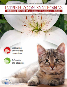
Scientific Journal of the Hellenic Companion Animal Veterinary Society (HCAVS)


Hellenic Journal of Companion Animal Medicine - Volume 2 - Issue 2 - 2013

Table of Contents

  • Bullet22 1

    Patellar luxation in dogs

  • Bullet22 2

    Drug toxicities

  • Bullet22 3

    Handling Histological Samples

  • Bullet22 4

    Whats your diagnosis
    Nictitating membrane prolapse and eye tumefaction in a dog.

  • Bullet22 4

    Whats your diagnosis
    Corneal lesion in a dog

  • Bullet22 9

    Instructions for authors


Editorial

I was honored to be invited by the board of the Journal to write this editorial for the 4th issue. This invitation will probably surprise the regular readers of the Journal, my colleagues who are general practitioner in companion animal medicine. Perhaps as a veterinarian who has worked exclusively in Bovine Medicine and Surgery for nearly 40 years, I may not be qualified to make use of this space, but I would like to thank the editorial board that made me the honour to express some views as the Head of the Department of Veterinary Medicine, in the School of Health Sciences at the Aristotle University of Thessaloniki.

Continue Reading

Drug Toxicities in Dogs and Cats

> Abstract
Drug toxicities are relatively common in dogs and cats and they can be classified into type Α or predictable which are caused by the pharmacological or the intrinsic toxic effects of the responsible drug and into type Β or non-predictable that are unrelated to the above. The appearance of type A drug toxicities depends on multiple factors that are related to the affected animal, the dosage regimen and the simultaneous administration of other drugs. Clinical manifestations most commonly originate from organ systems where the responsible drug accumulates or those that are characterized by an increased metabolic rate. In contrast, type B drug toxicities commonly affect organs presenting suitable proteins that after coupling with the drug or its metabolites (haptens) form complete antigens or organs that trap circulating immune complexes. Drugs most commonly responsible for toxicities in dogs and cats include aminoglycosides, macrocyclic lactones (avermectins and milbemycins), pyrethroids, non-steroidal anti-inflammatories, phenobarbital and diazepam.

Continue Reading

What’s your diagnosis?

Nictitating membrane prolapse and eye tumefaction in a dog.

A five years old, male Boxer presented with severe tumefaction of the left eye and depression (Figure.1 and 2). A nictitating membrane prolapse was recorded, lasting four days. The last two days the dog was depressed and inappetent.

Continue Reading

Instructions for authors

The Hellenic Journal of Companion Animal Medicine (H.J.C.A.M.) is a peer-reviewed, bilingual (Greek and English), publication of the Hellenic Companion Animal Veterinary Society (H.C.A.V.S.), which aims at the continuing education of the companion animal practitioners.

Manuscripts should be submitted for review, with the consent that they have not been submitted simultaneously or published in part or in full, to other journals.

Continue Reading

Retrospective study of 95 dogs with patellar luxation

> Abstract
The purpose of this study was the study of dogs with patellar luxation (PL) that were presented at the Companion Animal Clinic, School of Veterinary Medicine, Aristotle University of Thessaloniki, Greece during 2004 - 2010. Ninety-five dogs were included in the study. Among the information derived from the clinical examination records were breed, age, body weight, clinical signs upon presentation, site and grade of luxation, type of treatment and outcome of the dogs. Statistical analysis revealed that PL is observed more frequently in mixed breed (27.4%), small sized (61%) and female (52.6%) dogs. Regarding purebred dogs, PL has been more frequently diagnosed in Yorkshire Terriers, Poodles and Chihuahuas.

Continue Reading

Time for diagnostics...

Remember how...

Τissue handling by the practitioner; from collection to submission of the sample to the histopathology lab

> Introduction
Proper handling of tissue samples from their collection to their submission to the histopathology lab is crucial, so that autolysis (self-digestion) and technical errors, that would render the diagnosis difficult or even impossible, can be avoided. Furthermore, the information provided by the practitioner sometimes
contributes decisively to the interpretation of the histopathological findings. In any case, the best possible result is achieved when there is collaboration between the practitioner and the histopathologist, implying a common frame of reference in communication. Below is detailed information about the appropriate fixation/ preservation and submission/shipping of tissue samples that are obtained for histopathological examination.

Continue Reading

What’s your diagnosis?

Corneal lesion in a dog

A two-years old, male Rottweiler presented with blepharospasm and ocular discharge lasting one week. Nictitans gland had been removed at the age of five months.

Continue Reading

Quick Links

Social Media

Contact us

Hellenic Journal of
Companion Animal Medicine

Apollo Tower
64 Louizis Riankour Street,
115 23 Athens
Tel.: +30 210 7759727
Fax.: +30 210 7753460
iatrikizs@hcavs.gr

Sponsor

 
diagnovet